Ce ntral le ad adm inistratio n inhibits
wate r intake and so dium appe tite
in rats
1Departamento de Fisiologia, Instituto de Ciências da Saúde, and 2Departamento de Zoologia, Instituto de Biologia,
Universidade Federal da Bahia, Salvador, BA, Brasil E.J. De-Castro-e-Silva1,
L. Castro1, C.P. Luz1,
H. Ferreira1, A.K.S. Lima1,
F.S.F. Souza1, I. Maldonado1,
D.F. Macêdo1, M.G. Ferreira1,
I.P.V. Bandeira2, A.L.M. Amor2,
F.L.Q . Carvalho1, M.A. Rocha Jr.1
and J.B. Fregoneze2
Abstract
We have demonstrated that acute third ventricle injections of lead acetate (PbAc) exert a powerful antidipsogenic effect and induce a significant increase in renal sodium excretion. In the present study we confirm the antidipsogenic effect of lead and demonstrate that central administration of this metal, in minute amounts, significantly reduces salt intake both during dehydration and after central angiotensinergic stimulation. Adult male Wistar rats had the third ventricle cannulated seven days before the experiments. During this period they had free access to distilled water and hypertonic saline solution (1.5%). After a 24-h period of fluid deprivation, experimental animals received third ventricle injections of PbAc (0.3, N = 8 and 3.0 nmol/rat, N = 14) while controls received sodium acetate (NaAc; 3.0 nmol/rat, N = 10). Rats treated with PbAc at the highest dose showed a significant reduction (P<0.05) both in water and hypertonic saline intake when compared to controls. When the effect of lead administration on angiotensin II-induced water and salt intake was studied, normohy-drated animals received third ventricle injections of angiotensin II (9.6 nmol/rat) after pretreatment with 3.0 nmol/rat of PbAc (experimental group, N = 10) or NaAc (controls, N = 8). The group pretreated with PbAc presented a significant reduction (P<0.05) in both water and salt intake compared to controls. Thus, this study confirms the antidipso-genic effect of central lead injections and demonstrates that the presence of lead in the brain exerts a significant inhibition of sodium appetite.
Co rre spo nde nce
E.J. De-Castro-e-Silva Departamento de Fisiologia Instituto de Ciências da Saúde Universidade Federal da Bahia 40110-100 Salvador, BA Brasil
Fax: + 55-71-337-0195 E-mail: emilio@ svn.com.br
Research supported by CNPq (Nos. 300772/86-2 and 301099/92-8) and by the Bahia State Research Support Financial Agency (CADCT).
Received March 24, 1999 Accepted August 11, 1999
Ke y wo rds
·Lead
·Angiotensin II
·Water intake
·Sodium intake
·Rats
Lead, a non-physiological metal widely distributed in the environment, is still con-sidered an important developmental neuro-toxicant that induces cognitive deficits in humans even at low blood levels such as 10 µg/dl (1,2). Under experimental conditions, lead alters complex behaviors such as
effect of lead on animal behavior, as com-mented elsewhere (5).
We have demonstrated that acute third ventricle injections of lead in minute amounts induce a significant antidipsogenic effect on dehydrated rats, as well as in those whose drinking was induced by central angiotensi-nergic, cholinergic and ß-adrenergic stimu-lation (6,7). This thirst-inhibiting effect of central lead administration may be, at least in part, opioid-dependent, as we have also shown (8). Recently, we have demonstrated that central injection of lead, in the same amounts used to inhibit water intake, causes a significant increase in renal sodium and potassium excretion (9). Thus, central lead administration disturbs brain mechanisms related to several aspects of fluid and elec-trolyte control.
In the present study we investigate in adult rats the effects of third ventricle injec-tions of lead on sodium appetite, a further variable related to the homeostatic control of body fluids and electrolytes that is strictly and continuously controlled by the brain.
We used adult male Wistar rats kept un-der controlled light (lights on from 6:00 to 20:00 h) and temperature (26 ± 2o
C) condi-tions. The third ventricle of the animals was cannulated as described elsewhere (10) seven days before the experimental sessions. Briefly, the animals were anesthetized with sodium pentobarbital (40 mg/kg, intraperito-neally). After exposure of the skull, two screws were embedded into the bone to al-low the anchorage of the cannula with dental acrylic. The following coordinates were used: anteroposterior = 0.5 mm behind the bregma, lateral = just on the midline, and vertical = 8.5 mm below the skull. A mandril (28 gauge) was inserted into the cannula to avoid ob-struction. After the experiments, the animals were sacrificed and received third ventricle injections of Evans blue (2 µl, 5%); the brains were then removed and the position of the cannula was verified. Only animals whose cannulas were inserted exactly into
the third ventricle were considered.
The following drugs were used: angio-tensin II, lead acetate (PbAc) and sodium acetate (NaAc) obtained from Sigma Chem-ical Co., St. Louis, MO, USA. All drugs were dissolved in isotonic saline solution and injected into the third ventricle with a 10-µl Hamilton syringe connected to a Mizzy-Slide-Pak needle through polyethylene tub-ing. The volume injected into the third ven-tricle was 2 µl and the injections were made over a period of 60 s. Naive animals were used in all experiments (always performed between 8:00 and 11:00 a.m.).
During the days between stereotaxic can-nulation of the third ventricle and the exper-imental session all animals had free access to two bottles containing distilled water or hy-pertonic saline solution (1.5%). To study the effect of central injection of PbAc on sodium appetite in dehydrated animals, both bottles were removed from the cages 24 h before the experiments and were reintroduced into the cages immediately after PbAc injection, when the intake of both fluids began to be re-corded. To investigate the effect of PbAc on sodium appetite in normohydrated animals receiving angiotensin II, this peptide was injected into the third ventricle 30 min after PbAc injections. The intake of both fluids began to be recorded immediately after an-giotensin II injections. In both cases fluid intake was monitored for 120 min.
NaAc injection was used as control for groups receiving PbAc. We have shown that NaAc injection into the third ventricle does not modify water or hypertonic saline solu-tion intake when compared to animals receiving central injections of isotonic sa-line (6).
cumulative intake of water or hypertonic saline solution is presented as ml/100 g body weight (mean ± SEM).
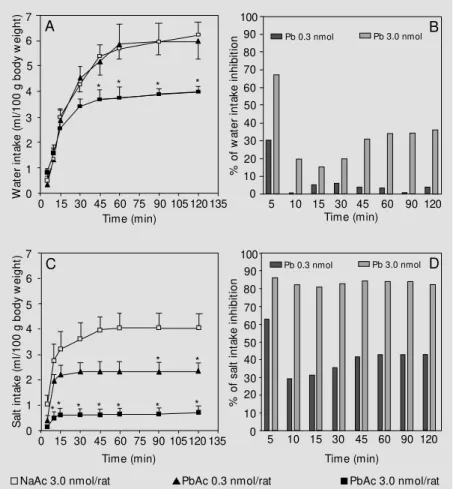
Figure 1 shows the effect of PbAc injec-tions into the third ventricle on water and hypertonic saline intake by dehydrated ani-mals. Panel A shows that, as expected, dehy-drated control animals receiving NaAc ex-hibited a high water intake. At the lowest dose employed (0.3 nmol/rat) PbAc was in-effective. At the highest dose employed (3.0 nmol/rat) PbAc administration induced a sig-nificant reduction in water intake after 45 min that lasted for the entire duration of the experiment. The percent inhibition from 45 min until the end of the experiment contin-ued to be about 35%, as seen in panel B. Panel C shows that even at the lowest dose employed (0.3 nmol/rat) PbAc generated a partial but significant blunting of salt intake after 90 to 120 min. At the highest dose employed (3.0 nmol/rat) a significant block-ade in hypertonic saline intake was evident after 10 min, persisting until the end of the experiment. The magnitude of this blockade was very high, corresponding to about 85% inhibition, as seen in panel D. Comparing panels B and D in Figure 1, it is easy to observe that PbAc more potently inhibited hypertonic saline intake than water intake.
Figure 2 shows the effect of PbAc on water and hypertonic saline intake by nor-mohydrated animals receiving angiotensin II, pretreated with PbAc (3.0 nmol/rat) or NaAc (3.0 nmol/rat). As seen in panel A, control animals receiving angiotensin II (9.6 nmol/rat) but pretreated with NaAc exhib-ited a high water intake, as expected. Pre-treatment with PbAc induced a significant blockade of water intake during the entire duration of the experiment. Normohydrated animals receiving NaAc presented a very low water intake as usual. The high percent-age of water intake inhibition is depicted in panel B. Control animals receiving angio-tensin II and pretreated with NaAc showed a high hypertonic saline intake, as seen in
panel C. Pretreatment with PbAc signifi-cantly blocked this natriorexigenic response. As expected, normohydrated animals receiv-ing NaAc alone displayed a very low intake of hypertonic saline. The percentage of inhi-bition of hypertonic saline intake is illus-trated in panel D.
The present study extends observations from our laboratory on the acute effects of heavy metals on central regulation of fluid and electrolytes in rats. We have previously shown that lead injections into the third ven-tricle induce a significant antidipsogenic
ef-Figure 1 - Cumulative w ater and salt intake (ml/100 g body w eight) by rats after a 24-h period of w ater deprivation. Absolute values of w ater intake are presented in panel A. The percentage of lead-induced w ater intake inhibition (as compared to NaAc-treated rats) is show n in panel B. Absolute values of hypertonic saline intake are presented in panel C. The percentage of lead-induced hypertonic saline intake inhibition (as compared to NaAc-treated rats) is show n in panel D. Absolute values are presented as means ± SEM . * P<0.05 compared to the control group receiving NaAc (ANOVA follow ed by Student-New man-Keuls test). Open squares (N = 10), NaAc 3.0 nmol/rat; triangles (N = 8), PbAc 0.3 nmol/rat, and closed squares (N = 14), PbAc 3.0 nmol/rat.
S a lt i n ta k e ( m l/ 1 0 0 g b o d y w e ig h t) 7 6 5 4 3 2 1 0
0 15 30 45 60 75 90 105 120 135
% o f s a lt i n ta k e i n h ib it io n 100 90 80 70 60 50 40 0 30 20 10
5 10 15 30 45 60 90 120
Time (min) W a te r in ta k e ( m l/ 1 0 0 g b o d y w e ig h t) 7 6 5 4 3 2 1 0
0 15 30 45 60 75 90 105 120 135 Time (min) % o f w a te r in ta k e i n h ib it io n 100 90 80 70 60 50 40 0 30 20 10
5 10 15 30 45 60
Time (min)
90 120
A B
C D
Pb 0.3 nmol Pb 3.0 nmol
Pb 0.3 nmol Pb 3.0 nmol
Time (min) * * * * * * * * * * * * *
fect in several physiological and pharmaco-logical situations (6,7), an effect due, at least partially, to a stimulation of central opioid pathways (8). More recently, we have dem-onstrated that acute intracerebroventricular injections of lead induce a powerful natri-uretic and kalinatri-uretic effect (9). Here, we confirm that central lead injections diminish water intake after dehydration and central angiotensinergic stimulation. More impor-tantly, we show that third ventricle injec-tions of lead significantly reduce sodium appetite.
No biological function has been demon-strated for lead. Contamination by lead may cause an endless sequence of pathological manifestations extensively reviewed else-where (4). The central nervous system may be especially sensitive to lead poisoning. Both the developing and the adult brain are especially prone to functional disturbances even when blood concentrations of the metal are very low. It is well established that lead alters the functional integrity of the blood-brain barrier, facilitating its entry into the brain (11). Once in the brain, lead presents a homogeneous distribution, being localized both in neurons and glia. By interfering with many biochemical processes, lead may alter a myriad of cellular events that potentially affect brain function. Neurotransmitter ki-netics derangement, voltage-dependent cal-cium channel blockade and alterations in calcium function by spurious binding with calcium-binding proteins are the most com-mon mechanisms by which lead distorts the cellular machinery (12).
The brain controls many aspects related to the homeostatic processes necessary to keep body fluid and electrolyte variables within the proper narrow limits. Fluctua-tions in these variables make the brain trig-ger regulatory mechanisms that include thirst, renal water and electrolyte excretion and sodium appetite. The central regions local-ized in the vicinity of the third ventricle are especially involved in these homeostatic pro-cedures, the most important being the hypo-thalamus, the subfornical organ, the organum vasculosum lamina terminalis and the anter-oventral third ventricle region (AV3V) (13). In this study lead was injected into the third ventricle, a route that provides fast access to these structures.
In the present study acute third ventricle injections of lead inhibited water intake after both dehydration and angiotensinergic stim-ulation. This effect had already been demon-strated in our laboratory, as mentioned above. We show here that sodium appetite, an
im-Figure 2 - Cumulative w ater and salt intake (ml/100 g body w eight) by rats after third ventricle injections of angiotensin II (AII; 9.6 nmol/rat). Absolute values of w ater intake are presented in panel A. The percentage of lead-induced w ater intake inhibition (as compared to NaAc-treated rats) is show n in panel B. Absolute values of hypertonic saline intake are presented in panel C. The percentage of lead-induced hypertonic saline intake inhibition (as compared to NaAc-treated rats) is show n in panel D. Absolute values are presented as means ± SEM . * P<0.05 for the group receiving NaAc + AII compared to the group receiving PbAc + AII (ANOVA follow ed by Student-New man-Keuls test). #P<0.05 for the group
receiving NaAc alone compared to both other groups. Squares (N = 9), NaAc 3.0 nmol/rat; lozenges (N = 8), NaAc + AII, and circles (N = 10), PbAc 3.0 nmol + AII/rat.
S a lt i n ta k e ( m l/ 1 0 0 g b o d y w e ig h t)7 6 5 4 3 2 1 0
0 15 30 45 60 75 90 105 120 135 Time (min)
Pb + AII
% o f s a lt i n ta k e i n h ib it io n 100 90 80 70 60 50 40 0 30 20 10
5 10 15 30 45 60 90 120 Time (min) W a te r in ta k e ( m l/ 1 0 0 g b o d y w e ig h t) 7 6 5 4 3 2 1 0
0 15 30 45 60 75 90 105120 135 Time (min) % o f w a te r in ta k e i n h ib it io n 100 90 80 70 60 50 40 0 30 20 10
5 10 15 30 45 60 Time (min)
90 120 Pb + AII
A B C D # * * * * * *
NaAc 0.3 + AII 9.6 nmol/rat PbAc 3.0 nmol + AII/rat NaAc 3.0 nmol/rat
portant contributory mechanism in the pro-cess of body fluid and electrolyte control, is also affected by central lead administration. Forebrain circumventricular structures play a facilitatory role in thirst and sodium appe-tite, in contrast to those located in the hind-brain that exert an inhibitory modulation of these behaviors. As we injected lead into the third ventricle, it is reasonable to suggest that, in this case, central lead disrupts both the thirst and salt intake triggering capacities of these forebrain regions. It is also clear that the structures involved in thirst generation and sodium intake induction are differen-tially affected by lead. Indeed, lead injec-tions block salt intake much more easily than water intake.
Our experimental protocol was specifi-cally designed to evaluate salt-intake behav-ior. We used hypertonic saline solution (1.5%) that is normally aversive to rats. Thus, ingestion of this solution means a salt-re-plenishing behavior. In other words, when rats drink hypertonic saline solution they are obeying a central salt-seeking command and this behavior does not represent a natriophilic hedonic behavior.
We have previously shown that central lead increases renal sodium excretion. An increase in sodium excretion coupled with a reduction in salt intake is a situation that favors efficient correction of hypernatremia and/or hyperosmolarity. Thus, it is reason-able to suggest that central lead injections may activate areas that normally trigger
com-pensatory mechanisms in response to these two conditions.
Central angiotensin II exerts powerful dipsogenic and natriorexigenic effects and central angiotensinergic activity increases during dehydration (14). Central lead injec-tions inhibited salt intake after both dehydra-tion and central angiotensinergic stimula-tion. Thus, it seems rational to suggest that the inhibitory effects of lead on salt intake rely on structures or components localized downstream of angiotensinergic compo-nents.
It is necessary to stress that the effects of lead demonstrated here were obtained with concentrations far below those normally pres-ent in the cpres-entral nervous system of intoxi-cated humans and laboratory animals, which range from 4.9 to 7 µg/g of tissue (15).
Taken together, the present data and those previously reported by our laboratory clearly show that lead in the brain may disturb the homeostatic control of body fluids in several ways. Indeed, it inhibits thirst, increases re-nal sodium excretion and reduces sodium appetite. Nephropathy may be one of the main consequences of lead poisoning and its clinical features could be further deterio-rated by these central actions of the metal.
Ackno wle dgm e nts
We are thankful to Mr. Vanilson Souza and Mr. José de Souza for their skilful tech-nical assistance.
Re fe re nce s
1. Boivin M J & Giordani B (1995). A risk evaluation of the neuropsychological ef-fects of childhood lead neurotoxicity. De-velopmental Neuropsychology, 11: 157-180.
2. Winneke G, Lillienthal H & Kramer U (1996). The neurobehavioral toxicology and teratology of lead. Archives of Toxi-cology, 18 (Suppl): 57-70.
3. Ferguson SA, Holson RR, Gazzara RA & Siitonen PH (1998). M inimal behavioral
effects from moderate postnatal lead treatment in rats. Neurotoxicology and Teratology, 20: 637-643.
4. Al-Saleh IAS (1994). The biochemical and clinical consequences of lead poisoning. M edicinal Research Review s, 14: 415-486.
5. Bellinger DC (1995). Interpreting the lit-erature on lead and child development: The neglected role of the “ experimental system” . Neurotoxicology and
Teratolo-gy, 17: 249-251.
6. Fregoneze JB, Cunha M , Bulcão C, Ferreira H & De Castro e Silva E (1994). Acute effect of intracerebroventricular ad-ministration of lead on the drinking behav-ior of rats induced by dehydration or cen-tral cholinergic and angiotensinergic stim-ulation. Physiology and Behavior, 56: 129-133.
V, Oliveira P, Nascimento T, Luz CP, Santana Jr P, De-Oliveira IR & De-Castro-e-Silva E (1997). Lead (Pb2+) and cadmium
(Cd2+) inhibit the dipsogenic action of
cen-tral beta-adrenergic stimulation by isopro-terenol. Brazilian Journal of M edical and Biological Research, 30: 419-423. 8. De-Castro-e-Silva E, Luz CP, Sarmento C,
Nascimento T, Gonzalez V, M arinho CA, Castro L, Oliveira P, Santana Jr P, De-Oliveira IR, De-Paula S, Lim a AKS & Fregoneze JB (1998). Opiatergic partici-pation in the thirst-inhibiting effect of acute third ventricle injections of cad-mium (Cd2+) and lead (Pb2+). Brazilian
Journal of M edical and Biological Re-search, 31: 805-810.
9. Fregoneze JB, Luz CP, Sarm ent o C, Gonzalez V, Oliveira P, Santana Jr P, M arinho CA, Castro L, Nascimento T, De Paula S, Lima AKS, Oliveira IR & De-Castro e Silva E (1998). Central lead ad-ministration induces natriuretic and kaliu-retic effects in rats. Physiology and Be-havior,65: 321-326.
10. Ant unes-Rodrigues J & M cCann SM (1970). Water, sodium and food intake induced by injections of cholinergic and adrenergic drugs into the third ventricle of rat brain. Proceedings of the Society for Experimental Biology and M edicine, 133: 1464-1470.
11. Goldstein GW (1994). Brain capillaries: A target for inorganic lead poisoning.
Neu-rotoxicology, 5: 167-176.
12. Goldstein GW (1990). Lead poisoning and brain cell function. Environmental Health Perspectives, 89: 91-94.
13. Johnson AK & Thunhorst RL (1997). The neuroendocrinology of thirst and salt ap-petite: Visceral sensory signals and mech-anisms of central integration. Frontiers in Neuroendocrinology, 18: 292-353. 14. Fitzsimons JT (1998). Angiotensin, thirst,
and sodium appetite. Physiological Re-view s, 78: 583-686.