INHIBITION OF CENTRAL ANGIOTENSIN II-INDUCED PRESSOR
RESPONSES BY HYDROGEN PEROXIDE
M. R. LAUAR, D. S. A. COLOMBARI, P. M. DE PAULA, E. COLOMBARI, L. M. CARDOSO, L. A. DE LUCA JR AND J. V. MENANI*
Department of Physiology and Pathology, Dentistry School, São Paulo State University (UNESP), Araraquara, São Paulo, Brazil
Abstract—Hydrogen peroxide (H2O2), important reactive ox-ygen species produced endogenously, may have different physiological actions. The superoxide anion (O2·ⴚ) is sug-gested to be part of the signaling mechanisms activated by angiotensin II (ANG II) and central virus-mediated overex-pression of the enzyme superoxide dismutase (that dismu-tates O2·ⴚ to H2O2) reduces pressor and dipsogenic re-sponses to central ANG II. Whether this result might reflect elevation of H2O2rather than depletion of O2
·ⴚhas not been addressed. Here we investigated the effects of H2O2injected intracerebroventricularly (i.c.v.) or ATZ (3-amino-1,2,4-tria-zole, a catalase inhibitor) injected intravenously (i.v.) or i.c.v. on the pressor responses induced by i.c.v. injections of ANG II. Normotensive male Holtzman rats (280 –320 g, nⴝ5–13/ group) with stainless steel cannulas implanted in the lateral ventricle were used. Prior injection of H2O2(5mol/1l) or ATZ (5 nmol/1 l) i.c.v. almost abolished the pressor re-sponses induced by ANG II (50 ng/1l) also injected i.c.v. (7ⴞ3 and 5ⴞ3 mm Hg, respectively, vs. control: 19ⴞ4 mm Hg). Injection of ATZ (3.6 mmol/kg b.wt.) i.v. also reduced central ANG II-induced pressor responses. Injections of H2O2 i.c.v. and ATZ i.c.v. or i.v. alone produced no effect on base-line arterial pressure. Central ANG II, H2O2or ATZ did not affect heart rate. The results show that central injections of H2O2and central or peripheral injections of ATZ reduced the pressor responses induced by i.c.v. ANG II, suggesting that exogenous or endogenous H2O2may inhibit central pressor mechanisms activated by ANG II. © 2010 IBRO. Published by Elsevier Ltd. All rights reserved.
Key words: hypertension, reactive oxygen species, superox-ide dismutase, arterial pressure, catalase inhibitor.
Superoxide anion (O2·⫺), hydroxyl radical (HO·) and hy-drogen peroxide (H2O2) known as reactive oxygen species (ROS) can be produced endogenously and act as cellular signaling molecules to regulate biological function (Adler et al., 1999; Chen et al., 2001; Zimmerman et al., 2002, 2004a; Rhee et al., 2003; Zimmerman and Davisson, 2004; Avshalumov et al., 2005; Bao et al., 2009). Super-oxide dismutase (SOD), an important enzyme in the me-tabolism of ROS catalyzing the dismutation of O2
·⫺to form
H2O2, is widely distributed in the CNS, where ROS are suggested to act as neuromodulators affecting neurotrans-mission and neuronal firing (Aizenman et al., 1989; Volt-erra et al., 1994; Zoccarato et al., 1995; Chen et al., 2001; Zimmerman et al., 2002; Zimmerman and Davisson, 2004; Avshalumov et al., 2005;Campese et al., 2007).
Angiotensin II (ANG II), the main peptide released by the activation of the renin-angiotensin system, acts cen-trally to produce pressor responses dependent on sympa-thetic activation and vasopressin secretion, as well as producing natriorexigenic and dipsogenic responses ( Hoff-man et al., 1977; Johnson et al., 1978; Johnson, 1985; Mahon et al., 1995; Fitzsimons, 1998). Previous studies have suggested that a decrease in ANG II-induced O2·⫺ formation by central adenovirus-mediated overexpression of SOD abolishes pressor and dipsogenic responses to central injections of ANG II, suggesting that O2·⫺is part of the signaling mechanisms activated by ANG II centrally (Zimmerman et al., 2002, 2004a; Zimmerman and Davis-son, 2004). The evidence that ANG II induces O2
·⫺
forma-tion is reinforced by studies showing that central ANG II increases dihydroethidium fluorescence, a standard probe selective for O2
·⫺and that ANG II induces calcium influx
dependent on O2
·⫺(Zimmerman et al., 2004b, 2005). In
addition, ANG II-induced ROS production is suggested to involve NADPH oxidase (Zimmerman et al., 2004a; Peter-son et al., 2009).
Hydrogen peroxide is a relatively stable and diffusible ROS that may act centrally through different mechanisms modulating neuronal synaptic transmission. Excitatory or inhibitory responses to H2O2 acting centrally have been reported (Sorg et al., 1997; Volterra et al., 1994; Zoccarato et al., 1995, 1999; Sah et al., 2002; Wehage et al., 2002; Bao et al., 2005; Avshalumov et al., 2005; Takahashi et al., 2007). Centrally, H2O2can block glutamate uptake by glial cells, which may result in an increase of extracellular glu-tamate levels enhancing neuronal excitability or even causing toxicity (Sorg et al., 1997; Volterra et al., 1994). On the other hand, it has also shown that H2O2inhibits gluta-mate and increases GABA release or acts at ion channels, especially ATP-sensitive potassium channels (KATP chan-nels) causing neuronal hyperpolarization and reducing neuronal excitability (Zoccarato et al., 1995, 1999; Sah et al., 2002; Takahashi et al., 2007; Bao et al., 2005; Avshalu-mov et al., 2005).
Studies have shown the importance of O2
·⫺as part of
the signaling mechanisms activated by ANG II, however, possible effects of other ROS, like H2O2, on ANG II-in-duced responses were not investigated yet. In spite of the controversies about a correlation between changes in
*Corresponding author. Tel:⫹55-16-33016486; fax:⫹55-16-33016488. E-mail address:menani@foar.unesp.br(J. V. Menani).
Abbreviations: ANG II, angiotensin II; ATZ, 3-amino-1,2,4-triazole; AV3V, anteroventral third ventricle; HO·, hydroxyl radical; H
2O2,
hy-drogen peroxide; HR, heart rate; i.c.v., intracerebroventricular; i.v., intravenous; MAP, mean arterial pressure; O2·⫺, superoxide anion;
ROS, reactive oxygen species; SOD, superoxide dismutase.
0306-4522/10 $ - see front matter © 2010 IBRO. Published by Elsevier Ltd. All rights reserved. doi:10.1016/j.neuroscience.2010.08.048
SOD activity and H2O2levels (Teixeira et al., 1998; Gard-ner et al., 2002; Chan et al., 2006; Kowald et al., 2006), central SOD overexpression might also reduce ANG II-induced responses due to increases in H2O2levels. Endo-genously, H2O2production may result from NADPH oxi-dase activity or mitochondrial respiration coupled to SOD pathway or monoamine oxidase activity (Maker et al., 1981; Zimmerman et al., 2004a; Peterson et al., 2009; Bao et al., 2005, 2009) and independently from the source, H2O2 might affect ANG II-induced responses through mechanisms that reduce neuronal excitability. Therefore, in the present study we investigated if exogenous H2O2 injected i.c.v. or the increase of endogenous H2O2 pro-duced by i.c.v. injections of the catalase inhibitor ATZ (3-amino-1,2,4-triazole) could modify the pressor re-sponses induced by i.c.v. ANG II. In addition, we also tested the effects of i.v. H2O2 or ATZ on the pressor response to ANG II i.c.v. or i.v.
EXPERIMENTAL PROCEDURES
Animals
Normotensive male Holtzman rats (baseline MAP: 109⫾1 mm Hg and baseline HR: 368⫾5 bpm) weighing 280 to 320 g were used. The animals were housed individually in stainless steel cages in a room with controlled temperature (23⫾2 °C) and humidity (55⫾10%). Lights were on from 7:00AMto 7:00PM. Guabi rat chow (Paulínia, SP, Brazil) and tap water were availablead libi-tum. The experimental protocols used in the present study were approved by the Ethical Committee for Animal Care and Use from Dentistry School of Araraquara, UNESP, Brazil.
Surgery for the implant of i.c.v. cannulas
Rats were anesthetized with ketamine (80 mg/kg of body weight, Cristalia, Itapira, SP, Brazil) combined with xylazine (7 mg/kg of body weight, Agener Uniao, Embu-Guacu, SP, Brazil) and placed in a stereotaxic frame (model 900, David Kopf Instruments, Tu-junga, CA, USA). Bregma and lambda were positioned at the same horizontal level. A stainless steel cannula (10⫻0.6 mm o.d.) was implanted into the lateral ventricle (LV) using the coordinates 0.3 mm caudal to bregma, 1.6 mm lateral to midline and 3.5 mm below of the skull bone. The cannulas were fixed to the cranium using dental acrylic resin and jeweler screws.
Rats were maintained in individual cages with free access to water and food pellets. Rats received a prophylactic dose of penicillin (30,000 IU) given i.m. and a s.c. injection of the analgesic Ketoflex (ketoprofen 1%, 0.03 ml/rat, Mundo Animal, Sao Paulo, SP, Brazil) post-surgically.
Arterial pressure and heart rate recordings
Mean arterial pressure (MAP) and heart rate (HR) were recorded in unanesthetized rats. Five days after brain surgery, rats were anesthetized again with ketamine (80 mg/kg of body weight) com-bined with xylazine (7 mg/kg of body weight) and a polyethylene tubing (PE-10 connected to a PE-50, Clay Adams, Parsippany, NJ, USA) was inserted into the abdominal aorta through the femoral artery. At the same time, in some rats, a polyethylene tubing was inserted into the femoral vein for drug administration. Venous and/or arterial catheters were tunneled s.c. and exposed on the back of the rat to allow access in unrestrained, freely moving rats. To record pulsatile arterial pressure, MAP and HR, the arterial catheter was connected to a Stathan Gould (P23 Db) pressure transducer (Sthatan Gould, Cleveland, OH, USA)
cou-pled to a pre-amplifier (model ETH-200 Bridge Bio Amplifier, CBSciences Inc., Dover, NH, USA) that was connected to a Powerlab computer data acquisition system (model Powerlab 16SP, ADInstruments, Castle Hill, NSW, Australia).
Central injections
The i.c.v. injections were made using 10 l Hamilton syringes
connected by polyethylene tubing (PE 10) to the injector needles that were 2.0 mm longer than the guide cannula implanted in the brain. The volume of i.c.v. injections was 1l.
Drugs
Hydrogen peroxide (H2O2, 5mol/1l), angiotensin II (ANG II, 50
ng/1l) and 3-amino-1,2,4-triazole (ATZ, 5 nmol/1l), purchased
from Sigma Chemical Co. (St. Louis, MO, USA), were injected i.c.v. The same doses of H2O2and ANG II in a volume of 0.1 ml of vehicle were injected i.v. ATZ at the dose of 3.6 mmol/kg of body weight was also injected i.v. Angiotensin II and ATZ were dissolved in saline and H2O2was diluted in phosphate buffered saline (PBS, pH 7.2). PBS or saline were injected i.c.v. or i.v. in control experiments. The doses of ANG II, H2O2and ATZ used in the present study were based on previous studies that tested the cardiovascular effects of these drugs injected central or peripher-ally (Menani et al., 1990; Aragon et al., 1991; Cardoso et al., 2006, 2009).
Histology
At the end of the experiments, 2% Evans blue solution (1l) was
injected i.c.v. Immediately after dye injection, the animals were deeply anesthetized with sodium thiopental (70 mg/kg of body weight, i.p., Cristalia, Itapira, SP, Brazil). Saline followed by 10% buffered formalin was perfused through the heart. The brains were removed, fixed in 10% buffered formalin, frozen, cut coronally (50
m sections), stained with Giemsa stain (that stains cell nuclei)
and analyzed by light microscopy to confirm the injections into the LV.
Statistical analysis
The results are reported as means⫾standard error of means (SEM). One-way analysis of variance (ANOVA) and Newman– Keuls tests were used for comparisons. Differences were consid-ered significant atP⬍0.05.
Experimental protocols
Cardiovascular responses produced by ANG II i.c.v. com-bined with H2O2i.c.v. MAP and HR were recorded one day after
the surgery for the implant the arterial catheter. Around 20 min after starting the recordings of MAP and HR, PBS (1l) or H2O2
(5mol/1l) was injected i.c.v. followed by an i.c.v. injection of
ANG II (50 ng/1l) 1 min later. MAP and HR recordings stopped
30 min after ANG II injection and started again 4 h later, when the same i.c.v. treatments were repeated in the same rats in a coun-terbalanced design.
Cardiovascular responses produced by ANG II i.c.v. com-bined with ATZ i.c.v. A protocol similar to that described above to test the effects of the combination of H2O2and ANG II i.c.v. was also used in a different group of rats to test the cardiovascular responses to the combination of ATZ (5 nmol/1l) and ANG II (50
ng/1l) i.c.v., except that ATZ instead of H2O2was injected i.c.v.
10 min before ANG II.
was also used in a different group of rats to test the cardiovascular responses to the combination of ATZ (3.6 mmol/kg of body weight) i.v. and ANG II (50 ng/0.1 ml) i.v.
In the same rats, 5 min after ANG II i.v., it was also injected ANG II (50 ng/1l) i.c.v.
Cardiovascular responses produced by i.v. ANG II combined with i.v. H2O2. A potentially confounding factor is that H2O2, a
mild oxidant, might damage the structure of the ANG II molecule, and thereby lead to a decrease in central ANG II-induced pressor responses. To exclude this possibility, a mix of ANG (50 ng/1l)
plus H2O2(5mol/1l) was prepared and maintained at room
temperature for at least 5 min. Then, the mix was diluted to have 50 ng of ANG II and 5mol of H2O2in a final volume of 0.1 ml,
which was injected i.v. The cardiovascular responses produced by the mix of ANG II⫹H2O2 injected i.v. were compared with the
responses produced by the same dose of ANG II injected i.v. alone 20 min before.
RESULTS
Histological analysis
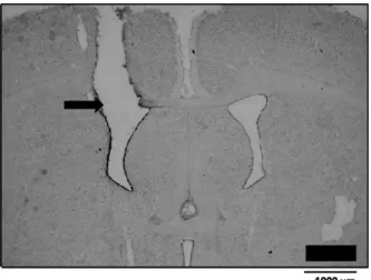
Fig 1shows the typical site of injections into the LV. As suggested by the histological analysis, i.c.v. injections of
H2O2produced no damage of the neural tissue that might affect ANG II responses. Besides the histological analysis, another reason to exclude any influence of neural damage on the effects of H2O2is the complete recovery of ANG II-induced pressor responses in rats that received a sec-ond injection of ANG II combined with saline i.c.v. (control) around 4 h after the first test in which they received ANG II combined with H2O2or ATZ i.c.v.
Cardiovascular responses produced by ANG II i.c.v. combined with H2O2or ATZ i.c.v.
Fig. 2shows representative recordings of pulsatile arterial pressure (PAP), MAP and HR in rats treated with vehicle, H2O2(5mol/1l) or ATZ (5 nmol/1 l) i.c.v. combined with ANG II (50 ng/1l) i.c.v.
The pressor response to ANG II (50 ng/1l) i.c.v. was reduced by the pre-treatment with H2O2(5mol/1l) i.c.v. (7⫾3 mm Hg vs. PBS: 19⫾4 mm Hg) [F(3,21)⫽14.3;
P⬍0.05] (Fig 3). Injections of H2O2 (5 mol/1 l) i.c.v. alone produced no change on MAP. Injection of H2O2and ANG II i.c.v. alone or combined produced no change on HR (Fig 3).
The pre-treatment with ATZ (5 nmol/1l) i.c.v. also reduced the pressor response to ANG II (50 ng/1l) i.c.v. (5⫾3 mm Hg vs. saline: 23⫾3 mm Hg) [F(3,36)⫽65.6;
P⬍0.05] (Fig 4). Injections of ATZ (5 nmol/1l) i.c.v. alone produced no change on MAP or HR. Injection of ANG II i.c.v. alone produced no change on HR, however, ANG II i.c.v. reduced HR after the pre-treatment with ATZ [F(3,36)⫽3.8;P⬍0.05] (Fig 4).
Cardiovascular responses produced by ANG II i.v. or i.c.v. combined with ATZ i.v.
Intravenous injections of ATZ (3.6 mmol/kg of body weight) reduced the pressor response to ANG II (50 ng/1l) i.c.v. (7⫾2 mm Hg, vs. saline: 21⫾1 mm Hg) [F(5,45)⫽65.5;
P⬍0.05] (Fig 5). However, ATZ (3.6 mmol/kg b.w.) i.v. did not affect the pressor response to ANG II (50 ng/0.1 ml/rat) i.v. (39⫾2 mm Hg, vs. saline: 41⫾3 mm Hg) (Fig 5). Fig. 1. Photomicrograh showing the typical site of injections into the
lateral ventricle (arrow).
Fig. 2. Representative recordings showing changes in PAP, MAP and HR induced by i.c.v. ANG II (50 ng/1l) after (A) vehicle i.c.v., (B) H2O2(5
ANG II i.v. alone or combined with ATZ i.v. reduced HR (Fig 5). ANG II i.c.v. alone or combined with ATZ i.v. produced no change on HR (Fig 5).
Injection of ATZ (3.6 mmol/kg b.w.) i.v. alone did not modify MAP or HR (Fig 5).
Cardiovascular responses produced by ANG II combined with H2O2i.v.
A potentially confounding factor is that H2O2, a mild oxi-dant, might damage the structure of the ANG II molecule, and thereby lead to a decrease in central ANG II-induced pressor responses. To exclude this possibility, the cardio-vascular effects of i.v. injection of a mix of ANG II⫹H2O2 were tested.
Similar pressor and bradycardic responses were pro-duced by i.v. injection of ANG II (50 ng/0.1 ml/rat) alone (48⫾1 mm Hg and⫺62⫾14 bpm, respectively,n⫽5 rats) or mixed with H2O2(5mol/0.1 ml/rat) i.v. (50⫾2 mm Hg and⫺62⫾10 bpm, respectively).
DISCUSSION
The present results show that i.c.v. injections of H2O2or ATZ strongly decrease the pressor responses to i.c.v. ANG
II, suggesting that increased levels of H2O2 in the brain from exogenous or endogenous origin impair central pres-sor mechanisms activated by ANG II. I.v. injections of a high dose of ATZ also almost abolished the pressor re-sponses to i.c.v. ANG II. However, i.v. ATZ or H2O2did not change the response to i.v. ANG II, suggesting that these treatments do not affect pressor mechanisms activated by ANG II acting on blood vessels. Injections of ANG II, H2O2 or ATZ alone or combined produced no consistent effects on HR.
I.v. injection of a mix of ANG II and H2O2 that was prepared at the same concentration tested centrally and maintained at room temperature for at least 5 min pro-duced pressor responses similar to those propro-duced by injections of ANG II alone. These results again suggest that H2O2 does not affect the pressor mechanisms acti-vated by ANG II acting peripherally and also that the effects of H2O2reducing ANG II responses are not related to any damage to the structure of ANG II molecule. Injec-tions of H2O2or ATZ i.c.v. at the doses tested also did not produce any neuronal destruction that might affect ANG II responses. The effect of i.c.v. H2O2or ATZ reducing the
Fig. 4. Changes in (A) MAP and (B) HR induced by i.c.v. injections of ANG II (50 ng/1l) combined with i.c.v. injections of saline or ATZ (5 nmol/1l). The results are represented as means⫾SEM,n⫽number of rats.
Fig. 3. Changes in (A) MAP and (B) HR induced by i.c.v. injections of ANG II (50 ng/1l) combined with i.c.v. injections of PBS or H2O2(5
pressor responses to ANG II is transitory and a complete recovery of ANG II-induced pressor responses occurs when rats received a second injection of ANG II combined with saline i.c.v. (control) around 4 h after the first test in which they received ANG II combined with H2O2or ATZ i.c.v. Therefore, any possible damage or destruction of ANG II receptors or neurons is also not the cause of the effects of H2O2.
The pressor response to central ANG II is strongly dependent on the activation of ANG II AT1 receptors, which are G-protein coupled and located in the neurons of the circumventricular organs that release facilitatory sig-nals to increase vasopressin secretion and sympathetic activation (Johnson et al., 1978; Johnson, 1985; Hoffman et al., 1977; Mahon et al., 1995). The circumventricular organs directly activated by i.c.v. ANG II are the subforni-cal organ (SFO), organum vasculoso of lamina terminalis
(OVLT) and median preoptic nucleus (MnPO). From these sites, facilitatory signals may reach the paraventricular nucleus of the hypothalamus (PVN) and the rostroventro-lateral medulla (RVLM) to increase sympathetic activity and PVN and supraoptic nucleus of the hypothalamus (SON) to increase vasopressin secretion (Hoffman et al., 1977; Johnson et al., 1978; Johnson, 1985; Mahon et al., 1995). Although H2O2may act in any of these nuclei to reduce the pressor response to i.c.v. ANG II, the main areas involved in the effects of H2O2 are probably the circumventricular organs easily reached by H2O2injected i.c.v. Therefore, without completely excluding other possi-bilities, H2O2i.c.v. may act at the same sites or even at the same neurons directly activated by ANG II i.c.v.
Preliminary results from our laboratory have shown that H2O2injected i.c.v. also reduces the pressor response induced by i.c.v. injections of the cholinergic agonist car-bachol or the dipsogenic response to i.c.v. ANG II. There-fore, the effects of H2O2injected centrally are not specific to ANG II-induced pressor responses. The pressor sponses to central carbachol, similar to the pressor re-sponses to central ANG II, depend on sympathetic activa-tion and vasopressin release (Imai et al., 1989; Kubo, 1998), which suggests that H2O2 reduces pressor re-sponses by reducing the activity of mechanisms that are common to ANG II and central cholinergic activation. Le-sions of the anteroventral third ventricle (AV3V) region that includes the OVLT, MnPO and the periventricular preoptic nuclei almost abolish the pressor and dipsogenic re-sponses to i.c.v. ANG II or carbachol, suggesting that important mechanisms for the pressor responses to icv ANG II or carbachol are present in this area (Brody et al., 1978; Johnson et al., 1978; Menani et al., 1990). Any substance injected into the lateral ventricle easily reaches the AV3V region and may affect responses dependent on the activity of this area. Therefore, H2O2 injected i.c.v. might reduce neuronal activity in the AV3V region that is essential for the pressor response to i.c.v. ANG II. It is important to note that injections of H2O2or ATZ into the lateral ventricle, similarly to AV3V lesions, reduce pressor responses to central ANG II, however, do not significantly modify baseline arterial pressure in normotensive rats, which again reinforces the suggestion that AV3V mecha-nisms might be those involved in the inhibition of ANG II-induced pressor responses by exogenous or endoge-nous H2O2acting centrally, that is, removing AV3V pressor mechanisms by lesions or central H2O2 action affects pressor responses to i.c.v. ANG II without changing base-line arterial pressure. Although the present results showed no significant effect of H2O2or ATZ injected i.v. or into the lateral ventricle on baseline MAP or HR in normotensive rats, previous studies showed that injections of H2O2into the fourth ventricle, even at doses lower than the doses used in the present study, produced pressor and brady-cardic responses, whereas injection of H2O2or ATZ also in low doses into the nucleus of the solitary tract (NTS) induced hypotensive and bradycardic responses, suggest-ing that H2O2may differently affect the mechanisms
volved in cardiovascular control depending on the specific sites of action in the brain (Cardoso et al., 2006, 2009).
Acting centrally, H2O2may affect neuronal excitability through different mechanisms which include changes in neurotransmitter release or ion channel activation. Studies have demonstrated that H2O2can block glutamate uptake by glial cells, which may result in an increase of extracel-lular glutamate levels and enhanced neuronal excitability (Sorg et al., 1997; Volterra et al., 1994). Conversely, H2O2 may also reduce neuronal excitability as a consequence of the inhibition of glutamate or increase in GABA release or by the activation of KATPchannels (Zoccarato et al., 1995, 1999; Sah et al., 2002; Takahashi et al., 2007; Bao et al., 2005; Avshalumov et al., 2005). The present results sug-gest that increased levels of H2O2 centrally reduce the facilitatory signals involved on central ANG II-induced pressor responses, however, they do not allow conclusion about which are the mechanisms activated by H2O2 cen-trally to reduce facilitatory signals produced by ANG II. As suggested above, H2O2may act decreasing the neuronal activity in the AV3V region to reduce the pressor response to central ANG II. Further studies are necessary to inves-tigate which are the central areas and mechanisms in-volved on H2O2-induced inhibition of ANG II responses.
The release of O2
·⫺by the activation of central AT1
receptors is considered part of the signaling mechanisms activated by ANG II and an essential step for the activation of central mechanisms involved in the pressor and dipso-genic responses induced by ANG II acting centrally ( Zim-merman et al., 2002, 2004a; ZimZim-merman and Davisson, 2004). Central SOD overexpression reduce O2
·⫺ levels
centrally and the pressor response to central ANG II ( Zim-merman et al., 2002, 2004a,b; ZimZim-merman and Davisson, 2004). Although changes in SOD activity do not necessar-ily affect H2O2levels (Teixeira et al., 1998; Gardner et al., 2002; Chan et al., 2006; Kowald et al., 2006), the effects of H2O2 on the pressor responses to central ANG II raise questions about a possible involvement of H2O2 in the inhibition of ANG II-induced pressor and dipsogenic re-sponses by SOD overexpression, which might be tested in future studies.
CONCLUSION
In summary, the present and previous results (Zimmerman et al., 2002, 2004a; Zimmerman and Davisson, 2004) sug-gest that O2·⫺ and H2O2have opposite roles on central ANG II-induced pressor response. Considering that SOD may control the levels of these two ROS, this enzyme might have an important role in the modulation of the central effects of ANG II. However, with the present results it is not possible to exclude alternative sources and mech-anisms for the inhibition of central ANG II-induced pressor responses by increased levels of H2O2centrally.
Acknowledgments—The authors thank Silas Pereira Barbosa, Reginaldo da Conceição Queiróz and Silvia Fóglia for expert technical assistance, Silvana A. D. Malavolta for secretarial as-sistance, and Ana V. de Oliveira and Pedro Vieira for animal care. This research was supported by funding from Brazilian public
research agencies (Fundação de Amparo à Pesquisa do Estado de São Paulo—FAPESP and Conselho Nacional de Desenvolvi-mento Científico e Tecnológico—CNPq).
REFERENCES
Adler V, Yin Z, Tew KD, Ronai Z (1999) Role of redox potential and reactive oxygen species in stress signaling. Oncogene 18:6104 – 6111.
Aizenman E, Lipton SA, Loring RH (1989) Selective modulation of NMDA responses by reduction and oxidation. Neuron 2:1257– 1263.
Aragon CM, Rogan F, Amit Z (1991) Dose- and time-dependent effect of an acute 3-amino-1,2,4-triazole injection on rat brain catalase activity. Biochem Pharmacol 42:699 –702.
Avshalumov MV, Chen BT, Kóos T, Tepper JM, Rice ME (2005) Endogenous hydrogen peroxide regulates the excitability of mid-brain dopamine neurons via ATP-sensitive potassium channels. J Neurosci 25:4222– 4231.
Bao L, Avshalumov MV, Patel JC, Lee CR, Miller EW, Chang CJ, Rice ME (2009) Mitochondria are the source of hydrogen peroxide for dynamic brain-cell signaling. J Neurosci 29:9002–9010. Bao L, Avshalumov MV, Rice ME (2005) Partial mitochondrial
inhibi-tion causes striatal dopamine release suppression and medium spiny neuron depolarization via H2O2elevation, not ATP depletion. J Neurosci 25:10029 –10040.
Brody MJ, Fink GD, Buggy J, Haywood JR, Gordon FJ, Johnson AK (1978) The role of the anteroventral third ventricle (AV3V) region in experimental hypertension. Circ Res 43:1–13.
Campese VM, Sindhu RK, Ye S, Bai Y, Vaziri ND, Jabbari B (2007) Regional expression of NO synthase, NAD(P)H oxidase and su-peroxide dismutase in the rat brain. Brain Res 1134:27–32. Cardoso LM, Colombari DSA, Menani JV, Chianca DA Jr., Colombari
E (2006) Cardiovascular responses produced by central injection of hydrogen peroxide in conscious rats. Brain Res Bull 71:37– 44. Cardoso LM, Colombari DS, Menani JV, Toney GM, Chianca DA Jr., Colombari E (2009) Cardiovascular responses to hydrogen perox-ide into the nucleus tractus solitarius. Am J Physiol 297:R462– R469.
Chan SH, Tai MH, Li CY, Chan JYH (2006) Reduction in molecular synthesis or enzyme activity of superoxide dismutases and cata-lase contributes to oxidative stress and neurogenic hypertension in spontaneously hypertensive rats. Free Radic Biol Med 40:2028 – 2039.
Chen BT, Avshalumov MV, Rice ME (2001) H2O2is a novel,
endog-enous modulator of synaptic dopamine release. J Neurophysiol 85:2468 –2476.
Fitzsimons JT (1998) Angiotensin, thirst, and sodium appetite. Physiol Rev 78:583– 686.
Gardner R, Salvador A, Moradas-Ferreira P (2002) Why does SOD overexpression sometimes enhance, sometimes decrease, hydro-gen peroxide production? A minimalist explanation. Free Radic Biol Med 32:1351–1357.
Hoffman WE, Philips MI, Schmid PG, Falcon J, Weet JF (1977) Antidiuretic hormone release and the pressor response to central angiotensin II and cholinergic stimulation. Neuropharmacology 16:463– 472.
Imai Y, Abe K, Sasaki N, Minami N, Munakata M, Yumita S, Nobunaga T, Sekino H, Yoshinaga K (1989) Role of vasopressin in cardio-vascular responses to central cholinergic stimulation in rats. Hy-pertension 13:549 –557.
Johnson AK, Hoffman WE, Buggy J (1978) Attenuated pressor re-sponses to intracanially injected stimuli and altered antidiuretic activity following preoptic hypothalamic periventricular ablations. Brain Res 157:161–166.
sys-tem involved in maintaining body fluid homeostasis. Brain Res Bull 15:595– 601.
Kowald A, Lehrach H, Klipp E (2006) Alternative pathways as mech-anism for the negative effects associated with overexpression of superoxide dismutase. J Theor Biol 238:828 – 840.
Kubo T (1998) Cholinergic mechanism and blood pressure regulation in the central nervous system. Brain Res Bull 46:475– 481. Mahon JM, Allen M, Herbert J, Fitzsimons JT (1995) The association
of thirst, sodium appetite and vasopressin release with c-fos ex-pression in the forebrain of the rat after intracerebroventricular injection of angiotensin II, angiotensin-(1-7) or carbachol. Neuro-science 69:199 –208.
Maker HS, Weiss C, Silides DJ, Cohen G (1981) Coupling of dopamine oxidation (monoamine oxidase activity) to glutathione oxidation via the generation of hydrogen peroxide in rat brain homogenates. J Neurochem 36:589 –593.
Menani JV, Saad WA, Camargo LAA, Renzi A, De Luca LA Jr., Colombari E (1990) The anteroventral third ventricle (AV3V) region is essential for pressor, dipsogenic and natriuretic responses to central carbacol. Neurosci Lett 113:339 –344.
Peterson JR, Burmeister MA, Tian X, Zhou Y, Guruju MR, Stupinski JA, Sharma RV, Davisson RL (2009) Genetic silencing of Nox2 and Nox4 reveals differential roles of these NADPH oxidase ho-mologues in the vasopressor and dipsogenic effects of brain an-giotensin II. Hypertension 54:1106 –1114.
Rhee SG, Chang TS, Bae YS, Lee SR, Kang SW (2003) Cellular regulation by hydrogen peroxide. J Am Soc Nephrol 14(Suppl 3):S211–S215.
Sah R, Galeffi F, Ahrens R, Jordan G, Schwartz-Bloom RD (2002) Modulation of the GABA(A)-gated chloride channel by reactive oxygen species. J Neurochem 80:383–391.
Sorg O, Horn TF, Yu N, Gruol DL, Bloom FE (1997) Inhibition of astrocyte glutamate uptake by reactive oxygen species: role of antioxidant enzymes. Mol Med 3:431– 440.
Takahashi A, Mikami M, Yang J (2007) Hydrogen peroxide increases GABAergic mIPSC through presynaptic release of calcium from IP3 receptor-sensitive stores in spinal cord substantia gelatinosa neurons. Eur J Neurosci 25:705–716.
Teixeira HD, Schumacher RI, Meneghini R (1998) Lower intracellular hydrogen peroxide levels in cells overexpressing CuZn-superoxide dismutase. Proc Natl Acad Sci U S A 95:7872–7875.
Volterra A, Trotti D, Tromba C, Floridi S, Racagni G (1994) Glutamate uptake inhibition by oxygen free radicals in rat cortical astrocytes. J Neurosci 14:2924 –2932.
Wehage E, Eisfeld J, Heiner I, Jüngling E, Zitt C, Lückhoff A (2002) Activation of the cation channel long transient receptor potential channel 2 (LTRPC2) by hydrogen peroxide. A splice variant re-veals a mode of activation independent of ADP-ribose. J Biol Chem 277:23150 –23156.
Zimmerman MC, Lazartigues E, Lang JA, Sinnayah P, Ahmad IM, Spitz DR, Davisson RL (2002) Superoxide mediates the actions of angiotensin II in the central nervous system. Circ Res 91: 1038 –1045.
Zimmerman MC, Davisson RL (2004) Redox signaling in central neu-ral regulation of cardiovascular function. Prog Biophys Mol Biol 84:125–149.
Zimmerman MC, Dunlay RP, Lazartigues E, Zhang Y, Sharma RV, Engelhardt JF, Davisson RL (2004a) Requirement for Rac1-de-pendent NADPH oxidase in the cardiovascular and dipsogenic actions of angiotensin II in the brain. Circ Res 95:532–539. Zimmerman MC, Lazartigues E, Sharma RV, Davisson RL (2004b)
Hypertension caused by angiotensin II infusion involves increased superoxide production in the central nervous system. Circ Res 95:210 –216.
Zimmerman MC, Sharma RV, Davisson RL (2005) Superoxide medi-ates angiotensin II-induced influx of extracellular calcium in neural cells. Hypertension 45:717–723.
Zoccarato F, Valente M, Alexandre A (1995) Hydrogen peroxide in-duces a long-lasting inhibition of the Ca(2⫹)-dependent glutamate release in cerebrocortical synaptosomes without interfering with cytosolic Ca2⫹. J Neurochem 64:2552–2558.
Zoccarato F, Cavallini L, Valente M, Alexandre A (1999) Modulation of glutamate exocytosis by redox changes of superficial thiol groups in rat cerebrocortical synaptosomes. Neurosci Lett 274: 107–110.