Effect of a combination of medium chain triglycerides, linoleic acid, soy lecithin and
vitamins A and E on wound healing in rats
1Efeito da combinação de triglicerídeos de cadeia média, ácido linoléico, lecitina de soja e
vitaminas A e E na cicatrização de ferida em ratos
Maria Sonia Felício MagalhãesI, Francisco Vagnaldo FechineII, Rafael Nogueira de MacedoIII, Diego Levi Silveira MonteiroIII,
Cecília Carvalho OliveiraIV, Gerly Anne de Castro BritoV, Maria Elisabate Amaral de MoraesVI, Manoel Odorico de MoraesVI
I Fellow PhD degree, Department of Surgery, Federal University of Ceará (UFC), Brazil. II PhD, Department of Physiology and Pharmacology, UFC, Brazil.
III Graduate students, School of Medicine, UFC, Brazil. IV Fellow Master degree, Pharmacology, UFC, Brazil.
V PhD, Associate Professor, Department of Morfology and Histology, UFC, Brazil. VI PhD, Associate Professor, Department of Physiology and Pharmacology, UFC, Brazil.
ABSTRACT
Purpose: The aim of the study was to determine the effect of a combination of medium chain triglycerides (caprylic, capric,
caproic and lauric acids), linoleic acid (essential fatty acid), vitamins A and E and soy lecithin, through a morphometric study, on the wound healing kinetics of experimental cutaneous ulcers. Methods: A total of 45 male Wistar rats were used, in which a skin flap of total thickness with an area of 4 cm2 was removed. The animals were divided randomly into 3 groups of 15 rats each,
Control, Reference and Test groups, which were treated topically with 0.9% NaCl, a preparation of clostebol combined with neomycin sulfate and the test formulation, respectively. The wound areas were measured by digital planimetry at days zero, 3, 7 and 12 postoperative. Based on the wound area, we determined the degree of tissue repair and mean rate of repair at different time intervals. Results: At day 3, an expansion of the wound area was observed in the Reference group and slight contraction in the Control and Test groups. On the subsequent days, the healing process, according to the degree of repair, proceeded in a linear manner, such that, at day 12, the healed area reached 77.95% of the initial ulcerated region in the Control group, 78.40% in the Reference group and 83.49% in the Test group, showing no significant differences. The overall mean rate of repair was equally similar at 12 days of treatment: 25.79 mm2/dia in the Control group, 25.42 mm2/dia in the Reference group and 27.38 mm2/dia in
the Test group. Conclusion: The test preparation, applied topically on the experimentally induced cutaneous ulcers in rats, did not accelerate the process of tissue repair by secondary union.
Key words: Triglycerides. Linoleic Acid. Wound Healing. Rats.
RESUMO
Objetivo: Avaliar o efeito da associação de triglicerídeos de cadeia média (ácidos caprílico, cáprico, capróico e láurico), ácido linoléico (ácido graxo essencial), vitaminas A e E e lecitina de soja, através de estudo morfométrico, na cinética de reparação de úlceras cutâneas experimentais. Métodos: Utilizaram-se 45 ratos, machos, da linhagem Wistar, nos quais foi removido um retalho cutâneo de espessura total com 4 cm2 de área. Os animais foram divididos aleatoriamente em 3 grupos constituídos de 15 ratos,
Controle, Referência e Teste, que foram tratados por via tópica respectivamente, com solução salina 0,9%, composto de clostebol associado a sulfato de neomicina e a formulação em teste. As áreas das feridas foram mensuradas por planimetria digital nos dias zero, 3, 7 e 12 de pós-operatório. A partir da área da ferida, calcularam-se ainda o grau de reparação e a taxa média de reparação em intervalo de tempo. Resultados: No 3o dia observou-se uma expansão da área da ferida no grupo referência e uma leve contração nos grupos controle e teste. Nos dias subseqüentes o processo de reparação, medido pela variável grau de reparação, evoluiu de forma linear, de modo que, no 12o dia, a área reparada alcançou 77,95% da região ulcerada inicial no grupo Controle,
78,40% no grupo Referência e 83,49% no grupo Teste, não sendo constatadas diferenças estatisticamente significante. Igualmente semelhantes foram os valores da taxa média de reparação referente aos 12 dias de tratamento: 25,79 mm2/dia no grupo Controle,
25,42 mm2/dia no grupo Referência e 27,38 mm2/dia no grupo Teste Conclusão: O composto em Teste, aplicado por via tópica
em úlceras cutâneas experimentalmente induzidas em ratos, não acelerou o processo de reparação recidual por segunda intenção.
Descritores: Triglicerídeos. Ácido Linoléico. Cicatrização de Feridas. Ratos.
Introduction
The phenomenon of wound healing permits surgical interventions to be performed in animals. In antiquity, humans observed that wounds sustained from injuries closed themselves after a period of time. Hippocrates conducted studies on wounds and noted that tissues have the power of natural wound heal-ing. Some studies were conducted on wound healing, but based on empirical observations in battle injuries. In the beginning of the XXth century, studies on laboratory animals were begun,
comparing wounds in humans. From then on, experimentations intensified and permitted a greater understanding of this sub-ject1.
The wound healing process consists of the perfect and coordinated cascade of cellular and molecular events that inter-act to bring about the repair and reconstitution of the tissue2.
This is a dynamic process that involves biochemical and physi-ological phenomena occurring in a harmonious manner to guar-antee tissue restoration.
The wound can be defined as any alteration in the ana-tomic integrity of the skin, resulting from any type of trauma, where it can even be classified as intentional (surgical incisions) or accidental3. Wound healing by secondary union are
submit-ted to the influence of various factors that can contribute to the delay of wound healing, resulting in the majority of cases in inflammation, edema and hypertrophic and unesthetic scars. All wounds, regardless of their etiology, are a disruption of conti-nuity in the tissue, which results in the interruption of blood flow, in the perturbation of sensitivity, in the accumulation of dead cell debris and in a variable degree of contamination (with or without infection). With the aim of restoring the integrity of the skin, the organism utilizes a complex mechanism called wound healing4.
The process of wound healing begins immediately af-ter the lesion and comprises the inflammatory, proliferative and maturation phases. The inflammatory phase is characterized by the recruitment of leukocytes, such as neutrophils and macroph-ages, to the lesion site. In the proliferative phase, the migration and proliferation of keratinocytes, fibroblasts and endothelial cells result in re-epithelization and formation of granulation tis-sue. Finally, in the maturation phase, the excess of collagen is degraded various proteolytic enzymes promoting complete tis-sue repair5.
The substance tested here is a oily formulation mar-keted as a hydrating oil and dermoprotector for topical use, with basic composition consisting of medium chain triglycerides (ca-prylic, capric, caproic and lauric acids) and linoleic acid (es-sential fatty acid), vitamins A and E and soy lecithin.
MCT (medium chain triglycerides) contain in their structure predominantly fatty acids with eight carbons (caprylic), ten carbons (capric), six carbons (caproic) and twelve carbons (lauric). The triacylglicerols of capric and caprylic acids de-serve special attention as esters. In being classified as MCT, they are useful as a nutritional source, solvent, and vehicle and stabilizer of products to be administered orally, topically or parenterally. They can have uses in the treatment and preven-tion of ammoniacal dermatitis and bedsores, by forming a pro-tective barrier for the skin, impeding maceration, besides being
of importance in processes of cellular inflammation, providing alleviaton after first application, and local cell nutrition, in ad-dition to having a great capacity of tissue regeneration. Me-dium chain fatty acids, such as monoacylglycerols, have been shown to have antimicrobial activity, reducing the formation of dental caries in a study conducted in laboratory animals6.
Fatty acids belong to a class of compounds formed by a long hydrocarbon chain and a terminal carboxylic group. They have three main functions: they are structural components of biological membranes, they act as precursors of intracellular messengers and they are oxidized producing ATP (adenosine triphosphate)7.
From the beginning of the 1970s, there have been stud-ies on the effects of fatty acids on the immune response. Such compounds interfere with various events of the inflammatory process, such as vascular contraction, chemotaxis, adhesion, diapedesis, activation and cell death, where the majority of these occur via arachidonic acids such as prostaglandins, leukotrienes, tromboxanes and lipoxins7.
PUFAs (polyunsaturated fatty acids) should be pointed out among the various fatty acids present in the plasma and in leukocytes. Besides their structural function, they can modu-late cell-cell interactions and intracellular signaling. Thus, the alteration of the composition of fatty acids of phospholipids in the cell membrana can modify fluidity and change the binding of cytokines to their receptors8.
Vitamin A interferes with wound healing causing lysis of lysosomal membranes stimulation fibroblasts and collagen deposition9. The biological activities of vitamin A have not
been completely elucidated, but it appears to act as an anti-inflammatory agent and its antioxidant properties suggest a pro-tective function for growing cells against oxygen radicals re-leased by leukocytes10.
Studies have shown that besides essential fatty acids, soy lecithin and vitamins A and E also contribute to the process of tissue repair. Soy lecithin, besides being a protective agent, provides maintenance of tissue hydration and helps in the pro-cess of wound healing of the skin9.
Linoleic acid is an essential fatty acid of 18 carbons. Through a desaturation process, it gives rise to arachidonic acid (20 carbons), a precursor of prostaglandins, leukotrienes, throm-boxanes and lipoxins, which in turn act as mediators of platelet function and of inflammatory, vascular, motor and sensory pro-cesses, among others2,7.
It has been observed that linoleic acid is capable of inhibiting the growth of Staphylococcus aureus by altering its protein synthesis, cell wall, nucleic acids and cell membrane during division11.
Linoleic acid has also been shown to participate in cell proliferation and inflammatory process, where in the latter it plays a role as a mediator of leukocyte function having chemo-tactic and stimulatory effects on neutrophils12.
of the decimal metric system in France.
The word morphometry is formed from the Greek pre-fix morphe which means shape, combined with the Greek suf-fix metrikos, or in Latin metricu, which means the act of mea-suring or the process of establishing dimensions13.
This term has a wide application in science, but in bio-medicine its meaning is measurement of anatomic structures. It is a method for the purpose of giving more objectivity and pre-cision to the collection, presentation and analysis of results in research and routine laboratory work, thereby permitting ana-tomical structures to be compared like functions. Some mor-phometric measurements are areas (surfaces), weights, volumes, lengths, angles, diameters and perimenters. These are measure-ments that can be determined in microscopic, mesoscopic and macroscopic structures13,14.
The measurement of areas can be performed with ac-curacy utilizing different planimetric methods, among which is planimetry by linear integration and by counting points. Such techniques are applicable only with flat images14.
The advent of computational systems for the process-ing of digital images made it possible to carry out planimetric measurements that are more objective, rapid and accurate13.
The aim of this work was to determine the effect of the combination of medium chain triglycerides (caprylic, ca-pric, caproic and lauric acids), linoleic acid (essential fatty acid), vitamins A and E and soy lecithin, on the tissue repair kinetics of experimental cutaneous ulcers, by means of a morphometric study.
Methods
The study was conducted in the Laboratory of Ex-perimental Surgery in the Department of Surgery of the Federal University of Ceara (UFC) following the protocol No 31/05,
approved by the Committee on Ethics in Animal Research (CEPA) of the same instituition, which was in accordance with the Ethical Principles in Animal Experimentation adopted by the Brazilian College of Animal Experimentation (COBEA).
A total of 45 male Wistar rats (Rattus norvegicus) were used, with a mean body weight of 149.69 g varying from 106 to 171g, obtained from the Central Animal Facility of UFC. The animals were previously examined with respect to general health conditions, and received an appropriate balanced ration for the species and water ad libitum. The animals were housed in indi-vidual cages without sawdust, so that it would not stick to the wounds; a paper bottom was provided which was changed daily. They were kept at room temperature with controled humidity and a 12-h photoperiod.
The surgical wound model utilized involved the re-moval of a skin flap of full thickness on the back of the animal. The rats were initially anesthesized with an intraperitoneal in-jection of ketamine hydrochoride (90 mg/kg) and xylazine hy-drochloride (10 mg/kg), and the dorsal was then shaved. After-ward, a 2x2 cm (4 cm2) area was outlined with a template made
of cellulose film, on the dorsal medial line, caudally correspond-ing to the forelimbs. Incisions were made on the skin and the square skin flap was removed.
The rats were divided randomly into 3 groups consist-ing of 15 animals each: Control, Reference and Test. From the first day postoperative up to the 12th day of treatment, the wounds
were cleaned daily with isotonic saline (0.9% NaCl) in the three groups. In the Reference group, the wound was treated with a daily topical application of a fine layer (0.5232 g) of a cream composed of clostebol acetate and neomycin sulfate. In the Test group, the wound was treated once a day with a topical applica-tion of 0.1435 g of test soluapplica-tion containing medium chain trig-lycerides, linoleic acid, vitamins A and E and soy lecithin.
Digital images of the ulcers were taken, in vivo, in a standardized manner, immediately after the surgical procedure (day zero) and on days 3, 7 and 12 postoperative. An analog video camera (Hitachi VCC-151, Japan) was used coupled to a surgical microscope (D.F. Vasconcellos M90, D.F. Vasconcellos S.A., São Paulo, Brazil), whose signal was transferred to a video capture plate (PixelView PV-TV304P, Prolink Microsystems Corp., Taiwan) installed in a microcomputer. The axis of the microscope was fixed perpendicular to the horizontal plane, keeping the objective at a distance of 33 cm from this plane. Such distance provided optimal focusing of the ulcer, with a magnification of 6X, when the animal was in the ventral decu-bitus position, on the horizontal plane, with the center of the ulcer coinciding with the center of the microscope field. The digitized images were stored in Windows Bitmap (BMP) for-mat, with the dimensions of 320x240 pixels, each pixel corre-sponding to 24 bits, based on the red, green, blue (RGB) color model.
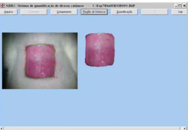
A computer program was developed specifically for the measurement of the area of the ulcer in digital images. The operator first outlined the borders of the ulcer with the mouse (Figure 1). Next, the software automatically cut out the out-lined region and calculated its area (A) in mm2, according to
the scale factor (number of pixels/mm with respect to the mag-nification and dimension utilized) previously determined. Sub-sequently, the region of the ulcer was reinserted into the origi-nal image, colored blue, so that the user could determine if the delimitation of the area had been performed properly (Figure 2).
FIGURE 2 – Quantification of the ulcer area
Results
In evaluating the temporal progression of tissue repair from a macroscopic point of view, it was seen that in the first days, there was an expansion of the ulcerated area in the Refer-ence group, while in the Control and Test groups the wound decreased in area. From the 3rd to 7th day, the presence of fibrin
was observed in the Reference group, a scab began to form in the Control group, and the wound remained moist with granu-lation tissue in the Test group. In the interval from the 7th to the
12th day, healing was more evident in the Control and Test group
with the presence of a scab, while there was an initial appear-ance of a tenuous scab in the Reference group but with a more enlarged area in relation to the other groups. Macroscopic evi-dence of better healing was only observed from the 7th to the
12th day, when the scab fell off in the Control and Test groups,
while in the Reference group, although the ulcerated area was still larger than in the other groups, wound healing was more homogeneous. On the 12th day, although complete wound
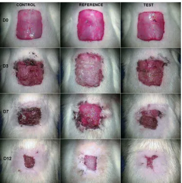
heal-ing was not demonstrated in any of the groups, the wound was smallest in the Test group (Figure 3).
The degree of wound repair (DR) of the ulcers was determined on days 3, 7 and 12, where it was calculated based on the initial area (measured on day zero) and expressed as a percentage, according to the following formula15,16.
where DRi denotes the degree of wound repair for day i and A0
and Aicorrespond to the area of the ulcer on days zero and i, respectively.
Also calculated was the mean rate of wound repair (RRm), which denotes how many mm2 the area of the ulcer
de-creased for a given time interval t1to t2, where it was expressed in mm2/day and defined by the quotient:
where A(t1) and A(t2) are the areas of the ulcera at times t1 and t2, respectively, and t2 > t1.
The efficacy (E) of a given treatment was calculated on day 12, where it was defined with respect to the Control group and expressed as a percentage based on the following formula:
where DR12(C) and DR12(T) correspond to the mean degree of healing determined on day 12 in the Control group and treated (Reference and Test) groups, respectively.
The data were initially evaluated by the Kolmogorov-Smirnov test to verify normality of the distribution. Since such requirement was observed for all the variables, we then calcu-lated for descriptive statistics the mean and standard deviation, and parametric tests were utilized for analysis of the data as well. Comparisons among the Control, Reference and Test groups, for a given day or interval, were performed by use of analysis of variance (ANOVA), combined with Tukey’s test for multiple comparisons, to determine differences among the groups two by two. The level of significance set at 5%. The statistics software GraphPad Prism® version 4.03 for Windows®
(GraphPad Software, San Diego, California, USA, 2005) was used for both the analysis of the data and the construction of the graphs.
100 . 0 0
A A A
DR i
i
1 2
1 2) ( ) (
t t
t A t A RRm
100 . ) (
) ( ) (
12 12 12
C DR
C DR T DR
E
FIGURE 3 – Appearance of the ulcer on days 0, 3, 7 and 12 of treat-ment in the Control, Reference and Test groups. Magnification, 6X
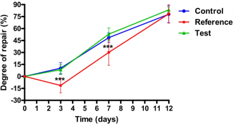
Figure 4 illustrates the temporal development of the tissue repair process according to degree of repair (DR). On the 3rd day, a small decrease in the area of the ulcer was noted in relation to the initial values in Control (10.2 ± 6.97%) and Test (8.45 ± 6.20%) groups. On the contrary, in the Reference group, there was an increase in the ulcerated area, denoted by the nega-tive DR value (-11.31 ± 9.22%), which was significantly lower (P < 0.001) than that observed in the other two groups. On the 7th day, the abrupt increase in DR in the three groups (Control:
groups. On the 12th day, however, there were no statistically
significant differences among the groups. In fact, a decrease in the slope of the DR curve was seen in Control (77.95 ± 10.71%) and Test (83.49 ± 3.50%) groups, indicating that in relation to day 7, the area of the ulcer regressed at a slower rate. On the other hand, in the Reference group, the tissue repair process proceded at the same intensity (78.40 ± 11.57%), as seen by the linear behavior of DR in the period between the 3rd and 12th day.
FIGURE 5 – Mean rate of wound repair (RRm) for the intervals be-tween days 0 and 3, 3 and 7, and 7 and 12. Data expressed as mean of measurements for 15 animals of each group. Analysis of variance was used to compare the three groups, followed by Tukey’s test to deter-mine differences between all pairs of groups. *** P < 0.001: Refer-ence lower than Control and Test; +++ P < 0.001. ReferRefer-ence higher than Control and Test (Tukey’s test)
FIGURE 4 – Temporal development of the tissue repair process in the Control, Reference and Test groups, based on the degree of tissue re-pair (DR). Data expressed as means and standard deviation of mea-surements for 15 animals of each group. Analysis of variance was used to compare the three groups, followed by Tukey’s test to determine differences between all pairs of groups. No statistically significant differences were found. *** P < 0.001. Reference lower than Control and Test (Tukey’s test)
0 1 2 3 4 5 6 7 8 9 10 11 12 -30
-15 0 15 30 45 60 75 90
Control
Test Reference
***
***
Time (days)
D
e
g
re
e
o
f r
e
p
a
ir
(%
)
The kinetics of tissue repair can be better understood by the analysis of the variable mean rate of wound repair (RRm) which represents the mean velocity of wound closing (Figure 5). In the period between days 0 and 3, as a result of the expan-sion of the ulcerated area, RRm for the Reference group was negative (-14.77 ± 12.59 mm2/day) and significantly lower (P <
0.001) than that of Control (13.72 ± 9.57 mm2/day) and Test
(11.33 ± 8.29 mm2/day) groups. In the interval between days 3
and 7, however, RRm of the Reference group increased mark-edly (40.61 ± 17.52 mm2/day), reaching values similar to that
observed in the Control (38.10 ± 8.68 mm2/day) and Test (43.86
± 6.89 mm2/day), such that no statistically significant
differ-ences were found. In this interval, in the three groups, RRm
reached their maximal values, characterizing the phase of great-est intensity of tissue repair. In the period between day 7 and 12, on the other hand, there was a deceleration in the repair of the ulcer in the Control (23.18 ± 8.04 mm2/day) and Test (23.83
± 4.38 mm2/day) groups, denoted by the diminution in the RRm
values. On the contrary, in the Reference group, RRm remained practically unchanged (37.37 ± 6.15 mm2/day), such that it was
significantly greater than the of the other groups (P < 0.001).
Although RRm by intervals revealed differences among the groups, RRm overall, that is, that refering to the period be-tween day 0 and 12 (Figure 6), was similar in the three groups: Control (25.79 ± 3.78 mm2/day), Reference (25.42 ± 4.76 mm2/
day), Test (27.38 ± 1.95 mm2/day). Therefore, there were no
statistically significant differences found (Figure 6).
FIGURE 6 – Mean rate of wound repair (RRm) for the interval be-tween days 0 and 12. Data expressed as mean and standard deviation of the measurements from 15 animals of each group. Analysis of vari-ance was used to compare the three groups, followed by Tukey’s test to determine differences between all pairs of groups. No statistically significant differences were found
0 1 2 3 4 5 6 7 8 9 10 11 12
-20 -10 0 10 20 30 40 50
Control Reference Test
***
+++
Time (days)
M
e
a
n
r
a
te
o
f r
e
p
a
ir
(m
m
2/d
ay)
Control Reference Test 0
10 20 30 40
M
e
a
n
r
a
te
of
r
e
pai
r
(m
m
2 /d
ay)
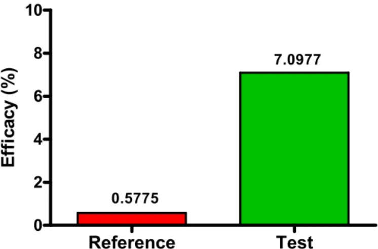
FIGURE 7 – Efficacy of the treatments calculated relative to the con-trol on the 12th day.
Discussion
In this study, the process of tissue repair of experi-mentally induced cutaneous wounds in rats was evaluated in vivo based on the kinetics of the regression of the ulcerated area over time. This was achieved by analyzing the variables that measure the degree of repair, which denotes the temporal contraction of the wound in relation to initial area, and mean rate of repair, that is, how many mm2 of the ulcer regressed per
day.
To determine the above parameters, it was necessary to calculate the area of the wounds using a planimetric tech-nique. Conventional planimetry consists of drawing the shape of the object of interest on graph paper and then counting the number of mm2 inside the demarcated region. However, we
chose to use digital planimetry, since it is a more objective, rapid and accurate method, whose use has become common-place5,13,14,16. Besides, digital images are a definitive
documen-tation of the tissue repair, such that they can be utilized for later review or in additional studies.
A computer program (written by the second author) was developed specifically for the planimetry of anatomic struc-tures. The proposal was to implement an easy-to-use software
that would be an alternative to complex image analyzing sys-tems of general use. The prior determination of the scale factor greatly facilitated the operation of the software, since it allowed actual measurements of the ulcerated region to be obtained, so that it was not necessary to place beside the wound an object such as a ruler or to measure on the animal one of the axes of the ulcer16.
The monitoring of the wound healing process in vivo
requires successive measurements in the same experimental ani-mal. In this way, the procedure of acquiring the digital images was standardized, in such a manner as to allow at different moments the capture of images of the wounds within the same frame. This strategy made it possible to detect, using the quan-tification software, the temporal variations in the area of the ulcer, even if minimal.
The image acquisition procedure combined with the quantification software constituted an accurate planimetric method which allowed the spatial and temporal monitoring of tissue repair in vivo. Such approach should be encouraged, for the purpose of dispensing with the sacrifice of animals, thereby requiring fewer experimental units to carry out assays.
Wound healing involves a complex and coordinated number of events which include inflammation, cell prolifera-tion, contraction of the wound and tissue remodeling17. Thus,
in this study, it was analyzed the effects of a combination of medium chain triglycerides (caprylic, capric, caproic and lau-ric acids), linoleic acid (essential fatty acid), soy lecithin and vitamins A and E, applied topically, on this process in rats, con-sidering the findings from earlier studies that demonstrated the efficacy of these agents in accelerating wound healing in hu-man patients9.
In this study, subcutaneous cellular tissue was removed, making it necessary for the formation of new tissue for repair of the lesion. The combined use of neomycin and clostebol as in the Reference group was to evaluate the repair of the lesion, unlike in a study conducted earlier which evaluated antimicro-bial activity18,19.
During the experiments, the protocol allowed us to pho-tograph the animals on the day of the surgical procedure (zero), and on the 3rd, 7th and 12th days of treatment. In this manner,
besides the objective quantification, it was also possible to per-form a subjective macroscopic clinical evaluation similar to that described in other studies carried out with the same test sub-stance in dogs9 and with linoleic acid in sheep15.
Up to the 3rd day, subjective observation indicated slow
retraction of the wound in the Control group, with hyperemia and scab formation. In the Test group, hyperemia and discrete formation of scab on the edges of the lesion were observed. Hyperemia, edema and fibrin were seen in the Reference group. In another study in which the effects of polyunsaturated fatty acids on the healing of cutaneous wounds were evaluated, a macroscopic difference in lesion repair was demonstrated in the first 48 h after the surgical procedure8, which was not
ob-served in our model.
The greater infiltration of polymorphonuclear cells and macrophages in the first three days can correspond to the phase of exudation and inflammation in the wounds treated with trig-lycerides as demonstrated in a study conducted in dogs.9 In the same period in the present study, there were no signs of exu-dates by macroscopic observation.
From the 3rd to the 7th day, there was progressive
re-pair of the wound area, which indicated development of granu-lation tissue in concordance with findings in the literature17.
The macroscopic appearance showed the wound area with the presence of fibrin and scab in the Reference group, complete scab formation in the Control group, and discrete hyperemia in the Test group. It is likely that the occurrence of discrete signs compatible with inflammation such as edema and hyperemia after the third day could have been associated with the antioxi-dant action of vitamin E, minimizing reperfusion lesions as a consequence of the release of toxic free radicals, in agreement with the study conducted in dogs9.
Reference
Test
0 2 4 6 8 10
0.5775
7.0977
E
ff
ica
cy (
%
The action of soy lecithin provides hydration of the tissues, constituting a favorable factor in the repair of the le-sion. Keeping the wound hydrated promotes autolytic debride-ment and contributes as a stimulus of epithelization, formation of granulation tissue and angiogenesis9. Thus, hydration
pro-moted by soy lecithin, complemented by normal saline solu-tion, made surgical debridement of the wounds unnecessary in this study. On the other hand, previous studies demonstrated that open and dried wounds undergo re-epitelization more slowly17.
From the 7th to 12th day, a greater rate of repair
oc-curred in the Reference group. The test preparation showed evident granulation tissue and greater tissue contraction around the edges of the wound, which had become irregular. In the Control group, gaps in the scab were seen on the 7th day, along
with irregular edges due to contraction of the wound, besides the presence of granulation tissue. The Reference group re-mained with the largest wound area and with irregular edges, which was also found in a study using linoleic acid15. The
treat-ments in the three groups contributed to the almost complete closing of the lesions, suggesting that growth factors were prob-ably responsible for the hyperplasia of the epithelium as re-ported in the literature17. In this period, there was agreement
with a study that considered cytokines as being important me-diators of neoangiogenesis, fibroplasia and maturation, which are released by cells such as platelets, neutrophils, macroph-ages, lymphocytes, mast cells and fibroblasts, making it easy to understand the importance of chemotactic properties of the test preparation in the repair of the lesions9.
A study that examined wound healing in sheep showed that linoleic acid constituted a powerful pro-inflammatory me-diator, being essential for the regulation of the biochemical events that precede fibroplasia in addition to stimulating growth factors and neovascularization15. It is possible that in this study
the presence of linoleic acid in the test preparation contributed to a similar event.
A study carried out with polyunsaturated fatty acids showed a tendency for the wound area to diminish in the first ten days of treatment, and demonstrated overall that PUFAs may play an important role in wound healing8.
On the 12th day, clinical observation showed a smaller
wound area characterized by the proximity of the edges (con-traction) with irregular outlines and better presence of granula-tion tissue, like that seen in the study with linoleic acid in sheep15.
Proliferation (fibroplasia and matrix formation) was demonstrated by scholars in the past as being extremely impor-tant in the formation granulation tissue. This depends on fibro-blasts which produce elastin, fibronectin, glucosaminoglycans and proteases17. In this phase, the presence of vitamin A in the
test preparation was important, because authors evaluating the same preparation found that it stimulates fibroblasts, the depo-sition of collagen and formation of connective tissue9.
The repair of wounds by secondary union showed that contraction could have been responsible for the reduction in wound area in the three groups, in concordance with a study in which contraction was shown to be able reduce the surface of the cutaneous defect by 62%17.
In the model presented here, triglycerides favored a greater repair from a macroscopic clinical viewpoint, where it contributed to the wound area being less on the 12th day of
treat-ment. However, compared to Control group, the efficacy of the test preparation was only 7.0977%, while the efficacy of the reference treatment was lower at only 0.5775%.
Conclusion
The test preparation applied topically to the surgical wound produced in this model did not accelerate significantly the process of tissue healing by secondary union.
References
1. Carrel A. The treatment of wounds. JAMA. 1910;55:2148-50.
2. Ortonne JP, Clévy JP. Physiology of cutaneous cicatrization. Rev Prat. 1994; 44(13):1733-7.
3. Araújo CFR, Souza Fo ZA, Greca FH, Guerreiro MHCPM, Leite AL, Mansur AE C, Kantor DC, Nassif AE. Effects of Agarol® and Trigliceril® on cutaneous wound healing:
experi-mental study in rats. Acta Cir Bras. 1998;13(4):232-7. 4. Banda MJ, Knighton DR, Hunt TK, Werb Z. Isolation of a nonmitogenic angiogenesis factor from wound fluid. Proc Nat Acad Sci. 1982;79:7773-7.
5. Mori, R.; Kondo, T.; Nishiie, T.; Ohshima, T.; Asano, M. Impairment of skin wound healing in ȕ -1,4-galactosyltransferase-deficient mice with reduced leukocyte recruitment. Am J Pathol. 2004;164(4):1303-14.
6. D’Agostini D. Obtenção de lipídíos estruturados por intereterificação de triacilglicerois de cadeia média e longa [Tese de Doutorado]. Universidade de São Paulo, Faculdade de Ciências Farmacêuticas; 2001.
7. Hatanaka E, Curi R. Fatty acids and wound healing: a re-view. RevRv Bras Farmacol. 2007;88(2):53-8.
8. Cardoso CRB, Souza MA, Ferro EAV, Favoreto S, Pena JDO. Influence of topical administration of n-3 and n-6 essential and n-9 nonessential fatty acids on the healing of cutaneous wounds. Wound Rep Reg. 2004;12:235-43.
9. De Nardi AB, Rodaski S, Sousa RS, Baudi DLK, Castro JHT. Secondary cicatrization in dermoepidermal wounds treated with essential fatty acids, vitamins A and E, soy lecithin and polynylpyrrolidone-iodine in dogs. Arc Vet Sci. 2004;9(1):1-16
10. Ehrlich HP , Hunt TK. Effects of cortisone and vitamin A on wound healing. Ann Surg. 1968;167:324-8.
11. Greenway DLA & Dyke KGH. Mechanism of the inhibi-tory action of linoleic acid on the growth of Staphylococcus aureus. J Gen Microbiol. 1979;115:233-45.
13. Teixeira VPA, Pereira SAL, Rodrigues DBR, Lino RSJ. Princípios básicos e aplicações da morfometria. Disponível em: h t t p : / / w w w . u f t m . e d u . b r / i n s t p u b / f m t m / p a t g e / morfometria01.htm. Acesso em: 17 jul.2007.
14. Mandarin-de-Lacerda, CA. Stereological tools in biomedi-cal reserarch. An Acad Bras Cienc. 2003;75(4):469-86. 15. Marques SR, Peixoto CA, Messias JB, Albuqurque AR, Silva Jr VA. The effects of topical application of sunflower-seed oil on open wound healing in lambs. Acta Cir Bras. 2004;19(3):196-209.
16.Martins NLP, Malafaia O, Ribas-Filho JM, Heibel M, Baldez RN, Vasconcelos PRL, Moreira H, Mazza M, Nassif PAN, Wallbach TZ. Healing process in cutaneous surgical wounds in
rats under the influence of Orbignya phalerata aqueous extract. Acta Cir Bras. 2006;21(supl 3):66-75.
17. Mandelbaum SH, Di Santis EP, Mandelbaum MHSA. Cica-trization: current and auxiliary resources-Part 1. An Bras Dermatol. 2003;78(4):393-410.
18. Rodrigues KL, Cardoso CC, Caputo LR, Carvalho JCT, Fiorini JE, Schneedorf JM. Cicatrizing and antimicrobial prop-erties of an ozonised oil from sunflower seeds. Inflammopharmacology. 2004;12(3):261-70.
19. Sakuma CH, Matera JM, Valente NS. Clinical study of skin flap application during oncologic surgery in dog. Bras J Vet Res Anim Sci. 2003;40 Suppl:32-7.
Conflict of interest: none Financial source: none
Correspondence:
Manoel Odorico de Moraes Rua Cel. Nunes de Melo, 1127 60430-270 Fortaleza – Ceará Brazil Phone: (55 85)3366-8201
Received: November 19, 2007 Review: January 23, 2008 Accepted: February 19, 2008
How to cite this article
Magalhães MSF, Fechine FV, Macedo RN, Monteiro DLS, Oliveira CC, Brito GAC, Moraes MEA, Moraes MO. Effect of a combination of medium chain triglycerides, linoleic acid, soy lecithin and vitamins A and E on wound healing in rats. Acta Cir Bras. [serial on the Internet] 2008 May-June;23(3). Available from URL: http://www.scielo.br/acb