Karyotype differentiation
in three
species of
Tripogandra
Raf.
(Commelinaceae) with different ploidy levels
André Marques, Fernando Roa and Marcelo Guerra
Laboratório de Citogenética Vegetal, Departamento de Botânica, Universidade Federal de Pernambuco,
Recife, PE, Brazil.
Abstract
Most species of the genusTripogandra (Commelinaceae) are taxonomically poorly circumscribed, in spite of having
a relatively stable basic numberx = 8. Aiming to estimate the cytological variation among Tripogandra species
carry-ing this base number, several structural karyotypic characters were investigated in the diploidT. glandulosa, the
hexaploidT. serrulata, and the octoploid T. diuretica. A careful evaluation of chromosome size and morphology did
not reveal clear chromosome homeologies among karyotypes. The mean chromosome size was strongly reduced in
the octoploid species, but not in the hexaploid species. They also differed largely in the CMA+
banding pattern and in the number of 5S and 45S rDNA sites per monoploid chromosome complement. All three species showed proximal
DAPI+
heterochromatin, although inT. serrulata this kind of heterochromatin was only visible after FISH. Further, the
meiosis inT. serrulata was highly irregular, suggesting that this species has a hybrid origin. The data indicate that, in
spite of the conservation of the base number, these species are karyologically quite different from each other. Key words: Tripogandra,cytotaxonomy, rDNA sites, CMA/DAPI bands, meiosis.
Received: December 4, 2009; Accepted: April 4, 2010.
The genusTripogandraRaf. (Commelinaceae, tribe Tradescantieae, subtribe Tradescantiinae) comprises ap-proximately 22 species, some of which are restricted to Mexico, while others are distributed throughout Mexico, Central America, and South America (Handlos, 1975). These species are characterized principally by the presence of dimorphic stamens and double cincinni (Handlos, 1975). Recently, molecular data have revealed that some
Tripogandraspecies are grouped into one of the clades of the genus Callisia (Bergamo, 2003; Evans et al., 2003; Burns, 2006), suggesting that Tripogandra is a poly-phyletic group. Cytotaxonomic analyses, mainly those that seek to identify the basic chromosome number, have con-tributed to the understanding of the phylogenetic relation-ships of many species groups (reviewed by Guerra, 2008), and can be useful in evaluating the relationships among species of this genus.
Within the family Commelinaceae, the subtribe Tradescantiinae, which includes Tripogandra, Callisia,
Tradescantia, andGibasis, stands out by its wide variation in basic numbers, chromosome morphology, and chromo-some sizes (Jones and Kenton, 1984; Pitrezet al., 2001). The haploid numbern = 8 and its euploid multiples
pre-dominate inTripogandra,withx= 8 largely accepted as the basic number of the genus, whilen= 5, 6, and 8 are encoun-tered inGibasis,n= 6, 7, and 8 inCallisia, andn= 6 or mul-tiples prevail inTradescantia (Jones and Jopling, 1972). AlthoughTripogandra,Callisia,Gibasis, and Tradescan-tiashare the basic numberx= 8, these genera show great di-versity in chromosome morphology and ploidy levels, hindering an understanding of the cytotaxonomy of the subtribe (Handlos, 1970).
Cytogenetic analyses using other karyotype parame-ters, such as C-banding and staining with fluorochromes, have also contributed to an understanding of the relation-ships among some Commelinaceae species (Jones and Kenton, 1984; Kenton, 1991; Sakurai and Ichikawa, 2001). Parokonnyet al.(1992) showed that some cryptic species of Gibasiscan be clearly distinguished using techniques such as fluorescencein situhybridization (FISH) and ge-nomicin situhybridization (GISH). However, this type of chromosome analysis is still restricted to just a few genera of the subtribe, and the cytology of a number of genera, such asCallisiaandTripogandra,has not been studied yet with any of these techniques.
In the present work several structural karyotype char-acteristics were analyzed in three species ofTripogandra
(T. diuretica,T. glandulosa,andT. serrulata) withx= 8, in order to understand the cytotaxonomical relationships among them. We also attempted to identify which struc-tural characteristic of these karyotypes was best conserved Send correspondence to Marcelo Guerra. Departamento de
Botâ-nica, Centro de Ciências Biológicas, Universidade Federal de Per-nambuco, Rua Nelson Chaves s/n, 50670-420 Recife, PE, Brazil. E-mail: [email protected].
and could represent a karyological synapomorphy of the genus. We investigated the structure of the interphase nu-clei, the distribution of heterochromatin by fluorochrome staining with chromomycin A3 (CMA) and 4’,6-diami-dino-2-phenylindole (DAPI), and the number and location of the 5S and 45S rDNA sites by in situ hybridization (FISH). Previous chromosome counts had reported 2n= 16 forT. glandulosa, 2n= 32, 39, 48, ca. 50 forT. serrulata, and 2n= 62 + 1B, 64 forT. diuretica(Simmonds, 1954; Celarier, 1955; Lewiset al., 1967; Handlos, 1970; Jones and Jopling, 1972; Pitrezet al., 2001). The meiotic behav-ior of the two polyploid species was also analyzed in order to evaluate the stability of these species.
Materials and Methods
Plant material
Samples of Tripogandra diuretica, T. glandulosa, andT. serrulatawere analyzed. All plant samples investi-gated grew in the experimental garden at the Department of Botany of the Federal University of Pernambuco, and the vouchers were deposited in the UFP herbarium of this uni-versity. Voucher numbers, provenances, and general cyto-logical data for each sample are presented in Table 1.
Slide preparation
Root tips were pre-treated with 8-hydroxyquinoline (0.002 M) for 5 h at 18 °C, fixed in absolute alcohol/glacial acetic acid (3:1, v/v) for 2-24 h at room temperature, and stored at -20 °C. Subsequently, they were washed three times in distilled water and digested in a 2% (w/v -0.02 U/mL) cellulase (Onozuka) and 20% (v/v - 120 U/mL) pectinase (Sigma) solution for 1-2 h at 37 °C. Meristems were squashed in a drop of 45% acetic acid and frozen in liquid nitrogen to remove the coverslip. The slides were stained with DAPI (2mg/mL):glycerol (1:1, v/v) solution to
permit selection of the best cells. Subsequently, the slides were destained in ethanol/glacial acetic acid (3:1, v/v) for 30 min at room temperature and transferred to absolute eth-anol overnight at 10 °C. Slides were air dried and aged for three days at room temperature for CMA/DAPI staining (Barros-e-Silva and Guerra, 2010).
For meiotic analysis, young inflorescences fromT. diureticaandT. serrulatawere fixed and stored as previ-ously described. Prior to slide preparation, the material was washed twice in distilled water, anthers were squashed in a drop of 45% acetic acid, and the coverslip was removed af-ter freezing in liquid nitrogen.
CMA/DAPI staining
For CMA/DAPI fluorochrome banding, aged slides were stained with 0.1 mg/mL CMA (Sigma) for 1 h and counterstained with 1mg/mL DAPI (Sigma) for 30 min.
The slides were mounted in 1:1 (v/v) McIlvaine’s buffer-glycerol with 2.5 mM MgCl2(pH 7.0) and kept in the dark Table
at room temperature for 3 days before analysis, as described by Barros-e-Silva and Guerra (2010). Well-spread cells were captured with a CCD Cohu camera on a Leica DMLB epifluorescence microscope using the Leica Q-FISH soft-ware. The slides were destained again and stored at -20 °C.
Fluorescencein situhybridization (FISH)
FISH procedure was based on Pedrosaet al.(2002) with minor modifications. Briefly, slides were treated with RNase at 37 °C for 1 h, washed three times, in 2x SSC for 5 min each , and digested with pepsin at 37 °C for 20 min. The preparations were post-fixed in 1% (para)formaldehyde in 1x PBS for 10 min and dehydrated in an ethanol series.
Two rDNA probes, D2 and R2, were used to locate the 5S and 45S rDNA sites, respectively. D2 is a 500-bp fragment of the 5S rDNA repeat unit fromLotus japonicus
(Pedrosaet al., 2002) and R2 is a 6.5-kb fragment that con-tains a 45S rDNA repeat unit fromArabidopsis thaliana
(Wanzenböcket al., 1997). The D2 probe was labeled with Cy3-dUTP (Invitrogen) and the R2 probe was labeled with digoxigenin-11-dUTP (Life Technologies) by nick transla-tion. The hybridization mixture contained 50% formamide (v/v), 10% dextran sulfate (w/v), 2x SSC, and 2-5 ng/mL of
probe. The probe mixture was denatured at 75 °C for 10 min and the chromosome denaturation was undertaken at 75 °C for 6 min. After overnight hybridization at 37 °C, slides were washed in the following order: 2x SSC at room temperature, 2x SSC, 0.1x SSC, and 2x SSC at 42 °C, and 2x SSC at room temperature, for 5 min each. The digo-xigenin probe was detected with sheep anti-digodigo-xigenin FITC conjugate (Dako) and amplified with rabbit anti-sheep FITC conjugate (Roche). All slides were mounted in DAPI (4mg/mL) that was diluted 1:1 (v/v) in Vectashield
(Vector). Metaphase images were acquired as indicated above and were later edited in Adobe Photoshop CS3.
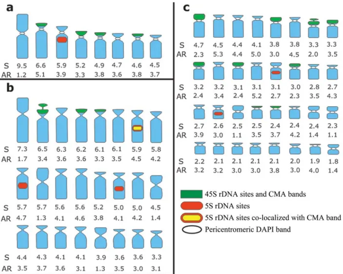
Chromosome measurements and idiograms
The total chromosome length (S) and the chromo-somes arm ratios (AR = long/short arm) were estimated using the Adobe Photoshop CS3 software. An idiogram summarizing most karyotype data for each species was constructed based on chromosome measurements of four well-spread cells, disregarding the length of the secondary constriction. The chromosomes were ordered by size from the largest to the smallest.
Results
Chromosome numbers, chromosome morphology, and nuclear structure
The karyotype parameters of theTripogandraspecies analyzed are shown in Table 1 including average chromo-some length.
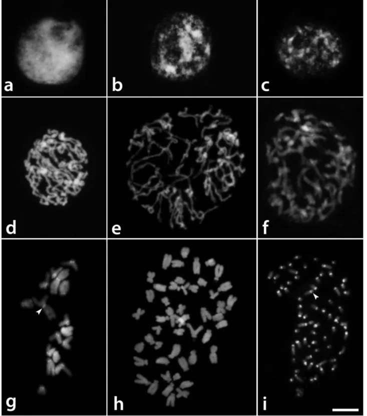
According to the nomenclature that was proposed by Delay (1949) for interphase nuclei,T. glandulosaexhibited
reticulate nuclei, with a dense and uniformly distributed chromatin structure, T. diuretica nuclei showed a semi-reticulate structure with irregularly distributed chromatin, and T. serrulata nuclei displayed an intermediary chro-matin structure between these two types (Figures 1a-c). Condensation of the prophase chromosomes was uniform along the chromosomes ofT. glandulosa(Figure 1d), while there were some regions that were less condensed than oth-ers inT. diureticaandT. serrulata(Figures 1e,f).
CMA/DAPI staining and FISH
The pericentromeric regions of all chromosomes of
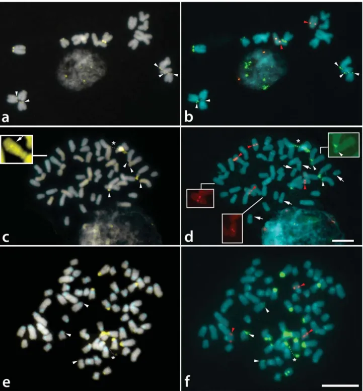
Tripogandra glandulosa(2n= 16) were generally observed as weak DAPI+ bands, which were better contrasted after FISH (Figure 1g). CMA+bands were observed on the short arms of the eight acrocentric chromosomes (Figure 2a), with a pair of them showing a much larger and brighter band. The distribution of the 45S rDNA sites coincided with the eight CMA+bands, while the 5S rDNA sites were localized in the interstitial/proximal regions of the long arms of two acrocentric chromosomes (Figure 2b).
The CMA+bands ofT. serrulata(2n= 48) were also co-localized with 45S rDNA sites on the short arms of six acrocentric chromosomes, four of which were brighter than the other two (Figure 2c,d). DAPI+ bands were not ob-served with direct CMA/DAPI staining (Figure 1h), although weak heterochromatic bands were sometimes ob-served after FISH in the interstitial and centromeric regions (not included in the idiograms) (Figure 2d). 5S rDNA sites were located in the interstitial region of three pairs of acrocentric chromosomes, one of them with very small sig-nals (inserts in Figure 2d). Exceptionally, one of these sites was found co-localized with a CMA+ band (see insert in Figure 2c).
Tripogandra diuretica(2n= 64) had DAPI+bands in the pericentromeric region of all chromosomes (Figure 1i), and CMA+bands co-localized with 45S rDNA sites on the short arms of 10 submetacentric and eight acrocentric chro-mosomes, three of them weakly stained (arrowheads in Fig-ures 2e,f). In some cells, three chromosome pairs with 45S rDNA sites appeared to have short arms slightly longer due to the decondensation of the nucleolar organizing regions. We observed 5S rDNA sites in the interstitial region of a pair of submetacentric chromosomes, and also in one small acrocentric chromosome (Figure 2f). The idiograms in Fig-ure 3 summarize these data. Chromosome pairs homozy-gous or heterozyhomozy-gous for bands or rDNA sites were repre-sented in the idiograms as a single chromosome having these marks.
Meiotic analysis
configura-tion was not clear. Bridges and laggard chromosomes were observed in most cells in anaphase I and II, and one to seven micronuclei were found in telophase II (Figures 4c,d).
Tripogandra diuretica, on the other hand, showed regular meiosis with the formation of 32 bivalent chromosomes in metaphase I and micronuclei in only 3% of the tetrads.
Discussion
The three species ofTripogandraanalyzed here had distinct karyotypes and different ploidy levels (2x, 6x, 8x). In addition to the chromosome number 2n= 48 (hexaploid) that was commonly observed in T. serrulata (Celarier, 1955; Lewiset al., 1967; and the present work), Handlos
(1970) reported some tetraploid individuals with 2n= 32. The chromosome numbers 2n= 39 and 2n= ca. 50 forT. serrulatareported by Lewiset al.(1967) and Simmonds (1954), respectively, are probably miscounts.
The karyotype formulae of the three species analyzed here [T. glandulosa, 2n = 16 (2M + 14A); T. serrulata, 2n= 48 (6M + 2SM + 40A);T. diuretica, 2n= 64 (8M +
Figure 2- Distribution of CMA/DAPI bands (a, c, e) and rDNA sites (b, d, f) inTripogandraspecies. a, b -Tripogandra glandulosa; c, d -T. serrulata; e, f -T. diuretica.White arrowheads indicate small CMA+bands and small 45S rDNA sites. Red arrowheads indicate 5S rDNA sites. Arrows in d point to DAPI+bands that appear only after FISH. Dots in c-f indicate secondary constrictions (asterisks). Insert in c shows CMA+band co-localized with 5S
12SM + 44A)] showed a number of metacentric chromo-somes that were proportional to those expected based on the karyotype formulae of the diploidT. glandulosa. How-ever, the metacentric chromosomes of the hexaploid T. serrulataand the octoploidT. diureticawere much smaller than those observed inT. glandulosa, suggesting that they may be non-homeologous. The divergence is even more ev-ident in the number of acrocentric chromosomes expected (56A) and observed (44A) in the octoploid. Therefore, these polyploids are probably not directly related to T. glandulosa.
The average haploid set length (HSL) of T. glandulosacomplement (45.6mm) andT. serrulata(123.8
mm) was proportional to the ploidy level and DNA amount
(T. glandulosa, 1C = 3.2 pg;T. serrulata, 1C = 9.75 pg; Bennett and Leitch, 1995). On the other hand, the HSL ofT. diureticawas much smaller than expected, based on the variation on ploidy level, a trend to genome downsizing more commonly found in old polyploids (Leitch and Ben-nett, 2004).No DNA content estimate was found for this
species.The difference in the average chromosome size of
T. serrulataandT. diureticasuggests that these polyploids represent different evolutionary lineages, withT. serrulata
being younger thanT. diuretica. Accordingly, these species differ in several morphological characters, such as the num-ber of stamens, style/ovary relative length, type of stigma, fruit form, and surface of seed testa (Handlos, 1970, 1975). In addition,T. diureticahas a South American distribution from Bolivia to Argentina, whereas T. serrulata occurs from Mexico to Peru and the Caribbean.
The two polyploids also differed in their meiotic be-havior.Tripogandra diureticashowed a completely regular meiosis, whereasT. serrulatadisplayed a series of meiotic irregularities, including an elevated number of univalents, anaphase bridges, and micronuclei. Curiously, Celarier (1955) reported a hexaploid specimen ofT. serrulatawith 24 bivalents and normal meiosis, suggesting the existence of different hexaploid cytotypes within this species. Thus,
T. serrulataseems to be a complex species, having popula-tions with normal meiosis as well as populapopula-tions with highly irregular meiosis.
Analyses of the interphase nuclear structure of the three species examined here revealed an additional differ-ence in their chromatin organization, which was reticulate in the diploid, semi-reticulate with chromocenters in the octoploid, and intermediate between these types in the hexaploid. This variation depends in part on the average chromosome size, which is generally larger in species with reticulate nuclei (Guerra, 1987). Interestingly, the differ-ences in the interphase nuclei were not reflected in the con-densation pattern of their prophase chromosomes, as has been observed in other species (Delay, 1949; Guerra, 1987).
The heterochromatin in these species is of two dis-tinct types: DAPI+, observed in the proximal regions ofT. glandulosaandT. diuretica, and CMA+, which was termi-nally located in all three species. The DAPI+ bands ob-served in T. serrulata only after FISH are probably not AT-rich and not similar to the DAPI+bands of the other two species (Barros-e-Silva and Guerra, 2010). The CMA+
heterochromatin was always co-localized with 45S rDNA sites, except in a chromosome ofT. serrulatain which it was co-localized with a 5S rDNA site. In general, only the 45S rDNA sites co-localize with CMA+bands (Moraeset al., 2007; Souzaet al., 2009), although in some species the CMA+ bands have been found co-localized with both 5S and 45S rDNA sites (Cabralet al., 2006). The number of CMA+bands and 45S rDNA sites varied broadly among the three species, without correlation to ploidy level, pointing once again to the instability of such chromosome land-marks (see Pedrosa-Harand et al., 2006; Kovarik et al., 2008).
The different karyotype parameters analyzed here varied among the three species, indicating that the basic number is the only cytological feature clearly shared by all three species. In general, T. glandulosa and T. serrulata were more similar to each other than to T. diureticain chromosome sizes, karyotype formulae, and number of 5S rDNA sites per monoploid chromosome complement.
Acknowledgments
The authors wish to thank the Brazilian agencies Conselho Nacional de Desenvolvimento Científico e Tec-nológico (CNPq) and Fundação de Amparo à Ciência e Tecnologia de Pernambuco (FACEPE) for their financial support, and Dr. Robert Faden (Smithsonian Institution), Mauro Grabiele, and Dr. Julio Daviña (Universidad Na-cional de Misiones) for their kind help in providing us with plant materials.
References
Barros-e-Silva AE and Guerra M (2010) The meaning of DAPI bands observed after C-banding and FISH procedures. Bio-tech Histochem 85:115-125.
Bennett MD and Leitch IJ (1995) Nuclear DNA amounts in angio-sperms. Ann Bot 76:113-176.
Cabral JS, Felix LP and Guerra M (2006) Heterochromatin diver-sity and its co-localization with 5S and 45S rDNA sites in chromosomes of fourMaxillariaspecies (Orchidaceae). Ge-net Mol Biol 29:659-664.
Celarier RP (1955) Cytology of the Tradescantieae. Bull Torrey Bot Club 82:30-38.
Delay C (1949) Recherches sur la structure dês noyaux quiescents chez les phanérogames. Rev Cyt Cytophysiol Vég 10:103-228.
Evans TM, Sytsma KJ, Faden RB and Givnish TJ (2003) Phylo-genetic relationships in the Commelinaceae: II. A cladistic analysis of rbcL sequences and morphology. Syst Bot 28:270-292.
Guerra M (1987) Cytogenetics of Rutaceae IV. Structure and sys-tematic significance of interphase nuclei. Cytologia 52:213-222.
Guerra M (2008) Chromosome numbers in plant cytotaxonomy: Concepts and implications. Cytogenet Genome Res 120:339-350.
Handlos WL (1970) Cytological investigations of some Comme-linaceae from Mexico. Baileya 17:6-33.
Handlos WL (1975) The taxonomy of Tripogandra (Comme-linaceae). Rhodora 77:218-319.
Jones K and Jopling C (1972) Chromosomes and the classification of the Commelinaceae. Bot J Linn Soc 65:129-162. Jones K and Kenton A (1984) Mechanisms of chromosome
chan-ge in the evolution of the tribe Tradescantieae (Commeli-naceae). In: Sharma A and Sharma AK (eds) Chromosomes in Evolution of Eukaryotic Groups II. CRC Press, Boca Raton, pp 143-168.
Kenton A (1991) Heterochromatin accumulation, disposition and diversity inGibasis karwinskyana(Commelinaceae). Chro-mosoma 100:467-478.
Kovarik A, Dadejova M, Lim YK, Chase MW, Clarkson JJ, Knapp S and Leitch AR (2008) Evolution of rDNA in Nicotiana allopolyploids: A potential link between rDNA homogenization and epigenetics. Ann Bot 101:815-823. Leitch IJ and Bennett MD (2004) Genome downsizing in
poly-ploid plants. Biol J Linn Soc 82:651-663.
Lewis WH, Suda Y and Oliver RL (1967) Chromosome numbers of phanerogams 2. Ann Missouri Bot Gard 54:178-180. Moraes AP, Soares Filho WS and Guerra M (2007) Karyotype
di-versity and the origin of grapefruit. Chromosome Res 15:115-121.
Parokonny AS, Kenton AY, Meredith L, Owens SJ and Bennett MD (1992) Genomic divergence of allopatric sibling species studied by molecular cytogenetics of their F1hybrids. Plant J
2:695-704.
Pedrosa A, Sandal N, Stougaard J, Schweizer D and Bachmair A (2002) Chromosomal map of the model legume Lotus japonicus. Genetics 161:1661-1672.
Pedrosa-Harand A, Almeida CCS, Mosiolek M, Blair MW Schweizer D and Guerra M (2006) Extensive ribosomal DNA amplication during Andean common bean (Phaseolus vulgarisL.) evolution. Theor Appl Genet 112:924-933. Pitrez SR, Felix LP, Barreto R and Guerra M (2001) Números
cromossômicos de espécies de Commelinaceae R. Br. ocor-rentes no nordeste do Brasil. Bol Bot Univ São Paulo 19:7-14 (Abstract in English).
Sakurai T and Ichikawa S (2001) Karyotypes and Giemsa C-ban-ding patterns ofZebrina pendula,Z. purpusiiandSetcreasea purpurea, compared with those ofTradescantia ohiensis. Genes Genet Syst 76:235-242.
Simmonds NW (1954) Chromosome behaviour in some tropical plants. Heredity 8:139-146.
Souza LGR, Crosa O, Winge H and Guerra M (2009) The karyo-type of Nothoscordum arenarium Herter (Gilliesioideae, Alliaceae): A populational and cytomolecular analysis. Genet Mol Biol 32:111-116.
Wanzenböck EM, Schöfer C, Schweizer D and Bachmair A (1997) Ribosomal transcription units integrated via T-DNA transformation associate with the nucleolus and do not re-quire upstream repeat sequences for activity inArabidopsis thaliana. Plant J 11:1007-1016.
Internet Resources
Bergamo S (2003) A phylogenetic evaluation ofCallisiaLoefl. (Commelinaceae) based on molecular data. PhD Thesis, The University of Georgia, USA.
https://getd.libs.uga.edu/pdfs/bergamo_stephanie_ 200312_phd/bergamo_stephanie_200312_phd.pdf (Febru-ary 26, 2010).
Burns JH (2006) A comparison of invasive and noninvasive Commelinaceae in a phylogenetic context. PhD Thesis. The Florida State University College of Arts and Sciences, USA. http://etd.lib.fsu.edu/theses/available/
etd-11092006-112630/unrestricted/BurnsMoriuchi_Disser-tation.pdf (February 26, 2010).
Editor: Angela M. Vianna-Morgante