Chromatin differentiation between
Theobroma cacao
L.
and
T. grandiflorum
Schum
Liliane G. Dantas and Marcelo Guerra
Laboratório de Citogenética Vegetal, Departamento de Botânica, Universidade Federal de Pernambuco,
Pernambuco, Recife, Brazil.
Abstract
A comparative analysis of mitotic chromosomes ofTheobroma cacao (cacao) and T. grandiflorum (cupuaçu) was performed aiming to identify cytological differences between the two most important species of this genus. Both spe-cies have symmetric karyotypes, with 2n = 20 metacentric chromosomes ranging in size from 2.00 to 1.19mm
(ca-cao) and from 2.21 to 1.15mm (cupuaçu). The interphase nuclei of both species were of the arreticulate type,
displaying up to 20 chromocentres, which were more regularly shaped in cacao than in cupuaçu. Prophase chromo-somes of both species were more condensed in the proximal region, sometimes including the whole short arm. Both species exhibited only one pair of terminal heterochromatic bands, positively stained with chromomycin A3, which
co-localized with the single 45S rDNA site. Each karyotype displayed a single 5S rDNA site in the proximal region of another chromosome pair. Heterochromatic bands were also observed on the centromeric/pericentromeric regions of all 20 chromosomes of cacao after C-banding followed by Giemsa or DAPI staining, whereas in cupuaçu they were never detected. These data suggest that the chromosomes of both species have been largely conserved and their pericentromeric chromatin is the only citologically differentiated region.
Key words:cacao, cupuaçu, heterochromatin, karyotype, rDNA sites,Theobroma.
Received: March 10, 2009; Accepted: July 16, 2009.
Introduction
Theobroma(Malvaceae) is a tropical genus native to South America, comprising some 22 species (Kennedy, 1995) and havingT. cacaoL. (cacao) as its most important representative. The second most important species in the genus isT. grandiflorumSchum. (cupuaçu), native of the Brazilian Amazon. It is largely cultivated and commercial-ized in Brazil where it is consumed as fruit juice, ice-cream, mousse, etc (Alveset al., 2007). A powder similar to cacao (“cupulate”) can also be obtained from cupuaçu seeds.
Cacao products are widely consumed around the world but large crop plantations are restricted to Brazil, Malaysia and a few countries in West Africa. In spite of its economic value, the cacao tree has received little attention as a crop and almost 70% of the currently cultivated plants have never been submitted to any kind of breeding program (Lockwood, 2003). Nevertheless, in the last 15 years a con-siderable effort has been put into the molecular mapping and genome sequencing of cacao, aiming to prevent the devastating agricultural effects of fungal diseases, like witches’ broom disease (Bennett, 2003).
In most crop plants, karyological analyses have been used to characterize cultivars, to integrate genetic and phys-ical maps, to investigate the origin of hybrids, etc (Jiang and Gill, 2006; Moraeset al., 2007). Nevertheless, rela-tively little is known about the chromosomes ofTheobroma
species. Analyses based on conventional techniques showed that allTheobromaspecies investigated presented the same diploid number (2n= 20) and chromosomes with similar morphology, ranging in size between 0.5 and 2.0mm (Carleto, 1946; Guerra, 1986; Kennedy, 1995).
Be-sides this apparent chromosome stability, meiotic analyses in some cultivars ofT. cacaohave revealed the occurrence of univalents and several multivalent associations, indicat-ing structural rearrangements (Opeke and Jacob, 1969; Carletto, 1974). Until now, the secondary constriction ob-served in one chromosome pair is the only chromosome landmark known for cacao (Glicenstein and Fritz, 1989).
In the present study, a detailed comparative analysis of mitotic chromosomes ofT. cacaoandT. grandiflorum
was performed to improve the karyotypes characterization and to identify possible differences between these two spe-cies. Four cytogenetic techniques were used: conventional staining of prophase and metaphase chromosomes, C-banding, staining with the fluorochromes chromomycin A3/4’-6-diamidino-2-phenylindole (CMA/DAPI) and
fluo-Send correspondence to Marcelo Guerra. Laboratório de Citoge-nética Vegetal, Departamento de Botânica, Universidade Federal de Pernambuco, 50670-420 Recife, PE, Brazil. E-mail: msfguerra@hotmail.com.
rescentin situhybridization (FISH). Conventional staining with Giemsa or DAPI has allowed the prophase/prometa-phase chromosome differentiation of several species, as rice (Fukuiet al., 2000) and cucumber (Kooet al., 2005). C-banding identifies most heterochromatin but does not re-late to the base pair composition. The fluorochrome CMA preferentially binds to GC-rich DNA sequences (Houet al., 2004), whereas DAPI preferentially binds to AT-rich se-quences (Kapuscinski, 1995). Chromosome double-staining with CMA/DAPI has allowed the identification of AT- and GC-rich heterochromatin fractions in many plant groups (reviewed by Guerra, 2000). FISH with 5S and 45S rDNA probes has also provided additional markers to dis-tinguish the karyotypes of species or cultivars of several angiosperms (Pedrosa-Harandet al., 2006; Moraeset al., 2007).
Material and Methods
Commercial seeds of both species were germinated in Petri dishes and cultivated in pots at the Experimental Gar-den of the Department of Botany. Chromosome prepara-tions were obtained from root tips pretreated with 8-hydroxyquinoline (0.002 M) at 18 °C for 4h30 min, fixed in 3:1 ethanol:acetic acid (v/v) at room temperature for 2-24 h and stored at -20 °C.
Fixed root tips were washed in distilled water, di-gested for 4-5 h at 37 °C in a mix containing 2% (w/v) cellulase (1 U/mg, Onozuka, Serva) and 20% (v/v) pecti-nase (625 U/mL, Sigma), incubated in 60% acetic acid for 10 min at room temperature and squashed in a drop of 45% acetic acid. Coverslips were removed by freezing in liquid nitrogen and the slides were briefly stained with a DAPI (2mg/mL): glycerol (1:1, v/v) solution. The best slides were
selected and subsequently destained in ethanol: acetic acid (3:1) for 30 min at room temperature, transferred to abso-lute ethanol and left overnight at 10 °C, air dried and stored at -20 °C.
Three days-old slides were stained with CMA (0.5 mg/mL, 1 h) and counterstained with DAPI (1mg/mL,
30 min) (Moraeset al., 2007). Cell images were acquired using a Leica DMLB epifluorescence microscope equipped with a Cohu CCD video camera and the Leica QFISH soft-ware. Some slides were destained and stored at -20 °C to be used in the FISH experiments. For the C-banding proce-dure, two days-old slides were hydrolyzed in 45% acetic acid at 60 °C for 10 min, denatured in a saturated barium hydroxide solution at room temperature for 10 min and in-cubated in 2x SSC at 60 °C for 120 min (Vanzela and Guerra, 2000). Slides were stained with 2% Giemsa and mounted in Entellan or, alternatively, they were stained with DAPI 1mg/mL for 30 min and mounted in McIlvaine
buffer pH 7.0: glycerol 1:1 (v/v). The images were acquired
and the best slides were destained and stored at -20 °C forin situhybridization.
The FISH procedure was based on Jianget al.(1995) with small modifications. R2, a 6.5 kb fragment containing an 18S-5.8S-25S rDNA repeat unit from Arabidopsis thaliana(Wanzenböcket al., 1997), and D2, a 500 bp frag-ment of 5S rDNA obtained fromLotus japonicus (Pedro-sa-Harandet al., 2006), were used as probes. They were labelled by nick translation with digoxigenin-11-dUTP (Roche) and Cy3-dUTP (GE Healthcare), respectively. The slides were denatured in 70% formamide at 90 °C for 7-10 min. The hybridization mixture, containing 60% for-mamide (v/v), 2x SSC, 5% dextran sulfate (w/v) and 5 ng/mL of probe, was denatured at 75 °C for 10 min. Each
slide received 10mL of the mix containing the probes. The
45S rDNA probe was detected with a sheep anti-digo-xigenin FITC conjugate (Roche) and amplified with a don-key anti-sheep FITC conjugate (Dako). All preparations were counterstained with DAPI (2mg/mL) and mounted in
Vectashield (Vector).
The total chromosome lengths (S) and chromosome arm ratios (AR) were estimated using the Adobe Photoshop CS2 version 9.0. Idiograms based on five metaphases were constructed using the Corel Draw version 11 software. The chromosomes were ordered in the idiograms according to the size of their short arms. The position of the CMA+bands and rDNA sites were additionally indicated.
Results and Discussion
The karyotypes were symmetric, consisting of 20 metacentric chromosomes, with arm ratios varying from 1.12 to 1.32 for cacao and from 1.10 to 1.30 for cupuaçu (Figure 1). A chromosome pair bearing a terminal second-ary constriction was often observed in metaphases of both species. Glicenstein and Fritz (1989) reported a single sat-ellited bivalent associated to the nucleolus in the meiosis of cacao, but this is the first report of a secondary constrictions in the mitotic chromosomes of cacao and cupuaçu.
The chromosome size ranged from 2.00 to 1.19mm
for cacao and from 2.21 to 1.15mm for cupuaçu (Figure 1).
In previous works, in which the authors analyzed histo-logical sections of untreated root tips (Carletto, 1946, 1971) or squashes of young leaves (Martinson, 1975), slightly dif-ferent sizes have been reported. The present data, using 8-hydroxyquinoline pretreated root tip cells, suggest that the chromosome size of cacao and cupuaçu display only a very small intra- and interspecific variation.
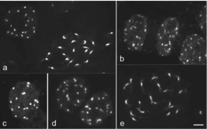
and C-banded prophase chromosomes of both species showed a higher condensation in the proximal region and decondensation at one or both chromosome termini (Figu-re 2a, e), as typically found in species with ar(Figu-reticulate nu-clei (Delay, 1949; Guerra, 1987). In general, species with smaller chromosomes tend to display more characteristic arreticulate nuclei (Barlow, 1977; Guerra, 1987), as ob-served in cacao. However, in this case, the difference in chromosome size between both species is insufficient to explain the different patterns found.
In both species, a CMA+/DAPI-band was present on the terminal region of the long arm of a single chromosome pair. This CMA+ band was frequently heteromorphic in size and distended in one or both homologues. Since most chromosome pairs were similar in morphology and size, it was not possible to precisely determine the position of this pair in the idiogram, although it was probably the second largest pair in both species (Figure 1; Figure 3a, b).
The analysis of the rDNA sites by FISH revealed a single 5S rDNA site in the proximal region of one of the three largest chromosome pairs (Figure 3d, g) and a single terminal 45S rDNA site co-localized with the CMA+band in both species (Figure 3e, h). The 45S rDNA sites exhib-ited the same size heteromorphism observed with the CMA+ bands. Heteromorphism for the 45S rDNA site is usually due to variation in the number of rDNA repeats in each homologue and has been often reported in other gen-era (see Pedrosa-Harandet al., 2006).
C-banding followed by either Giemsa or DAPI stain-ing revealed 20 well defined chromocentres in interphase nuclei of cacao (Figure 2c) and centromeric or proximal heterochromatic bands of similar size in all its 20 chromo-somes (Figure 3c). This heterochromatin distribution should be at least partially responsible for the proximal condensation pattern observed on prophase chromosomes of cacao after conventional staining (Figure 2a). Glicens-tein and Fritz (1989) tried unsuccessfully to obtain C-banded chromosomes in cacao using a different tech-nique. In cacao, as in some other genera (Bennettet al., 1995; Vanzela and Guerra, 2000), C-banding differentia-tion was better when the chromosomes were stained with
DAPI than with Giemsa. On the other hand, using the same C-banding technique for cupuaçu, no heterochromatin dif-ferentiation was found in metaphase or prophase chromo-somes, although the chromocentres were better contrasted after C-banding (Figure 2d) than after conventional stain-ing (Figure 2b). Furthermore, cacao chromosomes often exhibited pericentromeric bands after FISH, equivalent to those revealed by C-banding (Figure 3f), while in cupuaçu such a differentiation was never observed. Analyses of the pericentromeric chromatin in several plant species indicate that this region is prone to accumulate repetitive DNA se-quences and rapid differentiation (Lamb et al., 2007). Therefore, the difference between the proximal chromatin ofT. cacaoand ofT. grandiflorummay be related to the composition of the repetitive DNA sequences of this re-gion. Kawabe and Nasuda (2005) showed that the DNA se-quences of the proximal region can change very fast among
Arabidopsisspecies which may contribute to speciation. A similar process may have occurred since the beginning of the divergence between the cacao and the cupuaçu geno-mes. Hybrids between T. cacao andT. grandiflorumare sterile (Martinson, 1966) and it is possible that the different chromatin organization of their large pericentromeric
re-Figure 1- Idiograms ofTheobroma cacao(left) andT. grandiflorum(right). Chromosome pairs (P) are numbered at the top. Chromosome sizes (S) and arm ratios (AR) are indicated at the bottom. Hatched blocks = CMA+/45S rDNA; gray blocks = 5S rDNA; black blocks onT. cacaochromosomes =
C-bands.
gions may contribute to constrain the pairing between their homeologous chromosomes.
Acknowledgments
This work was supported by the Brazilian agencies Conselho Nacional de Desenvolvimento Científico e Tec-nológico - CNPq (476444/2006-3 and 134809/2006-8) and Fundação de Amparo à Ciência e Tecnologia do Estado de Pernambuco - FACEPE (EDT-0005-05.03/04).
References
Alves RM, Sebbenn AM, Artero AS, Clement C and Figueira A (2007) High levels of genetic divergence and inbreeding in populations of cupuassu (Theobroma grandiflorum). Tree Genet Genomes 3:289-298.
Barlow PW (1977) Determinants of nuclear chromatin structure in angiosperms. Ann Sci Nat 18:193-206.
Bennett AB (2003) Out of Amazon:Theobroma cacaoenters the genomic era. Trends Plant Sci 8:561-563.
Bennett ST, Leitch IJ and Bennett MD (1995) Chromosome iden-tification and mapping in the grassZingeria biebersteiniana (2n= 4) using fluorochromes. Chromosome Res 3:101-108. Carletto GM (1946) O número de cromosômios em cacaueiros.
Bol Técn Instituto de Cacau 6:32-46 (Abstract in English).
Carletto GM (1971) Morfologia dos cromossomos de cacaueiro “Catongo”. Rev Theobroma 1:11-14 (Abstract in English). Carletto GM (1974) Observações citológicas em células mães de
polem de cacaueiros. Rev Theobroma 4:34-40 (Abstract in English).
Delay C (1949) Recherches sur la structure des noyaux quiescents chez les phanérogames. Rev Cytol Cytophysiol Veg 10:103-228.
Fukui K, Ohmido N and Wako T (2000) Smallness: Gain and loss in plant chromosome research. Chromosomes Today 13:287-301.
Glicenstein LJ and Fritz PJ (1989) Ploidy level inTheobroma ca-caoL. J Hered 80:464-467.
Guerra MS (1986) Citogenética de angiospermas coletadas em Pernambuco. I. Rev Brasil Genet 9:21-40 (Abstract in Eng-lish).
Guerra MS (1987) Cytogenetics of Rutaceae IV. Structure and systematic significance of interphase nuclei. Cytologia 52:213-222.
Guerra M (2000) Patterns of heterochromatin distribution in plant chromosomes. Genet Mol Biol 23:1029-1041.
Hou M, Robinson H, Gao Y and Wang AH (2004) Crystal struc-ture of the [Mg2+-(chromomycin A3)2]-d(TTGGCCAA)2
complex reveals GGCCn binding specificity of the drug dimmer chelated by a metal ion. Nucleic Acids Res 32:2214-2222.
Figure 3- Heterochromatin and rDNA sites in the chromosomes of cacao and cupuaçu. Merged CMA/DAPI metaphase images of cacao (a) and cupuaçu (b). C-banded metaphase of cacao stained with DAPI (c). In the insert, one chromosome in higher magnification.In situhybridization of 5S rDNA (d) and 45S rDNA (e) in cacao. The insert in (e) shows a CMA+band co-localized with a 45S rDNA site. Metaphase chromosomes of cacao showing proximal bands after FISH (f).In situhybridization of 5S rDNA (g) and 45S rDNA (h) in cupuaçu. The insert in (g) shows chromosomes that were separated from the metaphase. The insert in (h) shows CMA+bands co-localized with 45S rDNA sites. Arrowheads and arrows indicate CMA+bands and rDNA sites,
Jiang J and Gill BS (2006) Current status and the future of fluores-cencein situhybridization (FISH) in plant genome research. Genome 49:1057-1068.
Jiang J, Gill BS, Wang G, Ronald PC and Ward DC (1995) Metaphase and interphase fluorescencein situhybridization mapping or the rice genome with bacterial artificial chromo-somes. Proc Natl Acad Sci USA 92:4487-4491.
Kapuscinski J (1995) DAPI: A DNA-specific fluorescent probe. Biotechnic Histochem 70:220-233.
Kawabe A and Nasuda S (2005) Structure and genomic organiza-tion of centromeric repeats in Arabidopsis species. Mol Genet Genomics 272:593-602.
Kennedy AJ (1995) Cacao. In: Smartt J and Simmonds NW (eds) Evolution of Crop Plants. 2nd edition. Longman Scientific and Technical, London, pp 472-475.
Koo D, Choi H, Cho J, Hur Y and Bang J (2005) A high-resolution karyotype of cucumber (Cucumis sativusL. ‘Winter-Long’) revealed by C-banding, pachytene analysis, and RAPD-aided fluorescencein situhybridization. Genome 48:534-540.
Lamb JC, Yu W, Han F and Birchler JA (2007) Plant chromo-somes from end to end: Telomeres, heterochromatin and centromeres. Curr Opin Plant Biol 10:116-122.
Lockwood R (2003) Who needs clothing? INGENIC Newsletter 8:2-5.
Martinson VA (1966) Hybridization of cacao andTheobroma grandiflora. J Hered 57:134-136.
Martinson VA (1975) Cytological studies of diploid and tetra-ploidTheobroma cacao. Genetica 45:341-348.
Moraes AP, Soares Filho WS and Guerra M (2007) Karyotype di-versity and the origin of grapefruit. Chromosome Res 15:115-121.
Opeke LK and Jacob VJ (1969) Cytological irregularities in Theobroma cacao L. In: 2ª Conferência Internacional de Pesquisas em Cacau, Salvador, pp 114-116.
Pedrosa-Harand A, Almeida CCS, Mosiolek M, Blair MW, Schweizer D and Guerra M (2006) Extensive ribossomal DNA amplification during Andean common bean (Phaseolus vulgaris L.) evolution. Theor Appl Genet 112:924-933.
Vanzela ALL and Guerra M (2000) Heterochromatin differentia-tion in holocentric chromosomes of Rhynchospora (Cyperaceae). Genet Mol Biol 23:453-456.
Wanzenböck EM, Schöfer C, Schweizer D and Bachmair A (1997) Ribosomal transcription units integrated via T-DNA transformation associate with the nucleolus and do not re-quire upstream repeat sequences for activity inArabidopsis thaliana. Plant J. 11:1007-1016.
Associate Editor: Yatiyo Yonenaga-Yassuda