Karyotype differentiation of four
Cestrum
species (Solanaceae)
revealed by fluorescent chromosome banding and FISH
Thiago Fernandes, Letícia do Nascimento Andrade de Almeida Rego, Mariana Nardy,
Priscila Mary Yuyama and André Luís Laforga Vanzela
Laboratório de Biodiversidade e Restauração de Ecossistemas, Centro de Ciências Biológicas,
Universidade Estadual de Londrina, Londrina, PR, Brazil.
Abstract
The karyotypes of four South American species ofCestrum (C. capsulare, C. corymbosum, C. laevigatum and C. megalophylum) were studied using conventional staining, C-CMA/DAPI chromosome banding and FISH with 45S and 5S rDNA probes. The karyotypes showed a chromosome number of 2n = 2x = 16, with metacentric chromo-somes, except for the eighth submeta- to acrocentric pair. Several types of heterochromatin were detected, which varied in size, number, distribution and base composition. The C-CMA+
bands and 45S rDNA were located predomi-nantly in terminal regions. The C-CMA+
/DAPI+
bands appeared in interstitial and terminal regions, and the C-DAPI+ bands were found in all chromosome regions. The 5S rDNA sites were observed on the long arm of pair 8 in all spe-cies exceptC. capsulare, where they were found in the paracentromeric region of the long arm of pair 4. The differ-ences in band patterns among the species studied here, along with data from other nine species reported in the literature, suggest that the bands are dispersed in an equilocal and non-equilocal manner and that structural rear-rangements can be responsible for internal karyotype diversification. However, it is important to point out that the structural changes involving repetitive segments did not culminate in substantial changes in the general karyotype structure concerning chromosome size and morphology.
Key words: Cestrum, chromosome banding, FISH, heterochromatin, physical maps, 45S and 5S rDNA.
Received: May 2, 2008; Accepted: September 8, 2008.
Introduction
The genera Cestrum L., Sessea Ruiz & Pav. and VestiaWilld. belong to the tribe Cestreae. Their representa-tives are trees, subtrees or bushes that are distributed in tropical and subtropical America (Hunziker, 1979). The fewCestrumspecies that have been cytogenetically studied presented the largest chromosomes of the family, up to about 14mm long (Fregoneziet al., 2006; Las Peñaset al.,
2006).Cestrumis the largest genus of the tribe (Juddet al., 1999), with 50 species in Brazil (Hunziker, 2001). Chromo-some banding studies have shown that in Cestrum heterochromatin varies in amount and type, and that the similarity observed among karyotypes regarding chromo-some size and morphology is apparent (see Berg and Grei-lhuber, 1992, 1993a, 1993b; Fregonezi et al., 2006). In spite of the small number of species studied,significant in-formation about the occurrence and distribution of different classes of repetitive DNA (45S and 5S rDNA,
micro-satellites and retroelements) has been reported (Berg and Greilhuber, 1992, 1993a, 1993b; Fregonezi et al., 2006, 2007; Las Peñas et al., 2006). Sykorová et al. (2003a, 2003b) described the absence of Arabidopsis (DC.) Heynh.-type (TTTAGGG)ntelomeres, which seem to be re-placed by an A/T-rich minisatellite family in Cestrum, Sessea and Vestia, besides an uncommon occurrence of 45S and 5S rDNA in B-chromosomes ofCestrum parqui L’Her. In two other studies, Fregoneziet al.(2006, 2007) described different numbers of 45S rDNA sites and the constancy in number and location of 5S rDNA sites in four species of Cestrum, plus the occurrence of a Ty3-gypsy retrotransposon family dispersed along chromosomes and forming blocks associated with NORs inC. intermedium Sendtn. andC. strigilatumRuiz & Pav.
The aim of this study was to extend the information about the occurrence and the physical location of some re-petitive DNAs in four South AmericanCestrumspecies. This work involved conventional karyotype determination and the physical location of different heterochromatin fam-ilies, as well as the 45S and 5S rDNA sites. Such informa-tion is useful in evoluinforma-tionary studies of this group and allows the development of new directions in the elucidation of karyotype differentiation inCestrum.
Send correspondence to André Luís Laforga Vanzela. Laboratório de Biodiversidade e Restauração de Ecossistemas, Centro de Ciências Biológicas, Universidade Estadual de Londrina, Caixa Postal 6001, 86051-990 Londrina, PR, Brazil. E-mail: [email protected].
Materials and Methods
Seeds of fourCestrumspecies were collected in five different localities, always from three individuals of each population, which included (i)C. capsulare Carvalho & Schnoor from Curiúva and Ventania, Paraná State, Brazil (24° 03’14” S, 50° 27’23” W), (ii)C. corymbosumSchltdl. from Tibagi, Paraná State, Brazil (24° 38’ 44” S, 50° 34’ 11” W), (iii)C. laevigatumSchltdl. from Misiones, Argen-tina (26° 48’ 25” S, 54° 44’ 22” W), and (iv) C. megalophylum Dunal from Salvador, Bahia State, Brazil (12° 52’ 51” S, 38° 21’ 41” W). Seeds were germinated and the seedlings were cultivated in the Laboratory of Biodiversity and Restoration of Ecosystems (LABRE) at the State University of Londrina (UEL). Vouchers are kept at the FUEL herbarium. For cytogenetic analysis, slides were prepared from root tips pretreated with 0.05% colchicine for 4 h, fixed in Farmer solution (ethanol/acetic acid 3:1, v:v) for up to 24 h, and stored at -20 °C. Roots were hydrolyzed in 1 M HCl for 10 min at 60 °C, dissected in a drop of 45% acetic acid and coverslipped. Coverslips were removed in liquid nitrogen, and the preparations were stained with 2% Giemsa and permanently mounted in Entellan (Merck). Chromosome measurements were made based on five metaphases, using the MicroMeasure 3.3 software (http://www.biology.colostate.edu), and the val-ues of total length, arm ratio and relative size were consid-ered for idiogram construction. Chromosomes were classified according to Guerra (1986).
Chromosome banding was performed as described by Schwarzacheret al.(1980), with minor modifications. Root tips were digested in an enzyme solution composed of 4% cellulase (w/v) and 40% pectinase (v/v) at 37 °C and dis-sected in a drop of 45% acetic acid. The slides were aged for three days and incubated in 45% acetic acid for 10 min at 60 °C, in 5% barium hydroxide for 10 min at room tem-perature, and in 2xSSC, pH 7.0, for 10 min at 60 °C. The samples were then washed in distilled water, air dried and stained with fluorochromes: 0.5 mg/mL chromomycin A3 (CMA) for 1.5 h, and 2 mg/mL
4-6-diamidino-2-phenylindole (DAPI) for 30 min. Slides were mounted with a medium composed of glycerol/McIlvaine buffer (pH 7.0) 1:1, plus 2.5 mM MgCl2. The option for CMA/DAPI stain-ing after chromosome bandstain-ing was to improve the contrast of the fine bands, due to greater retreat of the euchromatin and preservation of the heterochromatin.
Fluorescent in situ hybridization (FISH) was per-formed according to the procedures described by Heslop-Harrisonet al.(1991) and Cuadrado and Jouve (1994), with minor modifications. Slides were prepared as described for chromosome banding and immediately used for FISH. A wheat pTa71 probe containing the 45S rDNA sequence (Gerlach and Bedbrook, 1979) was labeled with digoxi-genin-11-dUTP by nick translation, and a pTa794 probe containing the 5S rDNA sequence (Gerlach and Dyer,
1980) was labeled with biotin-14-dATP, also by nick trans-lation. For each slide 34mL of a denatured hybridization
mixture was applied. This mixture was composed for 100 ng of labeled probe (4mL of each probe), 10 0%
formamide (15 mL), 50% polyethylene glycol (6 mL),
20xSSC (3mL), 100 ng of calf thymus DNA (1mL), and
10% SDS (1mL). Both slide and mixture were denatured at
90 °C for 10 min, and hybridization was performed over-night at 37 °C in a humidified chamber. Post-hybridization washes were carried out in 2xSSC, 20% formamide in 0.1xSSC, 0.1xSSC and 4xSSC/0.2% Tween 20, all at 42 °C. The probes were simultaneously detected with a so-lution composed of 5% BSA, avidin-FITC conjugate and antidigoxigenin-rhodamine conjugate (100:1:1, v:v:v), fol-lowed by post-detection baths in 4xSSC/0.2% Tween 20 at room temperature. Slides were mounted in 25mL of a
me-dium composed of 23mL of DABCO solution
[1,4-diaza-bicyclo (2.2.2)-octane (2.3%), 20 mM Tris HCl, pH 8.0, (2%) and glycerol (90%), in distilled water], 1 mL of
2mg/mL DAPI, and 1mL of 50 mM MgCl2.
All the images were acquired with a Leica DM 4500 B microscope equipped with a DFC 300FX camera and the Leica IM50 4.0 software, and optimized for best constrast and brightness with iGrafx Image software.
Results
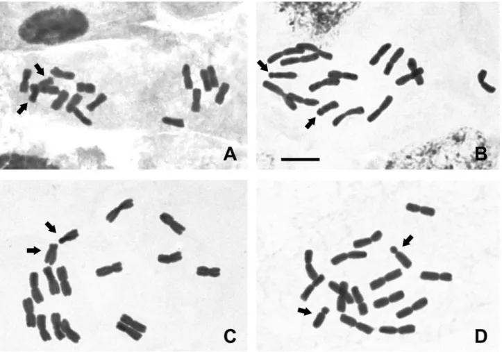
Conventional karyotype analysis of the fourCestrum species showed a constant chromosome number of 2n= 2x = 16 with metacentric chromosomes, except for the small-est pair that was from submetacentric to acrocentric (Fig-ures 1A, B, C and D and Figure 4). The interphase nuclei were also always of the reticulate type, without evident chromocenters when the nuclei were more condensed (Fig-ure 1A) and with some chromocenters when the nuclei were decondensed (Figure 1B). Differences were found in the karyotype formulae and haploid set sizes, being 7 m + 1 sm and 24mm inCestrum capsulare(Figures 1A and 4A),
7 m + 1 a in C. corymbosum, and 7 m + 1 sm in C. laevigatum, but both species presented 28mm of haploid set
sizes (Figures 1B and 4B and Figures 1C and 4C, respec-tively). An atypical idiogram was obtained for C. megalophylum, due to the presence of the heteromorphic pair 1 (Figures 1D and 4D), but the karyotype formula was 7 m + 1 a. The results of the chromosome measurements showed that C. megalophylum has the largest and C. capsulare the smallest haploid set size, while C. corymbosumandC. laevigatumshowed similar karyotype length.
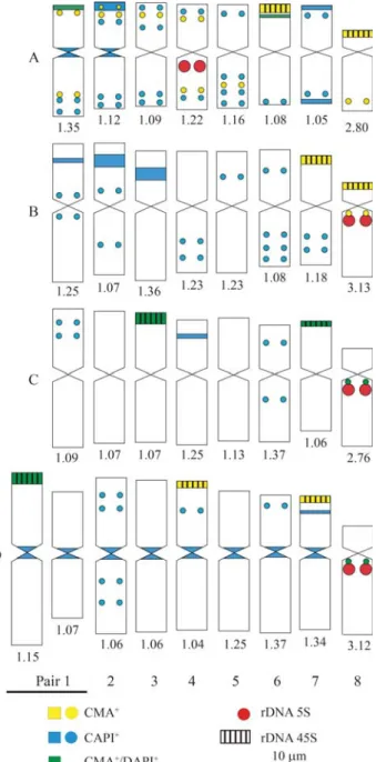
preferentially in the terminal and subterminal regions. Most of the smaller blocks or dots were observed in the intersti-tial regions (Figure 2), except forCestrum capsularethat exhibited a great number of C-DAPI+dots located in inter-stitial and terminal regions (Figure 4A). Cestrum corymbosumshowed the largest C-DAPI+bands in the in-terstitial region of the short arm of pairs 1, 2 and 3, and dots in the interstitial regions of pairs 1, 2, 4, 5, 6 and 7 (Figures 2C and 4B).Cestrum laevigatum presented the smallest number of C-DAPI+bands, located mainly in the interstitial regions of the short arms of pairs 1, 4 and 6. The only paracentromeric band was C-CMA+/DAPI+, observed on the long arm of pair 8 (Figures 2E and 4C). Cestrum megalophylum showed pericentromeric C-DAPI+ blocks on all chromosomes, except for pair 8, which was C-CMA+/DAPI+. It is important to point out that pericentromeric C-DAPI+bands were not strongly evident in all the preparations ofC. megalophylum.Larger intersti-tial C-DAPI+bands were found in pairs 3 and 7, besides in-terstitial dots in pairs 2, 4 and 6 (Figures 2G and 4D).
C-CMA+bands were usually present in the terminal regions of the short arms of meta-, submeta- and acro-centric chromosomes. InC. capsulare, besides the terminal blocks on pairs 6 and 8 associated with NORs, some
inter-stitial dots were found in subterminal regions, except in pairs 6 and 7. C-CMA+/DAPI+bands were observed in the terminal region of pair 1 and in interstitial positions on the short arms of pair 6 (Figures 2B and 4A). Cestrum corymbosumshowed terminal C-CMA+ blocks on pairs 7 and 8, in addition to a paracentromeric dot on the long arm of pair 8 (Figures 2D and 4B). In Cestrum laevigatum, C-CMA+/DAPI+ blocks were observed in the terminal re-gion of pairs 3 and 7, besides a paracentromeric dot on the long arm of pair 8 (Figures 2E, 2F and 4C). C-CMA+bands were located in the terminal region of five chromosomes in C. megalophylum, on the short arms of pairs 4 and 7 and of the larger chromosome of the heteromorphic pair 1, and in the paracentromeric region (dot) of the long arms of pair 8 (Figures 2H and 4D). Only the bands observed in pairs 1 and 8 were C-CMA+/DAPI+.
Figure 2- Chromosome banding with fluorochromes inCestrumspecies: C-DAPI banding inC. capsulare(A);C. corymbosum, incomplete metaphase (C);C. laevigatum(E); andC. megalophylum(G). C-CMA banding inC. capsulare(B),C. corymbosum(D),C. laevigatum(F) andC. megalophylum (H). The larger arrows indicate C- CMA+/DAPI+bands; the smaller arrows indicate C-CMA-/DAPI+or C-CMA+/DAPI-bands. Bar = 10
rDNA was associated with GC-rich bands. In C. laevigatum,signals were observed on pairs 3 and 7 (Figures 3C and 4C), but associated with C-CMA+/DAPI+bands. In C. megalophylum, hybridization signals were observed on pairs 4 and 7, associated with GC-rich bands, besides the larger chromosome of the heteromorphic pair 1, associated with C-CMA+/DAPI+ bands (Figures 3D and 4D). FISH with the 5S rDNA probe showed hybridization signals al-ways in the proximal region of pair 8, associated with GC-rich bands inC. corymbosum(Figures 3B and 4B) and associated with C-CMA+/DAPI+ bands in C. laevigatum andC. megalophylum(Figures 3C and 4C and Figures 3D and 4D, respectively).Cestrum capsulareshowed a hybrid-ization signal on pair 4, but also in the paracentromeric re-gion of the largest chromosome (Figures 3A and 4A).
Discussion
Cytogenetic analysis, performed for the first time in C. capsulare, C. corymbosum, C. laevigatum and C. megalophylum, showed a constant chromosome number (2n= 2x= 16), as previously described forCestrum(Berg and Greilhuber, 1992, 1993a, 1993b; Fregonezi et al., 2006). In addition to the numerical stability, other cytogenetic features were also constant, such as the pre-dominance of meta- and submetacentric chromosomes, with pair 8 always being acrocentric, and the reticulate interphase nuclei (Berg and Greilhuber, 1992, 1993a,
1993b; Fregoneziet al., 2006; Las Peñaset al., 2006). Al-though there was a predominance of metacentrics and two submetacentrics for pair 8 in this study, some arm ratio val-ues were very close to the limit between meta- and submetacentrics and submeta- and acrocentrics as defined by Guerra (1986) (see Figure 4). It is important to empha-size that the apparent constancy of chromosome number and karyotype morphology found inCestrumwas also ob-served in conventional cytogenetic studies of other Solanaceae, such as the generaCapsicumL. (Pozzobonet al., 2006),SolanumL. (Bernardello and Anderson, 1990) andLycopersicumHill (Pillenet al., 1996), all with 2n= 24.
Our results complement those obtained by Berg and Greilhuber (1992, 1993a, 1993b), Fregoneziet al.(2006) and Las Peñaset al.(2006), pointing out that there are some minor variations in the chromosome types regarding centromere position, chromosome size and karyotype sym-metry. Las Peñaset al.(2006) proposed that an increase in genome size is accompanied by a small karyotype asymme-try, common in the tribe Cestreae. According to these au-thors, Vestia species are the most plesiomorphic within Cestreae, with the most symmetrical karyotypes and the smallest chromosomes. On the other hand,Sesseaand Ces-trumspecies are more apomorphic, with more asymmetri-cal karyotypes and larger chromosomes. Our results partially support the ideas of Las Peñaset al.(2006), once C. capsulareshowed the most symmetrical karyotype and
had the smallest haploid set size, whileC. megalophylum presented the most asymmetrical karyotype and the largest haploid set size. This difference is sustained by taxonomic data, sinceC. capsularewas once classified into the genus Sessea(Sessea regnelliiTaub.) and was later reclassified to Cestrum(Carvalho and Schnoor, 1997). However, chromo-some banding (described below) showed thatC. capsulare accumulated more repetitive DNA than the other three spe-cies, which could produce an increase in karyotype differ-ences, including asymmetry.
Chromosome banding showed variation in the type, amount and distribution of heterochromatin among the four Cestrumspecies studied. However, we also found similari-ties, mainly with regard to the equilocal/equidistant and non- equilocal/equidistant distribution of bands (see Schweizer and Ehrendorfer, 1976; Schweizer and Loidl, 1987). C-CMA/DAPI banding allowed the identification of chromosomes and karyotypes, and the construction of idio-grams, as previously demonstrated by Berg and Greilhuber (1992, 1993a, 1993b) and Fregoneziet al.(2006).
The physical maps defined three “domains” for heterochromatin occurrence inCestrum. The first one was represented by the largest terminal C-CMA+ and C-CMA+/DAPI+bands. The terminal C-CMA+blocks were equilocal and equidistant, with interstitial dots on some heterologous chromosomes of C. capsulare. The second domain was represented by the largest interstitial/subtermi-nal C-DAPI+ bands, which are equilocal and equidistant, with C-DAPI+dots inC. corymbosumand less frequently in C. laevigatumandC. megalophylum.The third domain was represented by centromeric C-DAPI+ bands in Cestrum capsulareandC. megalophylum. The occurrence of these “domains” could be explained by the model of Schweizer and Loidl (1987), where different types of heterochromatin could begin preferentially in terminal regions and after-wards “contaminate” the interstitial regions of adjacent non-homologous chromosomes. Similar cases were re-ported inGibasis karwinskyana(Roem. & Schult.) Rohw., Commelinaceae (Kenton, 1991) and someCestrumspecies (Berg and Greilhuber, 1992, 1993a, 1993b; Fregonezi et al., 2006). This kind of dispersion was found here in C. corymbosumandC. megalophylum, and was previously re-ported forC. intermediumandC. strigilatum(Fregoneziet al., 2006).
The centromeric heterochromatin dispersion pattern detected by chromosome banding procedures can be con-sidered of little importance inCestrum, if compared with other plant groups, for exampleOphrysL. (Orchidaceae). D’Emericoet al. (2005) studied seven species ofOphrys and found evident centromeric C-bands (Giemsa) in all the chromosomes. Another example of repetitive DNA accu-mulation in the centromeric regions of plants was published by Valáriket al.(2002). These authors isolated and cloned several repetitive DNA sequences ofMusa acuminataL. (bananas) and, except for those associated with NORs, they appeared clustered in the centromeric regions of all chro-mosomes, as detected by FISH. These lines of evidence en-dorse the idea that there are specific positions in the chromosomes where arrays of repetitive DNA are accumu-lated, favored or tolerated. According to Flavell (1986), the selection process against or in favor of repetitive DNA ac-cumulation can occur in differential ways, and it could also explain the different physical maps generated forCestrum.
FISH with ribosomal probes showed a common pat-tern of 45S rDNA distribution in the four species studied Figure 4- Idiograms with physical location of repetitive segments in
here compared to otherCestrumspecies (Fregoneziet al., 2006), since their hybridization sites were always located in the terminal regions of the chromosomes. These segments were found in different numbers but always in terminal re-gions, as in other genera of Solanaceae, such asCapsicum (Mosconeet al., 1995) andNicotianaL. (Limet al., 2000). Cestrum capsulare, C. corymbosum and C. laevigatum showed four 45S rDNA sites, whereasC. megalophylum exhibited a heteromorphic pair with a terminal C-CMA+/NOR segment. NOR-associated heteromor-phisms can happen due to non-equal crossing-over, non-reciprocal translocations or deletions. However, our data did not allow us to determine responsible event for these heteromorphisms, as well as if this event was fixed in the population or not, because samples of few individuals were collected in a restricted area. Different numbers of 45S rDNA sites have been described forCestrum, varying from four to eight (Fregoneziet al., 2006). Thus, events in-volving the amplification of rDNA repeats or an increase in the number of sites do not seem to affect the adaptive suc-cess of Cestrumspecies. Additionally, it is possible that other events are responsible for the dispersion of 45S rDNA repeats. Fregoneziet al.(2007) showed that a representa-tive of theTy3-gypsyfamily appears to be associated with NORs ofC. intermediumandC. strigilatum, and that this retroelement could be involved in rDNA mobility among Cestrum species. Franz et al. (2000) suggested that in Arabidopsisthe various types of transposable elements as-sociated to NOR regions might favor rDNA mobility and affect both the evolution and the expression of rDNA loci.
The location of the 5S rDNA in paracentromeric re-gions of the long arm of pair 8 in C. corymbosum, C. laevigatumandC. megalophylumis in agreement with that described by Fregoneziet al.(2006) for otherCestrum spe-cies. Differences were found inCestrum capsulare, where its chromosome position is the same, but on a different chromosome (pair 4). It is possible that a translocation has moved the 5S rDNA segment from pair 8 to pair 4, since the long arm of pair 8 ofC. capsularedecreased in size and, ac-cording to measurements, this pair is not acrocentric as in other Cestrum species, but submetacentric. Another karyotype feature that attracted our attention was the differ-ential association of 5S rDNA with heterochromatin. In this regard, we observed three different conditions: (i) lack of association with heterochromatin, as inC. capsulare;(ii) association with C-CMA+ segments, as found in C. corymbosum; and (iii) association with C-CMA+/DAPI+ segments, as found inC. laevigatumandC. megalophylum. The co-location of 5S rDNA and heterochromatin has al-ready been described for other species. Xu and Earle (1996) showed that inLycopersicum(Solanaceae) the 5S rDNA was associated with heterochromatic bands, and Cerbahet al.(1998) and Ruaset al.(2005) reported this association in several species ofHypochoerisL. (Asteraceae). The occur-rence of paracentromeric 5S rDNA on the long arm of pair
8 is considered conserved inCestrum, but the discovery in C. capsulare, which had been earlier ascribed to the genus Sessea(S. regnellii), can indicate two possibilities: (i) with the increase in the number of species studied, this location cannot be considered a good chromosome marker for the genus; or (ii) our results may be in contradiction to the junc-tion of the generaCestrumandSessea, which was based on the diagnosis of fruit morphology, as proposed by Carvalho and Schnoor (1997). However, when we consider the gen-eral cytogenetic features ofC. capsulare, including large chromosomes, a great number of heterochromatic blocks, equilocal distribution and number and location of 45S rDNA,C. capsulareseems to be well bounded.
In conclusion, the physical mapping generated after fluorescent chromosome banding and FISH with 45S and 5S rDNA, when compared with the maps generated for otherCestrumspecies, reinforces the idea that the karyo-type changes inCestrumresulted mainly from a great varia-tion in the occurrence and distribuvaria-tion of different repetitive DNA segments, without major modifications in the karyotype formulae and chromosome size.
Acknowledgments
The authors thank the Brazilian agencies Fundação Araucária, IAP-SEMA and CNPq for financial support and Prof. Dr. José Marcelo D. Torezan for critical review and suggestions. Dr. A. Leyva provided English editing of the manuscript.
References
Berg C and Greilhuber J (1992) Cold-sensitive chromosome re-gions and their relation to constitutive heterochromatin in Cestrum parqui(Solanaceae). Genome 35:921-930. Berg C and Greilhuber J (1993a) Cold-sensitive chromosome
re-gions and heterochromatin in Cestrum (Solanaceae): C. strigilatum, C. fasciculatumandC. elegans. Plant Syst Evol 185:133-151.
Berg C and Greilhuber J (1993b) Cold-sensitive regions and heterochromatin in Cestrum aurantiacum (Solanaceae). Plant Syst Evol 185:259-273.
Bernardello LM and Anderson GJ (1990) Karyotypic studies in Solanum section Basarthrum (Solanaceae). Am J Bot 77:420-431.
Carvalho LAF and Schnoor A (1997)SesseaCarvalho et Schnoor – Nova seção para o gêneroCestrum(Solanaceae). Rodri-guésia 46:15-24.
Cerbah M, Coulaud J and Siljak-Yakovlev S (1998) rDNA orga-nization and evolutionary relationship in the genus Hypochaeris(Asteraceae). J Hered 89:312-318.
Cuadrado A and Jouve N (1994) Highly repetitive sequences in B chromosomes ofSecale cerealerevealed by fluorescencein situhybridization. Genome 37:709-712.
Flavell RB (1986) Repetitive DNA and chromosome evolution in plants. Phil Trans R Soc Lond 312:227-242.
Franz PF, Armstrong S, de Jong H, Parnell LD, Drunen CV, Dean C, Zabel P, Bisseling T and Jones G (2000) Integrated cytogenetic map of chromosome arm 4S of A. thaliana: Structural organization of heterochromatic knob and cen-tromere region. Cell 100:367-376.
Fregonezi JN, Fernandes T, Torezan JMD, Vieira AOS and Van-zela ALL (2006) Karyotype differentiation of fourCestrum species (Solanaceae) based on physical mapping of repeti-tive DNA. Genet Mol Biol 29:97-104.
Fregonezi JN, Vilas-Boas LA, Fungaro MHP, Gaeta ML and Vanzela ALL (2007) Distribution of aTy3/gypsy-like
retro-element on the A and B-chromosomes of Cestrum
strigilatumRuiz & Pav. andCestrum intermediumSendtn. (Solanaceae). Genet Mol Biol 30:599-604.
Gerlach WL and Bedbrook JR (1979) Cloning characterization of ribosomal RNA genes from wheat and barley. Nucleic Acids Res 7:1869-1885.
Gerlach WL and Dyer TA (1980) Sequence organization of the re-peating units in the nucleus of wheat which contain 5S rDNA genes. Nucleic Acids Res 8:4851-4865.
Guerra M (1986) Reviewing the chromosome nomenclature of Levanet al.Rev Bras Genet 9:741-743.
Heslop-Harrison JS, Schwarzacher T, Anamthawat-Jonsson K, Leitch AR, Shi M and Leitch IJ (1991)In situhybridization with automated chromosome denaturation. Technique 3:106-109.
Hunziker AT (1979) South AmericanSolanaceae, a synoptic sur-vey.In:Hawkes JG, Lester RN and Skelding AD (eds) The Biology and Taxonomy of theSolanaceae. 7th edition. Lin-nean Society Symposium Series. Academic Press, New York, pp 49-86.
Hunziker AT (2001) Genera Solanacearum: The Genera of Sola-naceae Illustrated, Arranged According to a New System.
Gantner Verlag K.-G., Liechtenstein, 500 pp.
The Genera of Solanaceae. A.R.G. Gantner Verlag K.-G., Liech-tenstein, 500 pp.
Judd WS, Campbell CS, Kellogg EA and Stevens PF (1999) Plant Systematics: A Phylogenetic Approach. Sinauer Associates, Sunderland, 464 pp.
Kenton A (1991) Heterochromatin accumulation, disposition and
diversity in Gibasis karwinskyana (Commelinaceae).
Chromosoma 100:467-478.
Las Peñas ML, Chiarini FE, Bernardello G and Benítez de Rojas C (2006) Karyotypes of some species ofCestrum,Sesseaand Vestia(tribe Cestreae, Solanaceae). Caryologia 59:131-137.
Lim KY, Matyasek R, Lichtenstein CP and Leitch AR (2000) Mo-lecular cytogenetic analyses and phylogenetic studies in the NicotianasectionTomentosae. Chromosoma 109:245-258. Moscone EA, Loidl J, Ehrendorfer F and Hunziker AT (1995)
Analysis of active nucleolus organizing regions inCapsicum (Solanaceae) by silver staining. Am J Bot 82:276-287. Pillen K, Pineda O, Lewis CB and Tanksley SD (1996) Status of
genome mapping tools in the taxon Solanaceae. In: Paterson AH and Landes RG (eds). Genome Mapping in Plants. Landes Company, Texas, pp 281-308.
Pozzobon MT, Schifino-Wittmann MT and Bianchetti LB (2006) Chromosome numbers in wild and semidomesticated Bra-zilian CapsicumL. (Solanaceae) species: Do x = 12 and
x = represent two evolutionary lines? Bot J Lin Soc
151:259-269.
Ruas CF, Vanzela ALL, Santos MO, Fregonezi JN, Ruas PM, Matzenbacher NI and Aguiar-Perecin MLR (2005) Chromo-somal organization and phylogenetic relationships in Hypochaerisspecies (Asteraceae) from Brazil. Genet Mol Biol 28:129-139.
Schwarzacher TP, Ambros P and Schweizer D (1980) Application of Giemsa banding to orchid karyotype analysis. Plant Syst Evol 134:293-297.
Schweizer D and Ehrendorfer FW (1976) Giemsa banded karyo-types, systematics, and evolution inAnacyclus (Asteraceae-Anthemideae). Plant Syst Evol 126:107-148.
Schweizer D and Loidl J (1987) A model for heterochromatin dis-persion and the evolution of C-banded patterns. Chrom To-day 9:61-74.
Sykorová E, Lim KY, Chase MW, Knapp S, Leitch IJ, Leitch AR and Fajkus J (2003a) The absence ofArabidopsis-type telo-meres in Cestrum and closely related genera Vestia and Sessea(Solanaceae); first evidence from eudicots. Plant J 34:283-291.
Sykorová E, Lim KY, Fakijus J and Leitch AR (2003b) The signa-ture of the Cestrumgenome suggests an evolutionary re-sponse to the loss of (TTTAGGG)ntelomeres. Chromosoma 112:164-172.
Valárik M, Simková H, Hribová E, Safár J, Dolezevolá M and Dolzel J (2002) Isolation, characterization and chromosome localization of repetitive DNA sequences in bananas (Musa spp.). Chromosome Res.10:89-100.
Xu J and Earle ED (1996) High-resolution physical mapping of 45S (5.8S, 18S and 25S) rDNA gene loci in the tomato ge-nome using a combination of karyotyping and FISH of pachytene chromosomes. Chromosoma 104:545-550.
Associate Editor: Marcelo Guerra