Dehydrocrotonin and its
h
-cyclodextrin complex: Cytotoxicity
in V79 fibroblasts and rat cultured hepatocytes
Daniel H.A. Correˆa
a,T
, Patricia S. Melo
a, Carlos A.A. de Carvalho
b,c,
Mariaˆngela B.M. de Azevedo
c, Nelson Dura´n
d,e, Marcela Haun
aa
Departamento de Bioquı´mica, Instituto de Biologia, Universidade Estadual de Campinas (UNICAMP), CP 6109, Campinas, SP 13083-970, Brazil b
Departamento de Quı´mica, Instituto de Cieˆncias Exatas e Biolo´gicas, Universidade Federal de Ouro Preto (UFOP), Ouro Preto, MG 35400-000, Brazil c
STQ Indu´stria e Come´rcio Ltda-ME-CIETEC, Cidade Universita´ria, Sa˜o Paulo, SP 05508-900, Brazil d
Laborato´rio de Quı´mica Biolo´gica, Instituto de Quı´mica, Universidade Estadual de Campinas (UNICAMP), CP 6154, Campinas, SP 13083-970, Brazil e
Nu´cleo de Cieˆncias Ambientais, Universidade de Mogi das Cruzes, Mogi das Cruzes, SP 08790-911, Brazil
Received 28 October 2004; received in revised form 10 January 2005; accepted 13 January 2005
Abstract
Trans-dehydrocrotonin has antiulcerogenic and antitumor activities. A complex ofh-cyclodextrin with dehydrocrotonin was developed to improve the delivery of dehydrocrotonin. Complex in solid state was evaluated using X-ray diffraction (XRD), differential scanning calorimetry (DSC), thermal gravimetric analysis (TGA) and scanning electron microscopy (SEM). X-ray diffraction and scanning electron
microscopy studies showed that dehydrocrotonin exists in a semicrystalline state in the complexed form withh-cyclodextrin. Differential
scanning calorimetry studies showed the existence of a complex of dehydrocrotonin withh-cyclodextrin. The thermal gravimetric analysis
studies confirmed the differential scanning calorimetry results of the complex. Free dehydrocrotonin and the dehydrocrotonin/h-cyclodextrin
inclusion complex were assayed in freshly isolated rat hepatocytes and in V79 cells. Cytotoxicity was determined using nucleic acid content, methylthiazoletetrazolium (MTT) reduction and neutral red uptake assays. In all assays, there was a large reduction (3.5–16.1-fold) in the
cytotoxicity of dehydrocrotonin in hepatocytes when complexed withh-cyclodextrin, whereas for V79 cells the decrease in cytotoxicity was
1.7- and 1.87-fold for MTT reduction and nucleic acid content assays, respectively. The lower cytotoxicity of the dehydrocrotonin/h
-cyclodextrin complex compared to free dehydrocrotonin in rat hepatocytes and V79 cells suggests that such a complex may be useful for the administration of dehydrocrotonin in vivo.
D2005 Elsevier B.V. All rights reserved.
Keywords:h-Cyclodextrin; Cytotoxicity; Dehydrocrotonin; Hepatocyte; V79 cell
1. Introduction
The primary purpose of drug delivery systems is to efficiently and precisely deliver the necessary amount of drug to the target site for a specific period of time. To design advanced forms of delivery; suitable carrier materials are used to overcome the undesirable properties of the drug molecules of interest (Hirayama and Uekama, 1999; Szente and Szejtli, 1999; Rajewski and Stella, 1996). Cyclodextrins
are a group of oligosaccharides obtained from the enzymatic degradation of starch and are known to modifying the physico-pharmaceutical properties of various drugs and components through the formation of inclusion complex (Bibby et al., 2000). Because of their ability to alter the physical, chemical, and biological properties of guest molecules in this way, cyclodextrins are potential candidates for drug delivery devices (Ren et al., 2002).
The a-, h-, and g-cyclodextrins are widely used
natural cyclodextrins, that consist of six, seven, and eight
d-glucopyranose residues, respectively, linked by a-1,4
glycosidic bonds into a macrocycle. Each cyclodextrin has its own characteristic ability to form inclusion complexes
0014-2999/$ - see front matterD2005 Elsevier B.V. All rights reserved. doi:10.1016/j.ejphar.2005.01.016
TCorresponding author. Tel.: +55 19 3788 6150; fax: +55 19 3788 6129. E-mail address:dcorrea@unicamp.br (D.H.A. Correˆa).
with specific guests that depends on a proper fit of the guest molecule into the hydrophobic cyclodextrin cavity (Stella and Rajewski, 1997; Loftsson, 1999). The most common pharmaceutical application of cyclodextrins is to enhance the solubility, stability and bioavailability of drug molecules (Loftsson, 1999).
The principal advantages of natural cyclodextrins as drug carriers are: (1) a well-defined chemical structure with many potential sites for chemical modification or conjugation, (2) the availability of cyclodextrins of different cavity size, (3) low toxicity and low pharmacological activity, (4) some degree of water solubility, and (5) protection of the included/conjugated drugs from biodegradation. h -Cyclo-dextrin, the most common natural cyclo-Cyclo-dextrin, has 21 hydroxyl groups that include 7 primary and 14 secondary hydroxyls. Topologically, this macrocycle can be described as a truncated cone in which the narrow rim (~6.42) bears the primary hydroxyl group whereas the wide rim (~15.42) bears the secondary OH groups. Since no hydroxyl group is present within the toroidal cavity of h-cyclodextrin, this zone of the molecule has a pronounced hydrophobic character (Thompson, 1997). Moreover, the inside of the
h-cyclodextrin molecule forms a hydrophobic cavity whereas the outer surface is hydrophilic; thereby enabling the molecule to act as a host for a wide variety of lipophilic drugs (Zhao et al., 1996).
Trans-dehydrocrotonin is a nor-clerodane diterpene lactone obtained fromCroton cajucara Benth (Euphorbia-ceae), a Brazilian medicinal plant known asbsacacaQthat is commonly used in folk medicine as an infusion in a powdered or dried pill form to treat a large number of disorders (Melo et al., 2002; Hiruma-Lima et al., 1999). Experimental studies with laboratory animals have shown that dehydrocrotonin has hypoglycemic, antigenotoxic, antiulcer, anti-inflammatory and antitumor effects (Agner et al., 2001; Souza-Brito et al., 1998; Grynberg et al., 1999). However, previous reports indicated that dehydrocrotonin induced hepatotoxicity and was cytotoxic to V79 fibroblasts and rat hepatocytes (Melo et al., 2002; Rodriguez and Haun, 1999).
Hepatotoxicity has been reported to be a possible limitation in the use of dehydrocrotonin (Rodriguez and Haun, 1999; Melo et al., 2002), and efforts have been made to develop new delivery systems to diminish the side effects of this compound. As part of a study to develop a suitable formulation for the delivery of dehydrocrotonin, in this work we investigated the formation of an inclusion complex between dehydrocrotonin and h-cyclodextrin using X-ray diffraction (XRD), differential scanning calorimetry (DSC), scanning electron microscopy (SEM) and thermal gravi-metric analysis (TGA). We also examined the cytotoxicity of this new formulation in primary cultures of rat hepatocytes and in V79 fibroblasts in order to assess its usefulness as a pharmaceutical drug and to explore the ability of the carrier to reduce the toxicity and enhance the pharmacological efficacy of dehydrocrotonin.
2. Materials and methods
2.1. Preparation of the inclusion complex of dehydrocroto-nin withb-cyclodextrin
Trans-dehydrocrotonin (5-(3-furanyl)-2V, 3V, 4, 4V, 4Va, 5, 8V, 8Va-octhydro-2V, 5V-dimethyl-[1Valpha(R*), 2Valpha, 4Va alpha, 8Va betha]spiro[furan-3(2H), 1V(7VH )-naphthalene]-2,7Vdione) was isolated and purified fromC. cajucara bark as previously described by Souza-Brito et al. (1998). The purity was over 99% as assessed by nuclear magnetic resonance, ultraviolet, infrared and mass spectroscopy techniques (Souza-Brito et al., 1998).h-Cyclodextrin was purchased from Sigma Chemical Co. (St. Louis, MO, USA) and was used without further purification.
Dehydrocrotonin and h-cyclodextrin were mixed in a molar ratio of 1:2 for the dehydrocrotonin/h-cyclodextrin solution in 1:1 organic–aqueous medium (ethanol/water), and stirred continuously with slight heating on a magnetic stirrer for 5 h. To obtain the solid inclusion complex, the solution was evaporated after preparation under vacuum in a rotating evaporator at 458C. A physical mixture with same molar ratio of 1:2 as used for the dehydrocrotonin/h -cyclodextrin complex was prepared by gently mixing the two compounds in a ceramic mortar for 2 min (De Azevedo et al., 2000).
2.2. X-ray diffraction studies
The powder X-ray diffraction (XRD) patterns of the samples were recorded using a Siemens D 5000-XRD X-ray diffractometer (Siemens Electronical Equipment, Toronto, ON, Canada) under the following conditions: target Cu, filter Ni, voltage 40 kV, current 30 mA, scanning speed 2 8C/min, chart speed 40 mm/min, and count range 1000 counts/s. The detector was a proportional counter with 1.7 kV detector voltage.
2.3. Differential scanning calorimetry (DSC) and thermal gravimetric analysis (TGA)
Differential scanning calorimetry (DSC) and thermal gravimetric analysis (TGA) were done using a DSC-STAR system (Mettler Toledo Inc., Columbus, OH, USA). Samples (2–5 mg) were heated in sealed aluminium pans under nitrogen flow (40–50 ml/min) at a heating rate of 108C/min, from 0 to 6008C.
2.4. Scanning electron microscopy
deposition current of 20 mA in a rarefied argon atmos-phere at 0.1–0.2 mbar for 60 s. The specimens were observed in a LEO 440i Scanning electron microscope (Leo Electron Microscopy Ltd., Cambridge, UK), using an acceleration voltage of 20 kV and an 18-mm working distance.
2.5. Animals
Male Wistar rats (200–250 g) were obtained from the Central Animal Facilities at UNICAMP (CEMIB/UNI-CAMP) and were housed under controlled conditions (22F18C, 55F5% relative humidity, and a 12 h light–dark
cycle), with free access to a standard chow (Nuvilab Cr-1-A, Nuvital, Sa˜o Paulo, Brazil) and tap water, for at least 1 week prior to use.
2.6. Isolation and culture of rat hepatocytes
Hepatocytes were isolated from 2-month-old male Wistar rats by a two-step collagenase perfusion method (Guguen-Guillouzo and Guillouzo, 1986). After assessing cell integrity by trypan blue exclusion, the cells were seeded at a density of 6105cells/ml in 96-well plates in 0.1 ml of DMEM supplemented with 10% foetal bovine serum, 0.2% bovine serum albumin, 0.1 IU of bovine insulin/ml, 10 6M dexamethasone, 50 IU of penicillin/ml and 50Ag of streptomycin/ml and then incubated at 37 8C in a 5% CO2humidified atmosphere. After 4 h incubation for cell attachment, the medium was changed to serum-free medium containing dehydrocrotonin, dehydrocroto-nin/h-cyclodextrin (1:2 ratio) or a physical mixture of the two compounds at eight concentrations (0 to 500 AM, relative to dehydrocrotonin) and the cells then incubated for 20 h. The compounds were initially dissolved in DMSO and then in supplemented DMEM without foetal bovine serum. The final concentration of DMSO in the test medium and controls was 1%. Each concentration was tested in six replicates in each of three separate experiments. At the end of the incubation, three endpoints for cytotoxicity (the nucleic acid content, MTT reduction and neutral red uptake) were evaluated as described below.
2.7. V79 fibroblast culture
The cytotoxicity of dehydrocrotonin and the dehydroc-rotonin/h-cyclodextrin complex was assessed in a perma-nent lung fibroblast cell line (V79) derived from Chinese hamsters. These cells are commonly used for cytotoxicity studies (Souza-Brito et al., 1998; Rodriguez and Haun, 1999). V79 fibroblasts were grown as monolayers in Dulbecco’s modified Eagle’s medium (DMEM) supple-mented with 10% foetal bovine serum, 100 IU of penicillin/ ml and 100Ag of streptomycin/ml in a humidified incubator with 5% CO2 in air at 37 8C. The cells were plated at a
density of 3104 cells/ml in 96-well plates. Forty-eight hours after cell seeding, semiconfluent cultures were exposed to dehydrocrotonin, dehydrocrotonin/h -cyclodex-trin (1:2 ratio) or a physical mixture of the two compounds at eight concentrations ranging from 0 to 500 AM (relative to dehydrocrotonin). The compounds were initially dis-solved in dimethyl sulfoxide (DMSO) and then in DMEM. The final concentration of DMSO in the test medium and controls was 0.2%. The cells were exposed for 24 h to the test medium with or without the compounds studied (control). Each concentration was tested in six replicates in each of three separate experiments. At the end of the incubation, three independent endpoints for cytotoxicity (the nucleic acid content, MTT reduction and neutral red uptake) were evaluated.
2.8. Endpoint tests for cytotoxicity
2.8.1. Nucleic acid content
The number of cells in control and treated wells was estimated from the total nucleic acid content (Cingi et al., 1991). After treatment, the cells were washed twice with cold phosphate-buffered saline (PBS) and a soluble nucleotide pool was extracted with cold ethanol. The cell monolayers were then digested in 0.5 M NaOH (0.1 ml/well) overnight at room temperature. The absorbance of the NaOH fraction was measured at 260 nm (UV–visible DUR 640B
Spec-trophotometer, Beckman Instruments, Inc., Fullerton, CA, USA) and was used as an index of cell number (Bianchi and Fortunati, 1990) and the results were expressed as a percentage of the 260 nm absorbance of the control wells.
2.8.2. Methylthiazoletetrazolium (MTT) reduction
The MTT reduction assay was done as described by Denizot and Lang (1986). Briefly, cells were washed once with PBS before adding 0.1 ml of serum-free medium containing 0.05% of MTT salt to each well. After incubation for 5 h, the culture medium was removed and 0.1 ml of ethanol was added to each well to solubilize the formazan formed. The plates were shaken gently for 10 min and the absorbance was measured at 570 nm (VersaMaxk, Tunable
Microplate Reader, Molecular Devices, Co., Sunnyvale, CA, USA).
2.8.3. Neutral red uptake
The neutral red uptake was measured by the method of Borefreund and Puerner (1984). Briefly, cells were washed once with PBS after removal of the culture medium. After 4 h of incubation with serum-free medium containing neutral red (50Ag/ml), the cells were washed in PBS and then 0.1 ml of a solution of 1% acetic acid and ethanol (50%) was added to each well to fix the cells and to remove the neutral red from the solution. The plates were then shaken gently for 20 min on a plate shaker and the absorbance of the solution was read at 540 nm (VersaMaxk, Tunable
2.9. Statistical analysis
The experiments were done three times (six replicates each) in separate experiments. To calculate the IC50 values (concentration that produced a 50% inhibitory effect on the evaluated parameter), the results were expressed as a percentage of the controls and were determined graphically from the concentration–response curves using the computer software package OriginR-Data Analysis and Technical
Graphics, version 6.0 (Copyright Software, Inc.).
3. Results
3.1. X-ray diffractograms
Fig. 1shows the X-ray diffraction patterns of dehydroc-rotonin,h-cyclodextrin alone, and the coevaporated mixture of dehydrocrotonin/h-cyclodextrin (1:2 molar ratio). The X-ray diffraction patterns showed the formation of a new crystalline phase indicated by the absence of some peaks present in the dehydrocrotonin diffraction patterns at 2h=8.08, 13.58, 16.58 and 248 (Fig. 1, line c) and in the
h-cyclodextrin diffraction patterns at 2h=8.58, 19.08and 218 (Fig. 1, line a). On the other hand, new peaks appeared in the inclusion compound diffraction pattern at 2h=12.08and 18.08 (Fig. 1, line b). The diffraction pattern of the 1:2 physical mixture (not shown) was simply a superposition of those of both components (dehydrocrotonin and h -cyclo-dextrin) alone.
3.2. Differential scanning calorimetry (DSC) and thermal gravimetric analysis (TGA)
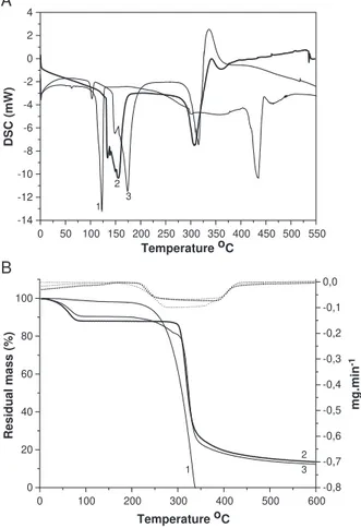
The differential scanning calorimetry thermograms (Fig. 2A) showed an endothermic peak at the dehydrocrotonin melting point (135.478C) and another endothermic peak at the dehydrocrotonin thermal collapse (416.17 8C). How-ever, these peaks were absent in the thermogram of the inclusion complex, suggesting that a new supramolecular
species had been obtained. The thermogram of the inclusion complex showed an endothermic peak at 117.49 8C that was not present in the dehydrocrotonin and h-cyclodextrin thermograms. The endothermic peak between 143.388C and 167.318C seen in the thermogram ofh-cyclodextrin, attributed to the loss of water molecules,
5 10 20 30 40 50 60
Scattering intensity (CPS)
Scattering angle, 2θ (degrees)
A
B
C
Fig. 1. X-ray diffractograms of (A)h-cyclodextrin alone, (B) coevaporate of dehydrocrotonin withh-cyclodextrin, (C) dehydrocrotonin alone.
A
B
0 50 100 150 200 250 300 350 400 450 500 550 -14
-12 -10 -8 -6 -4 -2 0 2 4
DSC (mW)
Temperature oC
1 2
3
0 100 200 300 400 500 600
0 20 40 60 80 100
-0,8 -0,7 -0,6 -0,5 -0,4 -0,3 -0,2 -0,1 0,0
Residual mass (%)
Temperature oC
mg.min
-1
1
2 3
appeared with a reduced area in the thermogram of the inclusion complex.
Thermal gravimetric analysis thermograms are shown in Fig. 2B. The thermal gravimetric analyses thermogram of dehydrocrotonin showed a single, continuous process of
thermal decomposition that started at 250 8C and reached 350 8C without the formation of any solid residue. In contrast, the thermal gravimetric analyses thermogram of the inclusion complex showed that the complex was thermally more stable than dehydrocrotonin, with the solid residue formed at 6008C corresponding to 17% of material analysed. The differential scanning calorimetry and thermal gravimetric analysis thermograms of the physical mixture were a superposition of the thermal events of free dehydrocrotonin andh-cyclodextrin (not shown).
The combination of these physicochemical techniques (X-ray diffraction, differential scanning calorimetry and thermal gravimetric analysis) suggested the formation of an inclusion complex.
3.3. Scanning electron microscopy (SEM)
The morphological appearance of the dehydrocrotonin crystals and the dehydrocrotonin/h-cyclodextrin inclusion complex is shown in Fig. 3. The crystalline aspect of dehydrocrotonin (Fig. 3A) disappeared in the inclusion complex (Fig. 3B), indicating a host–guest interaction.
3.4. Effects on cell viability
3.4.1. V79 fibroblasts
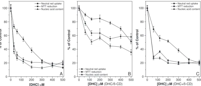
Fig. 4A, B shows the viability of V79 fibroblasts after treatment with free dehydrocrotonin and the dehydrocroto-nin/h-cyclodextrin complex for 24 h, respectively. The cytotoxicity of free dehydrocrotonin for the three endpoints assayed in this cell line were concentration-dependent (Fig. 4A). The dehydrocrotonin/h-cyclodextrin complex (Fig. 4B) and free dehydrocrotonin (Fig. 4A) showed a similar low cytotoxicity in the neutral red uptake assay (IC20of 400 AM and 480AM, respectively). In the MTT reduction assay,
Fig. 3. Scanning electron micrographs (SEM) of dehydrocrotonin (A) and the inclusion complex (B).
0 100 200 300 400 500 0
20 40 60 80 100
A
Neutral red uptake MTT reduction Nucleic acid content
% of Control
[DHC] µM
0 100 200 300 400 500 0
20 40 60 80 100
B
Neutral red uptake MTT reduction Nucleic acid content
% of Control
[DHC] µM (DHC/ß-CD)
0 100 200 300 400 500 0
20 40 60 80 100
C
Neutral red uptake MTT reduction Nucleic acid content
% of Control
[DHC] µM (DHC+ß-CD)
the dehydrocrotonin/h-cyclodextrin complex was less cyto-toxic (IC50 of 676 AM, estimated by extrapolating the plotted data) (Fig. 4B) than free dehydrocrotonin (IC50 of 400 AM) (Fig. 4A). A similar lower cytotoxicity for the dehydrocrotonin/h-cyclodextrin complex compared to free dehydrocrotonin was observed in the nucleic acid content assay (IC50of 420AM and 225AM, respectively) (Fig. 4B, A, respectively).
3.4.2. Rat hepatocytes
Fig. 5A, B shows the viability of recently isolated hepatocytes after treatment with dehydrocrotonin and the dehydrocrotonin/h-cyclodextrin complex for 20 h, respec-tively. The cytotoxicity of dehydrocrotonin on hepatocytes was be dependent on the concentrations used. IC50of 25AM and 254AM, and 144 AM and 500AM were obtained for free dehydrocrotonin (Fig. 5A) and the dehydrocrotonin/h -cyclodextrin complex (Fig. 5B) in the nucleic acid content and MTT assays, respectively. The dehydrocrotonin/h -cyclodextrin complex was less cytotoxic (IC50N500 AM) than free dehydrocrotonin (IC50of 31AM) in the neutral red uptake assay (Fig. 5B, A, respectively).
3.4.3. Possible steric hindrance between dehydrocrotonin andb-cyclodextrin
To investigate a possible steric hindrance between dehydrocrotonin and h-cyclodextrin, V79 fibroblasts and rat hepatocytes were incubated with a physical mixture of dehydrocrotonin and h-cyclodextrin (1:2 molar ratio) at dehydrocrotonin concentrations of 0 to 500 AM. Figs. 4C and 5Cshow the viability of V79 fibroblasts and recently isolated hepatocytes after treatment with a physical mixture of dehydrocrotonin andh-cyclodextrin. For V79 fibroblasts, there was no change in neutral red uptake assay when compared to the complex. In contrast, the MTT reduction
and nucleic acid content assays showed a significant reduction in the cytotoxicity of the physical mixture (IC20 of 500AM and IC50 of 350AM, respectively) compared to free dehydrocrotonin. The cytotoxicity of the physical mixture in hepatocytes cultures was similar to that of free dehydrocrotonin in the neutral red uptake and nucleic acid content assays and was slightly lower (1.48-fold) in the MTT reduction assay.
4. Discussion
In this work, the formation of a crystalline inclusion complex between dehydrocrotonin and h-cyclodextrin by coevaporation was analysed using differential scanning calorimetry, thermal gravimetric analysis and X-ray diffrac-tion. These three techniques allow a qualitative character-isation of the solid state of the systems by comparing their thermal and diffraction peaks, respectively (Mura et al., 1999). The X-ray diffraction data indicated the formation of a new crystalline phase based on a different peak that appeared in the diffraction pattern of the inclusion complex and the absence of some peaks present in the dehydrocro-tonin and h-cyclodextrin patterns.
The differential scanning calorimetry thermogram of the inclusion complex, when compared to those of dehydroc-rotonin and h-cyclodextrin, lacked the peak at 135.47 8C attributed to the melting point of dehydrocrotonin, which suggested that a new supramolecular species had been obtained. These findings were corroborated by the thermal gravimetric analysis thermograms for dehydrocrotonin and the inclusion complex. Under the conditions used here, dehydrocrotonin showed poor thermal stability when compared to the inclusion complex and started to decom-pose at 250 8C, with complete decomposition occurring at
0 100 200 300 400 500 0
20 40 60 80 100
A B C
Neutral red uptake MTT reduction Nucleic acid content
% of Control
[DHC] µM
0 100 200 300 400 500 0
20 40 60 80 100
Neutral red uptake MTT reduction Nucleic acid content
% of Control
[DHC] µM (DHC/ß-CD)
0 100 200 300 400 500 0
20 40 60 80 100
Neutral red uptake MTT reduction Nucleic acid content
% of Control
[DHC] µM (DHC+ß-CD)
350 8C without leaving any residue. In contrast, the inclusion complex formed a solid residue at 600 8C that corresponded to 17% of material analysed.
The lack of the endothermic peak at 416.17 8C in the differential scanning calorimetry thermogram of the inclu-sion complex, attributed to the thermal collapse of dehydrocrotonin, reinforced the findings presented of the thermal gravimetric analysis thermograms and indicated that the inclusion process involved stabilisation of the guest molecule. Further evidence for the formation of a new supramolecular species included the appearance of a new endothermic peak at 117.498C in the differential scanning calorimetry thermogram of the inclusion complex; this peak was not present in the other thermograms. The region between 143.38 8C and 167.31 8C in the h-cyclodextrin differential scanning calorimetry thermogram was attributed to the loss of water molecules and showed a reduced area in the differential scanning calorimetry thermogram of the inclusion complex. This reduction suggested an apolar– apolar interaction within theh-cyclodextrin cavity, with the substitution of some water molecules by the guest molecule. In an aqueous solution, the slightly apolar cyclodextrin cavity is occupied by water molecules that are energetically unfavored (polar–apolar interaction), and can therefore be readily substituted by appropriatebguest moleculesQthat are less polar than water (Szejtli, 1998). In most cases, the host– guest ratio is 1:1, although ratios of 2:1, 1:2 and 2:2, or more complex associations and higher order equilibrium almost always occur simultaneously (Szejtli, 1998). The results from differential scanning calorimetry, thermal gravimetric analysis and X-ray diffraction, combined with those of scanning electron microscopy, corroborated the formation of a new supramolecular species (De Azevedo et al., 2002).
Primary cultures of rat hepatocytes are routinely used to assess toxicity because of their capacity to maintain a sufficient level of xenobiotic metabolism and because the liver is one of the primary target organs for exposure to xenobiotics in vivo (Paillard et al., 1999). V79 fibroblasts do not have the cytochrome P450 system and are commonly used for cytotoxicity studies and to evaluate mutagenic effects (Rodriguez and Haun, 1999; Melo et al., 2002; Katzer et al., 2002). Melo et al. (2002) showed that the cytotoxicity of dehydrocrotonin in hepatocytes was induced by the greater inhibition of mitochondrial succinate dehy-drogenase. Indeed, the cytotoxicity in hepatocytes compared to V79 fibroblasts results from the bioactivation of dehydrocrotonin by the cytochrome P450 system to produce metabolites that are more toxic to hepatocytes than the parent compound (Melo et al., 2002; Rodriguez and Haun, 1999).
In our conditions, freeh-cyclodextrin (1 mM) was not cytotoxic to V79 fibroblasts or recently isolated hepato-cytes. Therefore, since free h-cyclodextrin showed no activity over the concentration range used in this study, the lower cytotoxicity of the dehydrocrotonin/h
-cyclodex-trin complex compared to free dehydrocrotonin was attributed to the formation of a host–guest complex between dehydrocrotonin and h-cyclodextrin, thereby avoiding the dehydrocrotonin side effects. In V79 fibroblasts, complexed dehydrocrotonin showed no significant change in cytotox-icity compared to free dehydrocrotonin in the neutral red uptake assay. In contrast, in the MTT reduction and nucleic acid content assays, the cytotoxicity was 1.7-fold and 1.87-fold lower than that of free dehydrocrotonin, respectively. The viability of recently isolated hepatocytes was increased by the presence of h-cyclodextrin. The cytotoxicity of the dehydrocrotonin/h-cyclodextrin complex was 3.5-fold, 10.2-fold and 16.1-fold lower than that of free dehydrocro-tonin in the MTT reduction, nucleic acid content and neutral red uptake assays, respectively.
Cyclodextrins accelerate or decelerate various types of reactions, with kinetic features similar to those of enzyme reactions, i.e., catalyst–substrate complex formation, com-petitive inhibition, saturation, and stereospecific catalysis (Szejtli, 1988). The most important primary consequence of the interaction between a poorly soluble guest and a cyclodextrin in aqueous solution that could explain the reduction in the cytotoxicity of dehydrocrotonin when complexed with h-cyclodextrin is a modification in the reactivity of the included molecule. In most cases, the reactivity decreases because of guest stabilisation, but in other cases the cyclodextrin behaves as an artificial enzyme that can accelerate and modify the reaction pathway (Szejtli, 1998). The rate of reaction is changed by the inclusion because the guest is transferred from the polar environment of water to a less polar one of the cyclodextrin cavity, i.e.; there is a microsolvent effect. The reaction rate increases when flexible guest molecules are forced to adjust to a reactive conformation and vice versa (Griffiths and Bender, 1973).
The cytotoxicity of the physical mixture in V79 fibroblasts showed no markedly difference from that of free dehydrocrotonin as assessed by the neutral red uptake assay. However, in the MTT reduction and nucleic acid content assays, a slightly lower cytotoxicity was observed (1.22-fold and 1.55-fold, respectively) compared to free dehydrocro-tonin. In contrast, when the hepatocytes was treated with the physical mixture, the cytotoxicity was 1.5-fold, 1.84-fold and 1.5-fold lower than for free dehydrocrotonin in the MTT reduction, nucleic acid content and neutral red uptake assays, respectively. These results indicate that the complex-ation of dehydrocrotonin with h-cyclodextrin altered its cytotoxicity since the physical mixture was not as effective in reducing the cytotoxicity.
Anazetti et al. (2004) showed that the dehydrocrotonin/
h-cyclodextrin modified the physicochemical characteristics of the guest molecule and diminished its cytotoxicity. Further experiments are needed to investigate the physico-pharmaceutical properties of the inclusion complex of dehydrocrotonin with h-cyclodextrin and to assess the extent to which the pharmacological activities are retained compared to free dehydrocrotonin.
5. Conclusions
The physicochemical results presented here and the decrease in the cytotoxicity of complexed dehydrocrotonin compared to free dehydrocrotonin indicate that dehydroc-rotonin/h-cyclodextrin inclusion complexes have a potential use in maintaining/enhancing the pharmacological activities of dehydrocrotonin and may have useful therapeutic applications.
Acknowledgements
The authors thank J. B. Fabrin Neto for technical assistance. This work was supported by CAPES, FAPESP, PRONEX and the Nanobiotechnology Network (MCT/CNPq).
References
Agner, A.R., Maciel, M.A., Pinto, A.C., Colus, I.M., 2001. Antigenotox-icity of trans-dehydrocrotonin, a clerodane diterpene from Croton cajucara. Planta Med. 67, 815 – 819.
Anazetti, M.C., Melo, P.S., De Azevedo, M.B.M., De Carvalho, C.A., Dura´n, N., Haun, M., 2004. h-Cyclodextrin complexed with dehy-drocrotonin induces apoptosis in HL60 cells as seen by flow cytometry. XII Congress of the Brazilian Society for Cell Biology and IX Ibero-American Congress of Cell Biology. Brazil: Campinas, I-012. Bianchi, V., Fortunati, E., 1990. Cellular effects of an anionic surfactant
detected in V79 fibroblasts by different cytotoxicity tests. Toxicol. In Vitro 4, 9 – 16.
Bibby, D., Davies, N.M., Tucker, I.G., 2000. Mechanisms by which cyclodextrins modify drug release from polymeric drug delivery systems. Int. J. Pharm. 197, 1 – 11.
Borefreund, E., Puerner, J.A., 1984. A simple quantitative procedure using monolayer cultures for cytotoxicity assays (HTD/NR 90). J. Tissue Cult. Methods 9, 7 – 9.
Cingi, M.R., De Angelis, I., Fortunati, E., Reggiani, D., Bianchi, V., Tiozzo, R., Zucco, F., 1991. Choice and standardization of test protocols in cytotoxicology: a multicentre approach. Toxicol. In Vitro 5, 119 – 125. De Azevedo, M.B.M., Alderete, J.B., Lino, A.C.S., Loh, W., Faljoni-Alario, A., Dura´n, N., 2000. Violacein/h-cyclodextrin inclusion complex formation studied by measurements of diffusion coefficient and circular dichroism. J. Ind. Phenom. Mol. Recognit. Chem. 37, 67 – 74. De Azevedo, M.B.M., Zullo, M.A.T., Alderete, J.B., De Azevedo, M.M.M.,
Dura´n, N., 2002. Characterization and properties of the inclusion
complex of 24-epibrassinolide with h-cyclodextrin. Plant Growth Regul. 37, 233 – 240.
Denizot, F., Lang, R., 1986. Rapid colorimetric assay for cell growth and survival. Modifications to the tetrazolium dye procedure giving improved sensitivity and reliability. J. Immunol. Methods 89, 271 – 277. Griffiths, D.W., Bender, M.L., 1973. Orientational catalysis by
cyclo-hexaamylose. J. Am. Chem. Soc. 95, 1679 – 1680.
Grynberg, N.F., Echevarria, A., Lima, J.E., Pamplona, S.S.R., Pinto, A.C., Maciel, M.A., 1999. Anti-tumour activity of two 19-nor-clerodane diterpenes, trans-dehydrocrotonin and trans-crotonin, from Croton cajucara. Planta Med. 65, 687 – 689.
Guguen-Guillouzo, C., Guillouzo, A., 1986. Methods for preparation of adult and fetal hepatocytes. In: Guillouzo, A., Guguen-Guillouzo, C. (Eds.), Research in isolated and cultured hepatocytes. Les Editions John Libbey Eurotext, London, UK, pp. 1 – 12.
Hirayama, F., Uekama, K., 1999. Cyclodextrin-based controlled drug release system. Adv. Drug Deliv. Rev. 36, 125 – 141.
Hiruma-Lima, C.A., Spadari-Bratfisch, R.C., Kassisse, D.M., Souza-Brito, A.R.M., 1999. Antiulcerogenic mechanisms of dehydrocrotonin, a diterpene lactone obtained from Croton cajucara. Planta Med. 65, 325 – 330.
Katzer, A., Marquardt, H., Westendorf, J., Wening, J.V., Von, F.G., 2002. Polyetheretherketone—cytotoxicity and mutagenicity in vitro. Bioma-terials 23, 1749 – 1759.
Loftsson, T., 1999. Pharmaceutical applications ofh-cyclodextrins. Pharm. Technol. Europe 11, 20 – 32.
Melo, P.S., Dura´n, N., Haun, M., 2002. Derivatives of dehydrocrotonin, a diterpene lactone isolated from Croton cajucara: cytotoxicity in rat cultured hepatocytes and in V79 cells. Human Exp. Toxicol. 21, 281 – 288.
Mura, P., Faucci, M.T., Parrini, P.L., Furlanetto, S., Pinzauti, S., 1999. Influence of the preparation method on the physicochemical properties of ketoprofen–cyclodextrin binary systems. Int. J. Pharm. 179, 117 – 128. Paillard, F., Finot, F., Mouche, I., Prenez, A., Vericat, J.A., 1999. Use of primary cultures of rat hepatocytes to predict toxicity in the early development of new chemical entities. Toxicol. In Vitro 13, 693 – 700. Rajewski, R.A., Stella, V.J., 1996. Pharmaceutical application of
cyclo-dextrins. 2. In vivo drug delivery. J. Pharm. Sci. 85, 1142 – 1169. Ren, X., Xue, Y., Liu, J., Zhang, K., Zheng, J., Luo, G., Guo, C., Mu, Y.,
Shen, J., 2002. A novel cyclodextrin-derived tellurium compound with glutathione peroxidase. ChemBioChem 3, 363 – 365.
Rodriguez, J.A., Haun, M., 1999. Cytotoxicity of trans-dehydrocrotonin fromCroton cajucaraon V79 cells and rat hepatocytes. Planta Med. 65, 522 – 526.
Souza-Brito, A.R.M., Rodriguez, J.A., Hiruma-Lima, C.A., Haun, M., Nunes, D.S., 1998. Antiulcerogenic activity oftrans-dehydrocrotonin fromCroton cajucara. Planta Med. 64, 126 – 129.
Stella, V.J., Rajewski, R.A., 1997. Cyclodextrins: their future in drug formulation and delivery. Pharm. Res. 14, 556 – 567.
Szejtli, J., 1988. Cyclodextrin technology. Kluwer Academic, Dordrecht, NL.
Szejtli, J., 1998. Introduction and general overview of cyclodextrin chemistry. Chem. Rev. 98, 1743 – 1754.
Szente, L., Szejtli, J., 1999. Highly soluble cyclodextrin derivatives: chemistry, properties, and trends in development. Adv. Drug Deliv. Rev. 36, 17 – 28.
Thompson, D.O., 1997. Cyclodextrins-enabling excipients: their present and future use in pharmaceuticals. Crit. Rev. Ther. Drug Carr. Syst. 14, 1 – 104.