Re sistance o f multice llular aggre gate s
to pharmo rubicin o bse rve d in human
he pato carcino ma ce lls
State Key Laboratory of Biomembrane and Membrane Biotechnology, Institute of Zoology, Chinese Academy of Sciences, Beijing, China Z. Jianmin,
W. Hongfang and F. Meifu
Abstract
The objective of the present study was to investigate the multicellular resistance of human hepatocarcinoma cells BEL-7402 to pharmorubi-cin. Cells (1 x 10⁴) and 200 microcarrier Cytodex-3 beads were seeded onto a 24-well plate and cultured in RPMI 1640 medium. After the formation of multicellular aggregates, morphology and cell viability were analyzed by scanning electron microscopy, transmission electron microscopy and flow cytometry, respectively. The IC50 was deter-mined by flow cytometry and MTT assay after the cells cultured in aggregates and monolayers were treated with pharmorubicin. The culture products exhibited structural characteristics somewhat similar to those of trabecular hepatocarcinoma in vivo. Among the microcar-riers, cells were organized into several layers. Intercellular spaces were 0.5-2.0 µm wide and filled with many microvilli. The percent of viable cells was 87%. The cells cultured as multicellular aggregates were resistant to pharmorubicin with IC50 4.5-fold and 7.7-fold that of monolayer culture as determined by flow cytometry and MTT assay, respectively. This three-dimensional culture model may be used to investigate the mechanisms of multicellular drug resistance of hepato-carcinoma and to screen new anticancer drugs.
Co rre spo nde nce
F. Meifu
State Key Laboratory of Biomembrane and Membrane Biotechnology Institute of Zoology Chinese Academy of Sciences Beijing, 100080
China
Fax: + 86-10-6257-1017 E-mail: fengmf@ panda.ioz.ac.cn
Research supported by a grant from Space Cell Culture.
Received June 11, 2001 Accepted O ctober 30, 2001
Ke y words
•Hepatocellular carcinoma
•Spheroids
•Drug resistance
•Microcarrier
•Epirubicin
Intro ductio n
Intrinsic or acquired resistance to chemo-therapeutic drugs is one of the major ob-stacles in the treatment of solid tumors. A number of possible mechanisms have been proposed to account for drug resistance (1). Previous research demonstrated that tumor cells cultured in three-dimensional (3-D) aggregates were resistant to cytotoxic drugs. The resistance was lost when the cells were disaggregated and cultured in monolayers.
Previously, 3-D culture of hepatocarcinoma was used to study the cytotoxic effects of alcohol and to determine the function of hepatoma cells. In the present study, human hepatocarcinoma cells (BEL-7402) were cultured with microcarrier beads (Cytodex-3) to establish a 3-D model and to investigate multicellular drug resistance to pharmorubi-cin.
Mate rial and Me thods
Ce ll line and ce ll culture
BEL-7402 cells were derived from a specimen obtained from a 53-year-old male patient with hepatocarcinoma in 1974. The cell line was established at the Shanghai Institute of Cell Biology, Academia Sinica. The cells were cultured in RPMI 1640 medi-um (Gibco BRL, Rockville, MD, USA) supplemented with 10% fetal calf serum (Hyclone, Logan, UT, USA), 100 U/ml peni-cillin and 100 U/ml streptomycin in the pres-ence of 5% CO2 and 95% air, at 37ºC (8).
Thre e -dime nsional culture
Microcarrier beads Cytodex-3 (Sigma, St. Louis, MO, USA) were allowed to swell in Ca2+- and Mg2+-free PBS and then steril-ized at 110ºC for 30 min. The beads were washed twice with sterile PBS, pH 7.4, be-fore culture. BEL-7402 cells were harvested freshly by treatment with 0.25% trypsin con-taining 2% EDTA. Cells (1 x 10⁴) and 200 microcarrier beads per well were seeded onto a 24-well plate and cultured in the same medium. The plate was coated with 10% poly(2-hydroxyethyl methacrylate) (Sigma) to prevent cell adhesion.
Scanning e le ctron microscopy
After a mean 10-day period of culture, the microcarrier beads were aggregated and covered with multilayer cells as observed
with the phase contrast microscope. A fur-ther 4-5 days later, samples were washed with PBS, pH 7.4, and fixed with 2.5% glu-taraldehyde (SPI-Chem, West Chester, PA, USA) in PBS, pH 7.4, for 1 h at room tem-perature. After three washes in PBS, they were post-fixed with 1.0% osmium tetroxide (Matthey, Materials Technology, Hertford-shire, England) in PBS for 1 h at room tem-perature followed by dehydration with a growing ethyl alcohol series (30, 50, 70, 80, 90 and 100%). The samples were then treated with iso-amylacetate for more than 2 h, dried to the critical point and coated with gold. In order to observe cross-sections of the aggre-gates, part of the aggregates were cleaved before being coated with gold. Finally, the samples were observed with a scanning elec-tron microscope (JEOL, TSM-5600LB, To-kyo, Japan).
Transmission e le ctron microscopy
Additional samples were fixed and dehy-drated as described for scanning electron microscopy and embedded in Epon812 ep-oxy resin (Epon812 kit, SPI-Chem). Thin sections were prepared and examined with a transmission electron microscope (JEM-1230, Tokyo, Japan).
Flow cytome try
ml) added). Samples stained only with propidium iodide or Annexin-V-FLUOS were used as single staining control. All samples were incubated at room tempera-ture for 10-15 min in the dark. A control without staining was also prepared. Incuba-tion buffer (400 µl) was added to the samples before flow cytometry. The data were ana-lyzed with the CellQuest software (Becton Dickinson).
Cells cultured in monolayers and the 3-D aggregates mentioned above were treated with 1.25, 2.5, 5, 10 or 20 µM pharmorubi-cin (Pharmacia & Upjohn S.P.A., Peapack, NJ, USA) in the above culture medium for 30 h. Then the apoptosis and necrosis of 5 x 10⁵ cells per sample were analyzed by the same method as above. The percentage of viable cells was determined and the IC50 was calculated by the regression method.
MTT assay
MTT (3-(4,5-dimethylthiazol-2-yl)-2,5-diphenyl tetrazolium bromide) (Sigma) was dissolved in PBS at 5 mg/ml and sterilized by filtration. After treatment with 1, 2, 4, 8, 16 or 32 µM pharmorubicin for 30 h, the cells were freshly disaggregated by enzymatic dis-sociation and the cell number was deter-mined with a hemocytometer. A cell suspen-sion (100 µl) of each sample was added to a 96-well plate. The cell number per well was within the range of the standard curve. Stock MTT solution (10 µl per 100 µl medium) was added to all wells. After incubation in the presence of 5% CO2 and 95% air at 37ºC for 6 h, the supernatant was discarded. Dis-solved solution (100 µl) (17% Triton X-100, 6.45 mol/l dimethylformamide, 0.2 mol/l cit-ric acid) was added to each well and mixed thoroughly to dissolve the dark blue crystals. After half an hour at room temperature to ensure that all crystals were dissolved, the plates were read with a MicroElisa reader at a wavelength of 570 nm. All samples were
read in triplicate. Finally the percentage of viable cells was determined and the IC50 was calculated by the regression method.
Statistical analysis
Data are reported as means ± SEM. The chi-square test was used for statistical anal-ysis (P<0.01).
Re sults
Morphology (scanning and transmission
e le ctron microscopy)
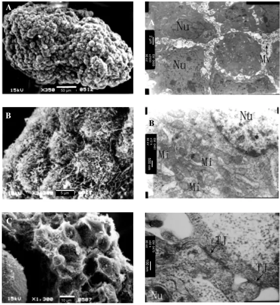
The aggregates were irregular with a di-ameter of up to 1.0-2.0 mm and composed of microcarriers and cells. The cells were oval spheroid or polyhedric with many microvilli on the surfaces and packed together densely. Cross-sections showed that cells were orga-nized densely in multilayers and distributed among the microcarriers (Figure 1). Trans-mission electron microscopy showed that cells were polygonal and arranged in several layers. Adjacent cells were simply apposed and tight junctions were observed. The inter-cellular space was 0.5-2.0 µm wide and con-tained many microvilli which enabled the exchange of liquid medium through the ag-gregates. Cytoplasm was full of mitochon-dria (Figure 2).
Ce ll viability
Apoptosis and necrosis of the 3-D aggre-gates described above were analyzed with a flow cytometer. Cell viability was 87.77 ± 1.76%. Apoptosis and necrosis were 2.77 ± 0.52 and 11.11 ± 3.37%, respectively.
Se nsitivity te st
cul-Figure 1. Scanning electron microscopy of 3-D culture of BEL-7402 cells. The 3-D structure lasted 4-5 days. A, The aggregates w ere irregular w ith a diameter up to 1.0-2.0 mm and covered w ith densely apposed cells (350X). B, Local amplification of aggregates (3,300X). The cells w ere spherical w ith many densely arranged microvilli. C, Cross-section of the aggregates (1,300X). The cells w ere polyhedric, arranged into multilayers and covered the microcarrier beads.
A
A
B
C
B
C
Figure 2. Transmission electron microscopy of 3-D cul-ture of BEL-7402 cells. The 3-D struccul-ture formed for 4-5 days. A, Cells arranged in multilayers (2,4-500X). The intercellular space w as 0.5-2.0 µm w ide and filled w ith many microvilli (M v). The nucleus (Nu) w as large and irregular. B, Cytoplasm w as rich in mitochondria (M i) (12,000X). C, Adjacent cells w ere connected by tight junctions (TJ) (50,000X).
tures treated with various concentrations of pharmorubicin was higher than that of mono-layer culture (P<0.01) (Figures 3 and 4). The IC50 for 3-D aggregates was 4.5-fold that of monolayer culture by flow cytometry and 7.7-fold by the MTT assay, indicating
that BEL-7402 cells cultured in 3-D became resistant to pharmorubicin (Table 1).
D iscussio n
Three-dimensional cell culture had been
2
µ
m
E
M
0
6
9
9
x
2
5
0
0
9
0
k
V
5
0
0
n
m
E
M
0
4
8
1
x
1
2
k
8
0
k
V
1
0
0
n
m
E
M
0
4
7
5
x
5
0
k
8
0
k
V
50 µm
5 µm
widely used in biomedical research (2,9-11). Many kinds of solid tumors in vivo and
tumor cells cultured in 3-D in vitro exhibit intrinsic or acquired resistance to cytotoxic drugs, which is one of the major obstacles to clinical treatment. Various factors are in-volved in multicellular drug resistance, such as alterations in drug transport (12), drug metabolism (13), drug target (14), cellular repair mechanisms (15) and a decreased sus-ceptibility to apoptosis (16). Understanding the mechanisms of multicellular drug resis-tance will contribute to the exploration of more efficient chemotherapy strategies. In this study, human hepatocarcinoma cells BEL-7402 were cultured with Cytodex-3 microcarrier beads to form 3-D aggregates. The results indicated that the cells were oval spheroid or polyhedric. Cells were arranged together densely in multilayers which were distributed among the microcarriers. In vivo,
trabecular hepatocarcinoma is characterized by multilayers of hepatocarcinoma cells that are separated by hepatocytes or sinuses (17). In this model the multilayers of hepatocarci-noma cells were separated by microcarrier beads but not by sinuses. Multilayers of cells were distributed among the microcarrier beads and covered the aggregates, thus re-sembling, to a certain extent, the morpho-logical characteristics of trabecular hepato-carcinoma in vivo. Tight junctions were found, and our previous study indicated that desmosomes were formed as well, proper-ties that are essential for the integrity of the 3-D aggregates. The percent of viable cells was 87%. The presence of many mitochon-dria and microvilli suggested that the cells were in good condition. The cells turned out to be less sensitive to pharmorubicin than the cells cultured in monolayers. Our previous study indicated that after the formation of 3-D aggregates the percent of cells in the G0-G1 phase increased, while the percent of cells in the G2-M phase decreased. This suggests that more cells shift into a quiescent state (data not shown). However, the
major-ity of conventional cytotoxic anticancer drugs preferentially kill cycling cells. The increase of the percent of quiescent cells and the decrease of efficiency of drug penetration into the center of the aggregates might result in a decreased sensitivity.
Understanding the mechanisms of multi-cellular drug resistance should improve clini-cal chemotherapeutic strategies. In terms of screening new drugs, if drugs demonstrate anticancer activity comparable to or greater than that seen in a monolayer culture, this could indicate more promising efficacy for
M
o
rt
a
lit
y
(
%
) 100
1.5 1.0
0.5 0
80 60 40 20 0
Concentration (log M )
M onolayer 3-D
Figure 3. Sensitivity of BEL-7402 cells to pharm orubicin deter-mined by flow cytometry. Cells cultured in monolayer and 3-D aggregates w ere treated w ith 1.25, 2.5, 5, 10 and 20 µM phar-morubicin for 30 h. Apoptosis and necrosis w ere then analyzed by cytometry and cell mortality w as determined. The cells in monolayer culture w ere more sensit ive t o pharm orubicin (P<0.01, chi-square test).
Table 1. IC50 (µM ) of pharmorubicin applied to BEL-7402 cells analyzed by flow cytometry and M TT assay.
M onolayer 3-D culture culture
IC50 flow cytometry 4.50 ± 0.68 20.16 ± 5.7 IC50 M TT assay 3.41 ± 0.63 26.30 ± 13.7
Data are reported as means ± SEM . 1.5 1.0 0.5 0
Concentration (log M )
M onolayer 3-D
M
o
rt
a
lit
y
(
%
) 10080
60
40 20 0
2.0
the treatment of solid tumors in vivo. Cells in
this 3-D model were organized into a multi-layer structure resembling, to a certain extent, the morphological characteristics of trabecular hepatocarcinoma in vivo. The cells cultured in 3-D aggregates were resistant to pharmorubicin compared with mono-layer culture, a result that may contribute to research on multicellular drug resis-tance and to the screening of new antican-cer drugs.
Ackno wle dgm e nts
We would like to offer special thanks to Hu Baihe and Zhao Xinrong, Peking Univer-sity Health Science Center, China, and to Gan Yaling, Li Shiwen and Li Wenhao, In-stitute of Zoology of the Chinese Academy of Sciences, for technical assistance with scanning and transmission electron micros-copy.
Re fe re nce s
1. Kessel D (1994). M odes of resistance to antitumor agents. In Vivo, 8: 829-834. 2. Graham CH, Kobayashi H, Stankiew icz KS,
M an S, Kapitain SJ & Kerbel RS (1994). Rapid acquisition of multicellular drug re-sistance after a single exposure of mam-mary tumor cells to antitumor alkylating agents. Journal of the National Cancer Institute, 86: 975-982.
3. Kobayashi H, M an S, Graham CH, Kapitain SJ, Teicher BA & Kerbel RS (1993). Ac-quired multicellular-mediated resistance to alkylating agents in cancer. Proceed-ings of the National Academy of Sciences, USA, 90: 3294-3298.
4. Spaulding GF, Jessup JM & Goodw in TJ (1993). Advances in cellular construction. Journal of Cellular Biochemistry, 51: 249-251.
5. Carlsson J & Yuhas JM (1984). Liquid-overlay culture of cellular spheroids. Re-cent Results in Cancer Research, 95: 1-23.
6. Santini M T, Indovina PL & Hausman RE (1987). Changes in myoblast membrane order during differentiation as measured by EPR. Biochimica et Biophysica Acta, 896: 19-25.
7. Sutherland RM & Durand RE (1984). Grow th and cellular characteristics of
mul-ticell spheroids. Recent Results in Cancer Research, 95: 24-49.
8. Chen J-M , Chu F-H & Yeh H-J (1978). The establishment and some characteristics of a human liver carcinoma cell line (BEL-7402) in vitro. Acta Biologicae Experimen-talis Sinica, 11: 37-47.
9. Inch WR, M cCredie JA & Sutherland RM (1970). Grow th of nodular carcinoma in rodents compared w ith multi-cell sphe-roids in tissue culture. Grow th, 34: 271-282.
10. Sutherland RM , M cCredie JA & Inch WR (1971). Grow th of multicellular spheroids in tissue cultures as a model of nodular carcinomas. Journal of the National Can-cer Institute, 46: 113-120.
11. Santini M T & Rainaldi G (1999). Three-dimensional spheroid model in tumor bi-ology. Pathobiology, 67: 148-157. 12. Kane SE, Pastan I & Gottesman M M
(1990). Genetic basis of multidrug resis-tance of tumor cells. Journal of Bioener-getics and Biomembranes, 22: 593-618. 13. Hoban PR, Robson CN, Davies SM , Hall
AG, Cattan AR, Hickson ID & Harris AL (1992). Reduced topoisomerase II and el-evated alpha class glutathione S-trans-ferase expression in a multidrug resistant CHO cell line highly cross-resistant to
mi-tomycin C. Biochemical Pharmacology, 43: 685-693.
14. M estdagh N, Pommery N, Saucier JM , Hecquet B, Fournier C, Slomianny C, Teis-sier E & Henichart JP (1994). Chemoresis-tance to doxorubicin and cisplatin in a murine cell line: analysis of P-glycoprotein topoisomerase II activity, glutathione and related enzymes. Anticancer Research, 14: 869-874.
15. Ishikaw a H, Kaw ano M M , Okada K, Tanaka H, Tanabe O, Sakai A, Asaoku H, Iw ato K, Nobuyoshi M & Kuramoto A (1993). Expression of DNA topoisomer-ase I and II gene and the genes possibly related to drug resistance in human my-eloma cells. British Journal of Haematolo-gy, 83: 68-74.
16. Kim R & Beck WT (1994). Differences betw een drug-sensitive and -resistant hu-man leukemic CEM cells in c-jun expres-sion, AP-1 DNA-binding activity, and for-mation of Jun/Fos family dimers, and their association w ith internucleosomal DNA ladders after treatment w ith VM -26. Can-cer Research, 54: 4958-4966.