A chro m ato graphic m e tho d fo r
the pro ductio n o f a hum an
im m uno glo bulin G so lutio n
fo r intrave no us use
Divisão de Produção e Desenvolvimento Industrial,
Fundação Pró-Sangue Hemocentro de São Paulo, São Paulo, SP, Brasil K. Tanaka, E. Sawatani,
E.M. Shigueoka, T.C.X.B. Campos, H.C. Nakao, G.A. Dias, R.K. Fujita and F. Arashiro
Abstract
Immunoglobulin G (IgG) of excellent quality for intravenous use was obtained from the cryosupernatant of human plasma by a chromato-graphic method based on a mixture of ion-exchange, DEAE-Sepharose FF and arginine Sepharose 4B affinity chromatography and a final purification step by Sephacryl S-300 HR gel filtration. The yield of 10 experimental batches produced was 3.5 g IgG per liter of plasma. A solvent/detergent combination of 1% Tri (n-butyl) phosphate and 1% Triton X-100 was used to inactivate lipid-coated viruses. Analysis of the final product (5% liquid IgG) based on the mean for 10 batches showed 94% monomers, 5.5% dimers and 0.5% polymers and aggre-gates. Anticomplementary activity was 0.3 CH50/mg IgG and
prekal-likrein activator levels were less than 5 IU/ml. Stability at 37oC for 30
days in the liquid state was satisfactory. IgG was stored in flasks (2.5 g/flask) at 4 to 8oC. All the characteristics of the product were
consistent with the requirements of the 1997 Pharmacopée Européenne.
Co rre spo nde nce
K. Tanaka
Divisão de Produção e Desenvolvimento Industrial Fundação Pró-Sangue Hemocentro de São Paulo
Av. Enéas C. Aguiar, 155, 1º andar 05403-000 São Paulo, SP Brasil
Publication supported by FAPESP.
Received April 28, 1998 Accepted August 18, 1998
Ke y wo rds
•Immunoglobulin G production
•Hemoderivate production •Intravenous gamma
globulin production •Industrial chromatography •Downstream process
Intro ductio n
Commercially available liquid or lyo-philized immunoglobulins G (IgG) are pro-duced from pooled human plasma from a large number of donors, usually more than one thousand, so that a wide variety of anti-bodies will be present in the product (1). Several production processes are employed, most of them based on the method of Cohn (2), i.e., fractionation with cold ethanol, with polyethyleneglycol (PEG) precipitation (3)
Thus, the major goal of IgG producers was to develop methods that would guaran-tee intravenous tolerance, eliminating or pre-venting the aggregation of molecules with-out affecting the activity of the antibodies present in the IgG preparation. The first process for the production of intravenous (iv) IgG was based on treatment of IgG with
a quantity of pepsin (10) or plasmin (11) that caused enzymatic cleavage of IgG molecules. This process, corresponding to the prepara-tion of first-generaprepara-tion iv IgG, is today
con-sidered to be obsolete (9).
Specific structural changes of IgG were introduced in the enzymatic methods to re-duce anticomplementary activity. The prod-ucts thus obtained usually have reduced in vivo survival times and their continuous use
may cause an antigen response depending on the enzyme used (12). The second-gen-eration iv IgG consisted of chemically
modi-fied preparations with more or less impaired Fc-related effector functions (9). From 1975 to 1989, the chemical modification of the protein was probably the most successful approach to the preparation of iv IgG (12).
The reagents used for this modification range from ß-propiolactone (13) to reducing/alky-lating agents (12,14), reducing/amidating agents (15) and reducing/sulfonating agents (16).
Today, all of these preparations are being replaced with third-generation products con-taining intact IgG molecules which retain effector functions. The latest generation in-cludes preparations that are free of comple-ment-activating aggregates thanks to the use of small or trace amounts of pepsin, pH 4.0, PEG and bulk adsorption with ion-exchange gel (9). This development was encouraged by the fact that the chromatographic method provides a safe and effective iv IgG product
meeting the 1982 requirements of the World Health Organization Committee (17). The choice of a third-generation preparation which exhibits all the functions of the IgG molecule will be determined on the basis of
safety in terms of viral transmission and absence of contaminants. Furthermore, the preparation should contain a normal distri-bution of IgG subclass molecules and have a half-life after infusion (9) which is in the physiological range of 21 to 36 days.
Mate rial and Me tho ds
Pro ductio n e quipme nt
Pharmacia liquid chromatography equip-ment was employed using a Bio Process controller (Uppsala, Sweden). The follow-ing columns were used: step 1, desaltfollow-ing, gel filtration on Sephadex G-25, 1 column, mo-del BPSS 400/600 (60 cm in height by 40 cm in diameter). Step 2, anion-exchange DEAE-Sepharose FF, 1 column, model PS-370/15 (15 cm in height and 37 cm in diameter). Step 3, affinity gel Arginine Sepharose 4B (40%) + anion-exchange DEAE-Sepharose FF (60%), 1 column, model PS-370/15 (15 cm in height by 37 cm in diameter) and cation-exchange CM-Sepharose FF, 1 col-umn, model Index 200/500 (15 cm in height and 20 cm in diameter). Step 4, cation-ex-change CM-Sepharose FF, 1 column, model Index 200/500. Step 5, gel filtration on Sephacryl S-300, 1 column, model BPG 200/ 950 (95 cm in height and 20 cm in diameter). Other instruments used were a tangential flow ultrafiltration Pellicon cassette system and a filtration system with a stainless steel sanitary filter holder, 293 mm in diameter (Millipore, Bedford, MA, USA), and a con-tinuous flow centrifuge model AG BKA-6 (Westphalia Separator, Oeld, Germany).
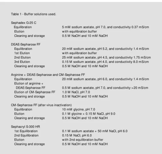
Details about the buffer solutions are given in Table 1.
Pre paratio n o f immuno glo bulin G
Approximately 1200 human plasma bags stored at -30oC were thawed at a temperature
of 2 to 4oC in order to form a 200-l pool.
obtain a cryoprecipitate to be used for the production of factor VIII. The supernatant of the cryoprecipitate was cleared by filtration through a 30-S depth filter (Zeta Plus, Cuno, Meriden, CT, USA) and desalted with coarse Sephadex G-25 filtration gel on a BPSS 400/ 600 column using 5 mM sodium acetate as elution solution. The pH was adjusted to 5.2 with 1 M CH3CO2H and the protein solution
(410 l) was allowed to stand overnight in a cold chamber at 4oC for euglobulin
precipi-tation. On the following day the preparation was centrifuged at 4oC to remove the
pre-cipitated euglobulins and the supernatant was cleared by filtration. The pH was read-justed to 5.2 with 1 M CH3CO2H and
con-ductivity was adjusted to 1.4 mS/cm with NaCl. The sample was applied to a PS-370/
15 column containing DEAE-Sepharose FF in order to separate gamma-globulin (unad-sorbed) from other plasma proteins such as albumin, etc.
The gamma-globulin fraction was sub-mitted to chromatography for the prepara-tion of IgG as described below. The albumin fraction was further purified by a chromato-graphic method (18). The pH of the gamma-globulin fraction (480 l) was adjusted to pH 6.0 with 1 M NaOH and conductivity was adjusted to 1.4 mS/cm. The fraction was then applied to two columns, PS-370/15 and Index 200/500, coupled in series. The first column was packed with a mixture of two gels, 40% arginine Sepharose 4B and 60% DEAE-Sepharose FF, and the second with 8 l of CM-Sepharose FF gel.
Table 1 - Buffer solutions used.
Sephadex G-25 C
Equilibration 5 mM sodium acetate, pH 7.0, and conductivity 0.37 mS/cm Elution w ith equilibration buffer
Cleaning and storage 0.5 M NaOH and 10 mM NaOH
DEAE-Sepharose FF
Equilibration 20 mM sodium acetate, pH 5.2, and conductivity 1.4 mS/cm 1st Elution w ith equilibration buffer
2nd Elution 25 mM sodium acetate, pH 4.5, and conductivity 1.75 mS/cm 3rd Elution 0.15 M sodium acetate, pH 4.0, and conductivity 8.0 mS/cm Cleaning and storage 0.5 M NaOH and 10 mM NaOH
Arginine + DEAE-Sepharose and CM -Sepharose FF
Equilibration 20 mM sodium acetate, pH 6.0, and conductivity 1.4 mS/cm Elution of arginine +
DEAE-Sepharose FF 0.5 M sodium acetate, pH 7.0, and conductivity >20 mS/cm Elution of CM -Sepharose FF 1.0 M NaCl, pH 7.0
Cleaning and storage 0.5 M NaOH and 10 mM NaOH
CM -Sepharose FF (after virus inactivation)
Equilibration 10 mM glycine, pH 7.0
Elution 0.1 M glycine + 0.15 M NaCl, pH 9.0 Cleaning and storage 0.5 M NaOH and 10 mM NaOH
Sephacryl S-300 HR
1st Equilibration 0.1 M sodium acetate + 50 mM NaCl, pH 6.0 2nd Equilibration 0.15 M NaCl, pH 6.0
Pellicon Cassette System 30,000 NMWL. The pH of the IgG solution (16 l) was ad-justed to 5.5 and the material was submitted to viral inactivation with a solvent/detergent combination (19), i.e., 1% Tri (n-butyl) phos-phate and 1% Triton X-100 at 35oC for 10 h.
After viral inactivation, the solvent/deter-gent combination was removed by ion-ex-change chromatography on CM-Sepharose FF in an Index 200/500 column containing 8 l of gel. The IgG solution was concentrated to 7%, pepsin was added (0.1 mg/g protein), pH 4.0, and the solution was heated at 37oC
for 10 h. The preparation was cooled to 20oC
and then applied to a BPG 200/950 column containing 20 l of Sephacryl S-300 HR fil-tration gel to remove the aggregated IgG molecules and pepsin. The eluted IgG solu-tion was concentrated to 6.5% and then for-mulated as follows: the pH was adjusted to 5.0 with 1 N HCl, the conductivity to 9 to 10 mS/cm with solid NaCl, and 7.5% (w/v) maltose and 0.1 M glycine were added as stabilizers (12,20). The material was steril-ized by filtration through a 0.22-µm mem-brane (Millipore) and the final product (14 l), containing 5% protein (w/v), was bottled in 50-ml type I (neutral) flasks, with 2.5 g IgG per flask and was stored at 4 to 8oC (see
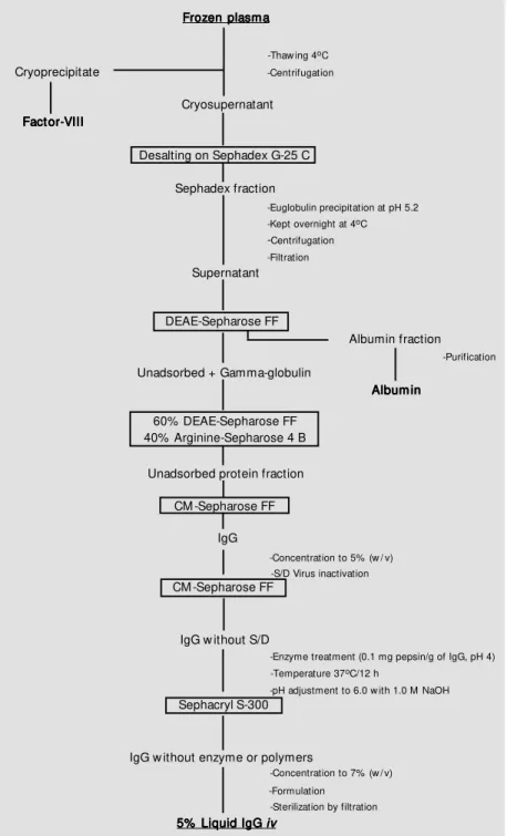
Figure 1).
Analytical me tho ds
The characteristics of normal liquid 5% IgG for intravenous use were evaluated by the methods described in the Pharmacopée Européenne (1) and in Regulation No. 2.419 of 12/17/1996 of the Brazilian Ministry of Health.
The immunochemical tests were carried out by cellulose acetate electrophoresis, by the micro-Ouchterlony method and by im-munoelectrophoresis using anti-total human protein and anti-animal protein antisera of domestic species such as horses, cows and sheep. Protein concentration was determined by the biuret method, pH was measured by
Cryoprecipitate
-Thaw ing 4oC
-Centrifugation
Cryosupernatant
Desalting on Sephadex G-25 C
Sephadex fraction
-Euglobulin precipitation at pH 5.2 -Kept overnight at 4oC
-Centrifugation -Filtration
Supernatant
DEAE-Sepharose FF
Unadsorbed + Gamma-globulin
Albumin fraction
Album in Album inAlbum in Album inAlbum in
-Purification
60% DEAE-Sepharose FF 40% Arginine-Sepharose 4 B
Unadsorbed protein fraction
CM -Sepharose FF
IgG
-Concentration to 5% (w / v) -S/D Virus inactivation
CM -Sepharose FF
IgG w ithout S/D
-Temperature 37oC/12 h
-pH adjustment to 6.0 w ith 1.0 M NaOH
Sephacryl S-300
IgG w ithout enzyme or polymers
-Concentration to 7% (w / v)
-Formulation -Sterilization by filtration
5% Liquid IgG 5% Liquid IgG 5% Liquid IgG 5% Liquid IgG 5% Liquid IgG iviviviviv
Figure 1 - Flow diagram for the production of liquid intravenous IgG.
The proteins not of interest for the pres-ent study, IgM, IgA, transferrin, etc, were adsorbed to the first column. IgG was adsorbed to the second column and eluted and concentrated to 5% (w/v) using the
-Enzyme treatment (0.1 mg pepsin/g of IgG, pH 4)
Frozen plasma Frozen plasmaFrozen plasma Frozen plasmaFrozen plasma
diluting to 1% in a 0.9% sodium chloride solution, and anti-A and anti-B hemaggluti-nins were determined by the indirect Coombs method. The presence of other plasma pro-teins such as IgA and IgM and of the IgG subclasses IgG1, IgG2, IgG3 and IgG4 was determined by radial immunodiffusion on Bindarid plates (The Binding Site Inc., San Diego, CA, USA).
Anticomplementary activity was deter-mined by the method of Mayer using guinea pig complement and sheep red cells. The prekallikrein activator was determined using the S2302 chromogenic substrate of Chro-mogenix (Mölndal, Sweden).
The biological safety of the product was evaluated by tests for the detection of anti-HIV, anti-HTLV-1 and 2, anti-HCV, and HB antigens at the Serology Laboratory of Fundação Pró-Sangue Hemocentro de São Paulo. Sterility was evaluated by the Steritest membrane method (Sterility Testing Sys-tem, Millipore). Pyrogenicity and toxicity were tested at Medlab, São Paulo, Brazil.
The distribution of monomers, dimers and polymers was evaluated by HPLC, and polio I, II and III, measles and anti-herpes activities were determined at Instituto Adolfo Lutz, São Paulo, Brazil. Anti-rubeola, anti-CMV and anti-streptolysin O activities were determined at the Immunology Labora-tory of IAMSPE, São Paulo, Brazil. Stability was evaluated by incubating the preparation at 57oC for 4 h and observing the presence of
jelling.
Re sults and D iscussio n
The size distribution of the product, evalu-ated by HPLC, indicevalu-ated 94% monomers, 5.5% dimers and 0.5% polymers, correspond-ing to standard values. The profile of the IgG molecule did not show any alterations when the IgG preparation was incubated at 37oC
for one month; the anticomplementary activ-ity was 0.3 CH50/mg IgG, in agreement with
the specifications that determine a value of
less than 1 CH50/mg IgG, and the functional
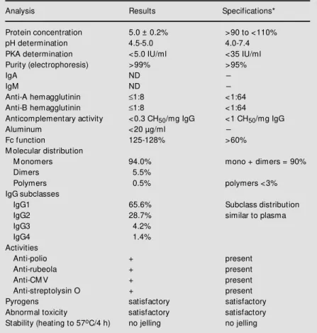
activity of Fc was unchanged, presenting an index of 125 to 128% of Fc, meeting the recommendation of Pharmacopée Europé-enne, which requires >60%. On the basis of these evaluations, we concluded that the prod-uct contained intact molecules. The sub-classes determined were IgG1 = 65.6%, IgG2 = 28.7%, IgG3 = 4.2%, and IgG4 = 1.4%, with no significant variations compared to normal plasma. For prekallikrein activator (PKA) determination we used a reference PKA from the FDA and the S2302 chro-mogenic substrate of Chromogenix. Values of less than 5 IU/ml 5% (w/v) IgG were obtained, well below the specification limit of 35 IU/ml of PKA. The remaining data which characterize the product are presented in Tables 2 and 3, and cellulose acetate
Table 2 - Characteristics of liquid intravenous IgG.
N = 10. ND, Not detected. * Pharmacopée Européenne (1).
Analysis Results Specifications*
Protein concentration 5.0 ± 0.2% >90 to <110%
pH determination 4.5-5.0 4.0-7.4
PKA determination <5.0 IU/ml <35 IU/ml Purity (electrophoresis) >99% >95%
IgA ND
-IgM ND
-Anti-A hemagglutinin ≤1:8 <1:64 Anti-B hemagglutinin ≤1:8 <1:64
Anticomplementary activity <0.3 CH50/mg IgG <1 CH50/mg IgG
Aluminum <20 µg/ml
-Fc function 125-128% >60%
M olecular distribution
M onomers 94.0% mono + dimers = 90%
Dimers 5.5%
Polymers 0.5% polymers <3%
IgG subclasses
IgG1 65.6% Subclass distribution
IgG2 28.7% similar to plasma
IgG3 4.2%
IgG4 1.4%
Activities
Anti-polio + present
Anti-rubeola + present
Anti-CM V + present
Anti-streptolysin O + present
Pyrogens satisfactory satisfactory
ing to 57oC for 4 h there was no jelling, and
in the quarantine test involving incubation in an oven at 37oC for 4 weeks, again there
were no alterations in the IgG molecule. Viral inactivation was performed with a solvent/detergent system (19), i.e., 1% Tri (n-butyl) phosphate and 1% Triton X-100, pH 5.5, at 35oC for 10 h, which was then
removed by the ion-exchange gel, CM-Sepharose FF. The protein aggregates gener-ated during the process were removed by gel filtration chromatography through Sephacryl S-300 HR. Thus, the product obtained con-sisted mainly of the monomer, presenting only trace amounts of polymers, consistent with low anticomplementary activity, i.e., less than 1 CH50/mg IgG.
Of the several stabilizing agents tested, glucose, sucrose and maltose, the last one at a 7.5% concentration, and 0.1 M glycine, pH 5.0 (12,20), were found to be the most ap-propriate. The IgG solution was limpid and transparent with no detectable molecular al-terations even when stored for more than 2 years at 5 to 8oC.
On the basis of in vitro and in vivo
labo-ratory tests, we conclude that the product fully satisfies all the requirements of the Pharmacopée Européenne (1), as well as the norms of the Brazilian Ministry of Health (Regulation No. 2419 of December 17, 1996). We are awaiting the results of clinical tests currently underway to liberate the product for use.
Ackno wle dgm e nts
The authors wish to thank Dr. Hiroyoshi Ito from the Japanese Red Cross, Chitose, Hokkaido, Dr. Komei Ohashi from Kaketsuken, Kumamoto, and Dr. Kentaro Nakamura from Green Cross, Osaka, for their helpful suggestions and the opportunity of training at their Plasma Fractionation Plants in Japan, and Dr. Erica Kitahara, Pharmacia Biotech, for supplying the re-agents.
Table 3 - Antibodies detected in liquid intravenous IgG.
N = 10.
Antibody M ethod Titer and/or unit
Anti-polio I Neutralization test 1:256 Anti-polio II Neutralization test 1:128 Anti-polio III Neutralization test 1:64 Anti-measles Hemagglutination inhibition 1:64
Anti-rubeola ELISA 756 IU/ml
Anti-herpes Immunofluorescence 1:128
Anti-cytomegalovirus ELISA 898 IU/ml
Anti-streptolysin O Nephelometry 716 IU/ml
Figure 3 - Immunoelectrophore-sis. a, Plasma pool (2 µl of 10 g/ l); b, immunoglobulin G (2 µl of 30 g/l). Protein w as detected w ith light green stain.
a
b
electrophoresis and immunoelectrophoresis data are shown in Figures 2 and 3, respec-tively.
We describe the preparation of intrave-nous 5% immunoglobulins in the liquid state from a pool of human plasma from 1200 donors by the chromatographic method. The analyses performed for quality control showed that the IgG met international speci-fications. In the stability test involving
heat-Figure 2 - Cellulose acetate electrophoresis. a, Plasma pool (25 µl of 10 g/l); b, immunoglo-bulin G (25 µl of 30 g/l). Protein w as detected w ith Ponceau S and the data are reported as percent of total densitometer units.
Plasma IgG
Serum protein Serum protein
Total protein: 30 g/l
100%
009 009
(+) (-)
Re fe re nce s
1. Pharmacopée Européenne (1997). 2nd edn. Part II. M aisonneuve S.A., Sainte Ruffine.
2. Cohn EJ, Strong LE, Hughes Jr WL, M ulford DJ, Ashw orth JN, M elin M & Tay-lor HL (1946). Preparation and properties of serum and plasma proteins. IV. A sys-tem for the separation into fractions of the protein and lipoprotein components of biological tissues and fluids. Journal of the American Chemical Society, 68: 459-475.
3. M ielka SI & Gozze I (1975). Anticomple-mentary activity of human immunoglobu-lin G. I. M echanism of the artifactual in-crease in anticomplementary activity of IgG during the assay. Vox Sanguinis, 29: 101-123.
4. Andersson I, Lindquist LO & Berglöf J (1994). An improved chromatography m et hod f or product ion of IgG f rom plasma. XXIII Congress of the Interna-tional Society of Blood Transfusion, The Netherlands, July 2-8.
5. Curling JM (1983). Separation of Plasma Proteins. Pharmacia Fine Chemicals AB, Uppsala.
6. Cohn EJ, Gurd FRN, Surgenor DM , Barnes BA, Brow n RK, Deronaux G, Gillespie JM , Kahnt FW, Lever WF, Liu CH, M ittelman D, M outon RF, Schmid K & Uroma E (1950). A system for the preparation of the components of human blood:
quanti-tative procedures for the separation of the protein components of human plasma. Journal of the American Chemical Soci-ety, 72: 465-474.
7. Oncley M , M elin DA, Richert JW , Cameron JW & Cross Jr PM (1949). The separation of the antibodies, isoaggluti-nins, prothrombin, plasminogen and ß1-lipoprotein into subfractions of human plasma. Journal of the American Chemi-cal Society, 71: 541-550.
8. Barandun S, Kistler P, Jeunet F & Isliker H (1962). Intravenous administration of hu-man γ-globulin. Vox Sanguinis, 7: 157-174. 9. Hässig A (1986). Intravenous immuno-globulins: pharmacological aspects and therapeutic use. Vox Sanguinis, 51: 10-17.
10. Schultze HE & Schw ick G (1962). Über neue M öglichkeiten intravenöser Gamma-globulin-Applikation. Deutsche M edizin-ische Wochenschrift, 87: 1643-1650. 11. Sgouris JT (1967). The preparation of
plas-min treated immune serum globulin for intravenous use. Vox Sanguinis, 13: 71-84.
12. Fernandes PM & Lundblad JL (1980). Preparation of a stable intravenous gam-ma-globulin: process design and scale-up. Vox Sanguinis, 39: 101-112.
13. Stephan W (1975). Undergraded human immunoglobulin for intravenous use. Vox Sanguinis, 28: 422-437.
14. Pappenhagen A, Lundblad J & Schroeder D (1975). Pharmaceutical compositions comprising intravenously injectable modi-fied serum globulin, its production and use. US Patent No. 3,903,262.
15. Schmidtberger R (1978). Amidated im-mune globulins and process for preparing them. US Patent No. 4,118,379. 16. M asuho Y, Tomibes S, M atsuzaw a K &
Ohtdu A (1977). Development of an intra-venous gamma-globulin w ith Fc activities. I. Preparation and characterization of S-sulfonated human gamma-globulin. Vox Sanguinis, 32: 175-181.
17. WHO Expert Committee on Biological Standardization (1982). Report of an infor-mal meeting on intravenous immuno-globulins (human), Geneva.
18. Tanaka K, Saw atani E, Nakao HC, Dias GA & Arashiro F (1996). An alternative col-umn chromatographic process for the pro-duction of human albumin. Brazilian Jour-nal of M edical and Biological Research, 29: 185-191.
19. Horow itz B, Wieb M E, Lippin A & Stryker H (1985). Inactivation of viruses in labile blood derivatives. Transfusion, 25: 516-522.
A PRIVATE BRAZILIAN
BIOTECHNOLOGY COMPANY
LEADER IN THE BRAZILIAN
INSULIN MARKET
World's fourth major insulin producer
Offers a complete line of insulins, i.e., a range of options from bovine/swine to human
insulin
Operational processes run under GMP (Good Manufacturing Practices)
F.D.A./USA inspected
Has over 450 people focusing their efforts on improving services to the diabetic patient Winner of a tender with international producers to supply human insulin to Russia
Produces a wide line of enzymes, peptones, insulin crystals, culture media and diagnostic products
Supports a strong in-house R&D
program and maintains several agreements for cooperation with research laboratories in universities and research centers