Oleanolic acid and ursolic acid inhibit peptidoglycan biosynthesis
in
Streptococcus mutans
UA159
Soon-Nang Park
1, Sug-Joon Ahn
2, Joong-Ki Kook
11
Korean Collection for Oral Microbiology and Department of Oral Biochemistry, School of Dentistry, Chosun University, Gwangju, Republic of Korea. 2
Dental Research Institute, School of Dentistry, Seoul National University, Seoul, Republic of Korea.
Submitted: February 23, 2013; Approved: October 29, 2014.
Abstract
In this study, we revealed that OA and UA significantly inhibited the expression of most genes re-lated to peptidoglycan biosynthesis inS. mutansUA159. To the best of our knowledge, this is the first report to introduce the antimicrobial mechanism of OA and UA againstS. mutans.
Key words:ursolic acid, oleanolic acid, peptidoglycan biosynthesis,Streptococcus mutansUA159.
Ursolic acid (UA, (3b)-hydroxy-urs-12-en-28-oic acid), oleanolic acid (OA, 3b -3-hydroxyolean-12-en-28-oic acid), and betulinic acid (BA, 3b -3-hydroxy-lup-20(29)-en-28-oic acid) are derivatives of triterpenoid sapo-nins (Liu, 1995; Fontanayet al., 2008). These compounds are naturally found in a large variety of vegetables, medici-nal herbs, and plants that have been investigated for anti-bacterial activity (Fontanayet al., 2008). The antimicrobial activity of OA and UA is stronger than that of BA (Fon-tanayet al., 2008). OA and UA inhibit growth of Gram-positive bacteria but not Gram-negative bacteria (Fontanay
et al., 2008), but their antimicrobial mechanism is un-known. Previously, we reported that OA and UA have strong antimicrobial activity againstStreptococcus mutans
(Kimet al., 2010; Kim et al., 2011) but BA had no anti-microbial activity againstS. mutans(data not shown). One of the characteristic differences between Gram-positive and Gram-negative bacteria is the thickness of the peptido-glycan layer. Considering this difference, it has been sug-gested that the mechanism of OA and UA antimicrobial ac-tivity may be related to inhibition of peptidoglycan biosynthesis (Horiuchiet al., 2007). Thus, the objective of this study was to investigate the effect of BA, OA, and UA on peptidoglycan biosynthesis inS. mutansUA 159 using the quantitative real-time polymerase chain reaction (qPCR) method to identify the antimicrobial mechanism againstS. mutans.
S. mutansUA159 was a kind gift from Dr. Robert A. Burne, Department of Oral Biology, College of Dentistry, University of Florida. The strain was cultured in brain heart infusion (BHI) broth (Difco, Lab., Detroit, MI, USA) or on BHI agar plates in a 37 °C incubator.
Overnight cultures (1 mL) ofS. mutansUA 159 were transferred to 9 mL BHI broth and grown at 37 °C to the mid-log phase (OD600= 0.35). UA (Sigma, St. Louis, MO, USA), OA (Sigma), or BA (Sigma) solutions were added to each tube to a final concentration of 64mg/mL. The bacte-rial culture solutions were incubated in a 37 °C incubator for 90 min and then harvested by centrifugation at 10,000 x
gfor 10 min at 4 °C. After discarding the supernatant, liquid nitrogen was immediately added to the tube. The frozen bacterial pellets were homogenized using a mortar and pes-tle (Smile Science, Seoul, Korea). Total RNAs were ex-tracted from the homogenized bacteria following the manu-facturer’s instructions of the RNAqueous® kit (Ambion, Austin, TX, USA). DNase I treatment was conducted to completely remove the bacterial genomic DNAs using a TURBO DNA-freeTMKit (Ambion) according to the manu-facturer’s protocol. RNA concentration was determined at 260 nm 280 nm with a UV spectrophotometer (Ultrospec 2000, Pharmacia Biotech., UK).
We consulted the KEGG pathway database to deter-mine the target genes related toS. mutansUA159 peptido-glycan biosynthesis (KEGG pathway). qPCR primers were designed based on the nucleotide sequences of the target
Send correspondence to J.-K. Kook. Korean Collection for Oral Microbiology and Department of Oral Biochemistry, School of Dentistry, Chosun Uni-versity, 309 Pilmun-daero, 501-759 Dong-Gu, Gwangju, Republic of Korea. E-mail: [email protected].
genes (GenBank accession no. AE014133.2) using the MegAlign and PrimerSelect programs (LasergeneTM 8.0, DNAStar, Inc., Madison, WI, USA) (Table 1). The qPCR primers were synthesized by Bioneer Co. (Daejeon, Ko-rea).
First-strand cDNAs synthesis was performed with 4mg of total RNA, random hexamers, and anAccuPower® RocketScriptTMRT PreMix (Bioneer Co., Deajeon, Korea) using theExicyclerTM96 Real-Time Quantitative Thermal Block (Bioneer Co.) and following the manufacturer’s pro-tocol. qPCR was performed with the AccuPower®
GreenStarTM qPCR PreMix (Bioneer Co.) using the
ExicyclerTM 96 Real-Time Quantitative Thermal Block. Each PCR reaction was performed in a total volume of 20mL containing 1 mL each of the forward and reverse primers (final concentration, 500 nM each), 0.63 mL cDNA, and the appropriate dose of sterilized DNase-RNase-free water in PreMix PCR tubes. The qPCR condi-tions were initial denaturation at 95 °C for 3 min, 40 cycles of denaturation at 95 °C for 15 sec, primer annealing and extension at 55 °C for 15 sec, and final cooling at 25 °C for 1 min. Each qPCR reaction was performed in triplicate.
The expression levels of each target gene in the group were normalized with those of 16S rDNA. The normalized expression of each gene (N) was determined as:
N = 2DCt = 2(Ct value of the target gene - Ct value of 16S rDNA)
Relative expression of the target genes between each experimental group (BA, OA, or UA) and the negative con-trol group (1% DMSO) was calculated as [N of each experi-mental group (BA, OA, or UA)/N of the negative control group].
Data are expressed as mean±standard deviation. In-dependent sample two-tailedt-tests were conducted to cal-culate thep-values using SPSS version 12 software (SPSS Inc., USA). A p-value£0.05 was considered statistically significant.
We determined the BA, OA, and UA concentration of 64mg/mL by investigating the antimicrobial mechanism againstS. mutansUA159 by calculating the growth curve at 16, 32, and 64mg/mL BA, OA, and UA for 60, 90, 120, and 180 min in S. mutans UA159 at the mid-log phase (OD600= 0.35). The growth ofS. mutansUA159 was in a static (inhibited) state at concentrations of 64 and 32mg/mL of OA and UA, but in the exponential state at concentra-tions of 64 and 32mg/mL in the BA-treated and control groups (data not shown). We decided to further explore us-ing 64mg/mL BA, OA, and UA.
The synthesis of UDP-N-acetylglucosamine (UDP-GlcNAc) and UDP-N-actetylmuramic acid (UDP-MurNAc), the building blocks of peptidoglycan, is the first phase of first stage (stage I) of intracellular peptidoglycan assembly (Bugg and Walsh, 1992). UDP-GlcNAc is syn-thesized from GlcNAc-1-P by UDP-N-acetylglucosamine
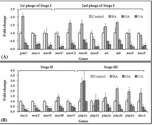
pyrophosphorylase (GlmU). UDP-MurNAc is synthesized from UDP-GlcNAc by UDP-N-acetylglucosamine 1-car-boxyvinyltransferase (MurA) and UDP-N -acetylenol-pyruvoylglucosamine reductase (MurB) (Bugg and Walsh, 1992). The data showed that UA and OA, but not BA, sig-nificantly downregulatedglmU,murA, andmurB expres-sion (Figure 1A). Interestingly,glmU was overexpressed by a factor of two in the BA-treated group compared to that in the control group. The second phase of stage I of peptido-glycan biosynthesis is the assembly of the UDP-MurNAc-pentapeptide (Bugg and Walsh, 1992). The D-amino acids, D-alanine, D-glutamate, and DL-diaminopimelate, occur in peptidoglycan (Bugg and Walsh, 1992). ForS. mutans
UA159, D-alanine and D-glutamate are catalyzed by ala-nine racemase (Alr) and glutamate racemase (MurI), re-spectively (the KEEG pathway). Biosynthesis of DL-dia-minopimelate (DL-DAP) is catalyzed by DAP epimerase or meso-DAP D-dehydrogenase (Bugg and Walsh, 1992). These genes are missing inS. mutansUA159, even though UDP-N-acetylmuramyl tripeptide synthetase (MurC2) which catalyzes adding DL-DAP to UDP-MurNAc-L-ala-nine-D-glutamate occurs (KEEG Pathway). The reason for the discrepancy is unclear, but it is possible that DL-DAP is synthesized through an unknown pathway or that DL-DAP is not used in peptidoglycan synthesis inS. mutansUA159. Generally, DL-DAP is added as the third residue in the pentapeptide of negative bacteria and some positive bacteria, whereas lysine is added in other Gram-positive bacteria (http://www.enzyme-database.org/reac-tion/polysacc/PepGly1.html). Using these D-amino acids and D-Ala-D-Ala formed by D-alanine-D-alanine ligase (DdI), two types of MurNAc-pentapeptide, UDP-MurNAc-L-Ala-g-D-Glu-DL-DAP-D-Ala-D-Ala (UDP-MurNAc-pentapeptide I) and UDP-MurNAc-L-Ala-g -D-Glu-DL-DAP-D-Ala-D-Ala (UDP-MurNAc-pentapeptide II) are synthesized by UDP-N-acetyl muramate-alanine ligase (MurC), UDP-N -acetylmuramoyl-L-alanyl-D-glu-tamate synthetase (MurD), MurC2 or UDP-N -acetylmura-moyl-L-alanyl-D-glutamate-L-lysine ligase (MurE), and UDP-N-acetylmuramoyl-tripeptide-D-alanyl-D-alanine ligase (MurF) (the KEGG Pathway). The data showed that second stage-related genes were downregulated by OA and UA, butmurEwas downregulated only by BA (Figure 1A). These results reveal that OA and UA inhibited expression from the first stages ofS. mutans UA159 peptidoglycan biosynthesis, whereas BA induced overexpression of UDP-GlcNAc, the first substrate for peptidoglycan synthe-sis.
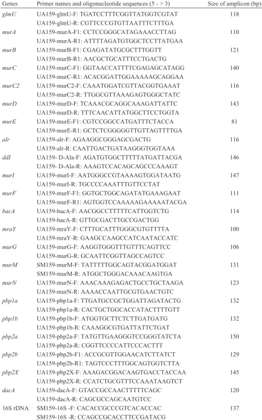
Table 1- Quantitative real-time polymerase chain reaction (qPCR) primers used in this study.
Genes Primer names and oligonucleotide sequences (5 - > 3) Size of amplicon (bp)
glmU UA159-glmU-F: TGATCCTTTCGGTTATGGTCGTAT 118 UA159-glmU-R: CGTTCCCGTGTTAATTTCTTTGA
murA UA159-murA-F1: CCTCCGGGCATAGAAACCTTAG 110 UA159-murA-R1: ATTTTAGATGTGGCTCCTTATGAA
murB UA159-murB-F1: CGAGATATGCGCTTTGGTT 121
UA159-murB-R1: AACGCTGCATTTCCTGACTG
murC UA159-murC-F1: GGTAACCATTTTCGAGAGCATAGG 140 UA159-murC-R1: ACACGGATTGGAAAAAGCAGGAA
murC2 UA159-murC2-F: CAAATGGATCGTTACGGTGAAAT 116 UA159-murC2-R: TTGGCGTTAAAGAGTGGGCTATC
murD UA159-murD-F: TCAAACGCAGGCAAAGATTATTC 143 UA159-murD-R: TTTCAACATTATGGCTTCCTGGTA
murE UA159-murE-F1: CGTCCGGCCATGATTTCTACCA 81 UA159-murE-R1: GCTCTCGGGGGTTGTTAGTTTTGA
alr UA159-alr-F: AGAAGGCGGGAGCGACTG 116
UA159-alr-R: CAATTGACTGATAAGGGTGGTAAA
ddI UA159- D-Ala-F: AGATGTGGCTTTTTATGATTACGA 146 UA159- D-Ala-R: AAAGTCCACAGCAGCCCAAAGT
murI UA159-murI-F: AATGGGCCGTAAAAGTGGATAATG 147 UA159-murI-R: TGCCCCAAATTTGTTCCTAT
murF UA159-murF-F1: GGTGCTGGCAGATATGAAAGAAT 111 UA159-murF-R1: AGTGGTCCAAAAAGAAAAATACGA
bacA UA159-bacA-F: AACGGCCTTTTTCATTGGTCTG 114 UA159-bacA-R: GTTGCGACTTGCCGACTGG
mraY UA159-mraY-F: CTTTGCATTTGGGCGTGTTTTA 100 UA159-mraY-R: GAAGCCAAGCCATCAATACCATC
murG UA159-murG-F: AAGGTGGGTTTGTTTCAGTTCC 106 UA159-murG-R: GCAATTCGGTTAGCCAGTCC
murM SM159-murM-F: TATTTTTGGCAGTACGGATGGAT 131 SM159-murM-R: ATGGCTGGGACAAACAAGTGA
murN UA159-murN-F: AAACAAAGAGACTGCCTGCTAAGA 123 UA159-murN-R: AAAACCAATTGCGTGAACTGTC
pbp1a UA159-pbp1a-F: TTGATGCCGCTGGATTAGATACTG 132 UA159-pbp1a-R: CACTGCTGGCACCATACTTTTGTT
pbp1b UA159-pbp1b-F: ATGGTGCTTCTCTTGATGATG 132 UA159-pbp1b-R: CAAAGGCGTGATTATTCTGAT
pbp2a UA159-pbp2a-F: TATGTTGAAGGGTCCGGGTATCTA 150 UA159-pbp2a-R: CGGTTCCCCATTCCCACTTT
pbp2b UA159-pbp2b-F1: ACCGCGTTGGAACATCTTATCT 129 UA159-pbp2b-R1: TAGTCCCTTTGGCAGTGGTCTTA
pbp2X UA159-pbp2X-F: AAAGACGGACAAGTGACCTACCAA 145 UA159-pbp2X-R: CCATCTGCGTTTCCAAATAAGTCT
dacA UA159-dacA-F: GTACCGCCAACTTTTTCAGC 120
UA159-dacA-R: CAGCGCCAGCAATGTCC
16S rDNA SM159-16S -F: CACACCGCCCGTCACACCAC 137 SM159-16S -R: CCAGCCGCACCTTCCGATACG
glmU, UDP-N-acetylglucosamine pyrophosphorylase gene; murA, UDP-N-acetylglucosamine 1-carboxyvinyltransferase gene; murB, UDP-N-acetylenolpyruvoylglucosamine reductase gene;murC, UDP-N-acetyl muramate-alanine ligase gene;murC2, UDP-N-acetylmuramyl tripeptide synthetase gene; murD, UDP-N-acetylmuramoyl-L-alanyl-D-glutamate synthetase gene; murE, UDP-N-acetylmuramoyl-L-alanyl-D-glutamate-L-lysine ligase gene;alr, alanine racemase gene;ddI, D-alanine-D-alanine ligase gene;murI, glutamate racemase gene;murF, UDP-N-acetylmuramoyl-tripeptide—D-alanyl-D-alanine ligase gene;bacA, undecaprenyl pyrophosphate phosphatase gene;
2008). The coupling of UDP-MurNAc-pentapeptide to UP to produce UPP-MurNAc-pentapeptide (Lipid I) and UMP is catalyzed by phospho-N -acetylmuramoyl-pentapeptide-transferase (MraY). Then, another glucosamine residue, GlcNAc, is attached to Lipid I by undecaprenyldiphospho-muramoylpentapeptide beta-N -acetylglucosaminyltransfe-rase (MurG) and forms UPP-MurNAc-(GlcNAc)-penta-peptide (Lipid II). 2 L-Alanine (L-Ala) is added to UDP-MurNAc-(GlcNAc)-L-Ala-g-D-Glu-L-Lys-D-Ala-D-Ala by UDP-N-acetylmuramoylpentapeptide-lysine N6-alanyl-transferase (MurM) and another alanine-adding enzyme (MurN) to form UDP-MurNAc-(GlcNAc)-L-Ala-g -D-Glu-L-Lys-(L-Ala)2-D-Ala-D-Ala (the KEGG pathway). The data showed that OA and UA significantly inhibited expression ofbacA,mraY,murG,murM, andmurNand that BA also downregulated these genes, except murM and
murN(Figure 1B). These findings reveal that OA, UA, and BA inhibited translocation of the MurNAc-pentapeptide, an intermediate or monomer of peptidoglycan in the cell wall.
The last stage (stage III) of peptidoglycan biosyn-thesis is transglycosylation, transpeptidation, and trimming of the peptidoglycan, which are catalyzed by penicillin-binding proteins (PBPs) and transpeptidase (Pinhoet al., 2001; Leeet al., 2003; Mainardiet al., 2005; Fanet al., 2007).S. mutans UA159 has five PBPs such as PBP1a, PBP1b, PBP2a, PBP2b, and PBP2X, as well as serine-type D-Ala-D-Ala carboxypeptidase (DacA), which is involved in the last stage of peptidoglycan biosynthesis (the KEEG pathway). The data showed that these genes, except PBP1a and PBP1b, were downregulated 52-85% and 31-56% in the OA and UA groups compared to those in the control group. Our qPCR data showed that OA and UA signifi-cantly downregulated the PBP genes, except pbp1a and
pbp1b, whereas BA significantly upregulated these genes, exceptpbp1bandpbp2a(Figure 1B). These results indicate that OA and UA inhibit polymerization between a newly synthesized peptidoglycan monomer and the existing pepti-doglycan, and that BA increased polymerization in S. mutansUA159.
In summary, OA and UA inhibited the expression of peptidoglycan biosynthesis-related genes of S. mutans
Figure 1- Relative expression of genes related to peptidoglycan biosynthesis, (A) stage I and (B) stage II and III, inStreptococcus mutansUA159 follow-ing treatment with betulinic acid (BA), oleanolic acid (OA), or ursolic acid (UA).glmU, UDP-N-acetylglucosamine pyrophosphorylase; murA, UDP-N-acetylglucosamine 1-carboxyvinyltransferase; murB, UDP-N-acetylenolpyruvoylglucosamine reductase; murC, UDP-N-acetyl muramate-alanine ligase;murC2, UDP-N-acetylmuramyl tripeptide synthetase;murD, UDP-N-acetylmuramoyl-L-alanyl-D-glutamate synthetase;murE, UDP-N-acetylmuramoyl-L-alanyl-D-glutamate-L-lysine ligase; alr, alanine racemase;ddI, D-alanine-D-alanine ligase;murI, glutamate racemase; murF, UDP-N-acetylmuramoyl-tripeptide—D-alanyl-D-alanine ligase;bacA, undecaprenyl pyrophosphate phosphatase;mraY, phospho-N-acetylmuramoyl-pentapeptide-transferase;murG, undecaprenyldiphospho-muramoylpentapeptide beta-N-acetylglucosaminyltransferase; murM, UDP-N-acetylmura-moylpentapeptide-lysine N6-alanyltransferase;murN, alanine adding enzyme;pbp1a, penicillin binding protein 1a;pbp1b, penicillin binding protein 1b;
UA159 at the transcriptional level which might be the one of the antimicrobial mechanism of OA and UA againstS. mutans.
Acknowledgments
This study was supported by a grant from the Korea Healthcare Technology R&D Project, Ministry for Health, Welfare and Family Affairs, Republic of Korea (A091074).
References
Bouhss A, Trunkfield AE, Bugg TDet al.(2008) The biosynthesis of peptidoglycan lipid-linked intermediates. FEMS Micro-biol Rev 32:208-233.
Bugg TD, Walsh CT (1992) Intracellular steps of bacterial cell wall peptidoglycan biosynthesis: enzymology, antibiotics, and antibiotic resistance. Nat Prod Rep 9:199-215. Fan X, Liu Y, Smith Det al. (2007) Diversity of
penicillin-binding proteins. Resistance factor FmtA ofStaphylococcus aureus. J Biol Chem 282:35143-35152.
Fontanay S, Grare M, Mayer Jet al.(2008) Ursolic, oleanolic and betulinic acids: antibacterial spectra and selectivity indexes. J Ethnopharmacol 120:272-276.
Higashi Y, Strominger JL, Sweeley CC (1970) Biosynthesis of the peptidoglycan of bacterial cell walls. XXI. Isolation of free C55-isoprenoid alcohol and of lipid intermediates in peptidoglycan synthesis fromStaphylococcus aureus. J Biol Chem 245:3697-3702.
Horiuchi K, Shiota S, Hatano Tet al.(2007) Antimicrobial activ-ity of oleanolic acid from Salvia officinalis and related
com-pounds on vancomycin-resistant enterococci (VRE). Biol Pharm Bull 30:1147-1149.
KEGG Pathway (2013) Peptidoglycan biosynthesis -
Streptococ-cus mutans UA159. Available at:
http://www.ge-nome.jp/kegg-bin/show_pathway?smu00550. Accessed 5 March 2014.
Kim MJ, Kim CS, Ha WHet al.(2010) Antimicrobial effects of oleanolic acid againstStreptococcus mutansand Streptococ-cus sobrinusisolated from a Korean population. Int J Oral Biol 35:191-195.
Kim MJ, Kim CS, Park JYet al.(2011) Antimicrobial effects of ursolic acid against mutans streptococci isolated from Kore-ans. Int J Oral Biol 36:7-11.
Lee M, Hesek D, Suvorov Met al.(2003) A mechanism-based in-hibitor targeting the DD-transpeptidase activity of bacterial penicillin-binding proteins. J Am Chem Soc 125:16322-16326.
Liu J (1995) Pharmacology of oleanolic acid and ursolic acid. J Ethnopharmacol 49:57-68.
Mainardi JL, Fourgeaud M, Hugonnet JEet al.(2005) A novel peptidoglycan cross-linking enzyme for a beta-lactam-resis-tant transpeptidation pathway. J Biol Chem 280:38146-38152.
Pinho MG, Filipe SR, de Lencastre Het al. (2001) Comple-mentation of the essential peptidoglycan transpeptidase function of penicillin-binding protein 2 (PBP2) by the drug resistance protein PBP2A inStaphylococcus aureus. J Bac-teriol 183:6525-6531.
Associate Editor: Luis Henrique Souza Guimarães