* Corresponding author.
E-mail: b069508@dac.unicamp.br (B.A. Lima).
0102-695X/$ - see front matter © 2014 Sociedade Brasileira de Farmacognosia. Published by Elsevier Editora Ltda. All rights reserved. http://dx.doi.org/10.1016/j.bjp.2014.11.002
Original article
Halistanol sulfate A and rodriguesines A and B are antimicrobial
and antibiofilm agents against the cariogenic bacterium
Streptococcus mutans
Bruna de A. Lima
a,b,*, Simone P. de Lira
c, Miriam H. Kossuga
d, Reginaldo B. Gonçalves
e,
Roberto G.S. Berlinck
d, Regianne U. Kamiya
faFaculdade de Odontologia de Piracicaba, Universidade Estadual de Campinas, Piracicaba, SP, Brazil bInstituto de Biologia, Universidade Estadual de Campinas, Campinas, SP, Brazil
cEscola Superior de Agronomia Luiz de Queiroz, Universidade de São Paulo, Piracicaba, SP, Brazil dInstituto de Química de São Carlos, Universidade de São Paulo, São Carlos, SP, Brazil
eFaculté de Médicine Dentaire, Université Laval, Québec, Canada
fInstituto de Ciências Biológicas e da Saúde, Universidade Federal de Alagoas, Maceió, AL, Brazil
A RT I C L E I N F O
Article history: Received 17 June 2014 Accepted 17 November 2014
Keywords: Biofilms
Molecular oral microbiology Dental caries
Streptococcus
Marine natural products
A B S T R A C T
In the present investigation we report the antibacterial activity of halistanol sulfate A iso-lated from the sponge Petromica ciocalyptoides, as well as of rodriguesines A and B isolated from the ascidian Didemnum sp., against the caries etiologic agent Streptococcus mutans. The transcription levels of S. mutans virulence genes gtfB, gtfCand gbpB, as well as of house-keeping genes groEL and 16S, were evaluated by sqRT-PCR analysis of S. mutans planktonic cells. There were no alterations in the expression levels of groEL and 16S after antimicrobial treatment with halistanol sulfate A and with rodriguesines A and B, but the expression of the genes gtfB, gtfC and gbpB was down-regulated. Halistanol sulfate A displayed the most potent antimicrobial effect against S. mutans, with inhibition of biofilm formation and re-duction of biofilm-associated gene expression in planktonic cells. Halistanol sulfate A also inhibited the initial oral bacteria colonizers, such as Streptococcus sanguinis, but at much higher concentrations. The results obtained indicate that halistanol sulfate A may be con-sidered a potential scaffold for drug development in Streptococcus mutans antibiofilm thera-py, the main etiologic agent of human dental caries.
© 2014 Sociedade Brasileira de Farmacognosia. Published by Elsevier Editora Ltda. All rights reserved.
Introduction
Caries is an infectious disease caused by the accumulation of biofilm on the tooth surface. Streptococcus mutans is considered the main etiologic agent of human dental caries (Whiley and
promoting acid-mediated tooth demineralization and acid tolerance. S. mutans is adapted to the biofilm lifestyle, which is essential for the pathogenesis of dental caries (Stipp et al., 2008).
One of the strategies for preventing the formation and development of dental caries is to control the growth and adherence of mutans streptococci on the dental surface (Smith, 2002) by inhibiting cell-cell signaling (Lonn-Stensrud et al., 2007) and/or acid production. Several S. mutans virulence genes and corresponding functions have been identified. For example, glucosyltransferases GtfB and GtfC catalyze the extracellular synthesis of a water-insoluble glucan matrix from sucrose, and are essential for the accumulation of bacteria in the dental biofilm. GbpB (Glucan Binding Proteins), an essential protein of S. mutans, might also mediate cell surface interaction with glucan, increasing the adherence ability of the bacterial pathogen in the dental biofilm (Mattos-Graner et al., 2001). Inhibitors of these virulence factors represent important candidates for antibiofilm therapy.
Fluoride, chlorhexidine and their combinations have been used to control dental caries (Bader et al., 2001). Additionally, natural product-based treatments, such as propolis and plant extracts, have been successfully used against dental caries (Koo et al., 2000, 2006; Galvão et al., 2012). Examples of anticariogenic natural products include anthraquinones, apigenin, tt-farnesol, chitosan, 7-epiclusianone, 2-aminoimidazole and 2-aminobenzimidazole, several of which have shown antibiofilm activity against Streptococcus mutans (Koo et al., 2003; Coenye et al., 2007; Murata et al., 2008; Pasquantonio et al., 2008; Liu et al., 2011). However, very few natural products isolated from marine organisms have been evaluated as inhibitors of bacterial oral pathogens.
Marine natural products isolated from invertebrates, such as sponges, ascidians, octocorals and bryozoans, as well as from associated microorganisms, such as bacteria, fungi, cyanobacteria and dinoflagellates, are considered very promising sources of new drug candidates, including antibiotics (Seleghim et al., 2007; Liu et al., 2011; Mai et al., 2011; Kossuga et al., 2012; Mayer et al., 2013; Butler et al., 2014; Blunt et al., 2014).
The aim of this investigation was to evaluate the antimicrobial activity of halistanol sulfate A and of rodriguesines A and B against three species of oral bacterial and three species of pathogens. The same compounds were further evaluated for their effect on the transcription levels of known biofilm-associated genes, including gtfB, gtfC and gbpB, of two Streptococcus mutans strains. In addition, the transcription levels of groEL and 16S genes were also evaluated on Streptococcus mutans.
Material and methods
Isolation of halistanol sulfate A and of rodriguesines A and B
Halistanol sulfate A (1) was obtained from the sponge Petromica ciocalyptoides as previously described (Kossuga et al., 2007). Rodriguesines A (2) and B (3) were obtained from an ascidian of the genus Didemnum as an inseparable mixture of homologues,
as previously described (Kossuga et al., 2009) and tested as such. Both halistanol sulfate A and rodriguesines A and B have been identified by a complete analysis of spectroscopic data, as previously reported (Kossuga et al., 2007, Kossuga et al., 2009).
Microorganisms
Pure compounds obtained from marine organisms were tested against Streptococcus mutans UA 159, Streptococcus mutans CI (clinical isolate), Streptococcus sanguinis ATCC 15300, Streptococcus sobrinus ATCC 27607, Staphylococcus aureus ATCC 6538, Enterococcus faecalis ATCC 14506 and Escherichia coli NCTC 8196. Streptococcus mutans CI was isolated from an active caries individual, as previously described (Klein et al., 2004).
Antimicrobial assays
The minimal inhibitory concentration (MIC) test or broth microdilution assay was carried out using microplates (96 wells), following the procedure described by Oliveira et al. (2006), with modifications. Aliquots of pure compounds were diluted in 1% (v/v) dimethylsulfoxide (DMSO - 1250 μg/ ml). Serial dilutions of antimicrobial agents were prepared in 80 μl of BHI (Brain Heart Infusion) medium, and 20 μl of bacterial suspension (approximately 1 × 10⁸ CFU/ml) were added to the wells containing different final concentrations of the antimicrobial agents ranging from 0.05 to 500 µg/ml, at a final volume of 100 μl/well. Microplates were incubated at 37°C and 10% CO2 for 24 h. The bacterial growth was measured by absorbance at OD550-560 nm in an automated microplate reader (Molecular Devices, Versa Max Program). The MIC values were defined as the lowest antimicrobial concentration that inhibited 90 to 100% of bacterial growth compared to a positive control. Microbial cultures grown in medium (1% DMSO v/v) without antimicrobial samples and with doxycycline (0.05 to 5 µg/ml) were used as the negative and positive controls, respectively. All samples were assayed in triplicate.
concentrations ≥ MIC was inoculated on BHA medium. The plates were incubated at 37°C and 10% CO2 for 24 to 48 h. The MBC values were defined as the lowest concentration of a pure compound that promoted no visible bacterial growth in the tested medium. To determine the nature of the antibacterial effect of the samples, the bacterial MBC/MIC ratio was calculated. When the MBC/MIC ratio was between 1 and 2, the extract or pure compound was considered as bactericidal against the tested pathogen. When the MBC/MIC ratio was higher than 2, the active extract or pure compound was considered as bacteriostatic (Galvão et al., 2012).
Inhibitory activity against biofilm formation
The selected microorganisms were Streptococcus mutans UA 159 and Streptococcus mutans CI. The samples of halistanol sulfate A (1) and of rodriguesines A (2) and B (3) that displayed broad-spectrum antimicrobial activity against cariogenic species were selected based on data obtained after the MIC and MBC testing. Serial dilutions of the samples were added to wells of sterile polystyrene U-bottom microtiter plates. Final concentrations between 0.05 to 62.5 µg/ml of the antimicrobial agents in 0.5% (w/v) sucrose-containing THB medium (Toddy Hewitt Broth) were separately added to inocula (approximately 108 CFU/ ml) of Streptococcus mutans UA 159 or Streptococcus mutans CI. The microplates were incubated in 10% CO2 at 37°C for 18 h. Biofilm growth was examined and quantified using the crystal violet staining method by measuring the absorbance at OD575 nm (O’Toole and Kolter, 1998). Briefly, after 18 h of incubation, the spent medium was aspirated, non-adhering cells were removed, the wells were washed three times with sterile distilled water, and the plates were dried for 45 min. The wells were stained with crystal violet at 1% (w/v) for 30 min, washed three times with distilled water and destained with 98% ethanol (v/v) before carrying out the biofilm quantification in an automated microplate reader (Molecular Devices, Versa Max Program). Doxycycline in concentrations between 0.05 and 1.0 µg/ml was used as a positive control. DMSO 1% (v/v) was used as a negative control.
The MIC of biofilm inhibition was determined as the lowest antimicrobial concentration capable of inhibiting biofilm formation without interfering with planktonic growth. Tests were performed in quadruplicate. Planktonic cultures in the wells containing biofilms at the MIC of biofilm inhibition, as well as 1% (v/v) DMSO vehicle, were serially diluted and plated on BHA (Brain Heart Agar). The number of viable cells (in CFU/ml) at the MIC of biofilm inhibition, relative to control, was measured to confirm the minimal interference of the antimicrobial activity on planktonic growth.
RNA extraction, synthesis of cDNA and semi-quantitative RT-PCR
RNA extraction of planktonic cell cultures was performed. Growth curves of two Streptococcus mutans strains were determined to establish the middle log phase (D.O. ≅ 0.5). Briefly, frozen Streptococcus mutans cultures were reactivated in 5 ml TSB (Trypticase Soy Broth) and incubated at 37°C in 10% CO2 for 10 to 12 h. Standard inocula of the strains
(approximately 108 CFU/ml, at OD
550 nm) were transferred to tubes containing 5 ml of fresh pre-warmed BHI medium and incubated in the same conditions described above for the determination of the growth curves. The optical density (O.D.) was measured spectrophometrically at OD550-560 nm every 1-2 h. Bacterial cell cultures in the middle log phase (O.D. ≅
0.5) were submitted to pure compounds, doxycycline or 1% (v/v) DMSO control. Antimicrobial samples were evaluated for biofilm inhibition for 1 min (named T1) and 30 min (named T30) (Koo et al., 2006) with treated cells, which were collected by centrifugation (10,000 x g for 2 min, at 2 °C) and then frozen at -70 °C for RNA extraction. The experiment was conducted in duplicate, using two distinct cultures for each strain. RNA extraction of the bacterial cells was achieved using a Qiagen RNeasy kit according to the manufacturer’s protocol. Cells were lysed by incubating them with lysozyme (20 µg/ml) in a water bath at 37°C for 30 min and vortexing the sample in 5 min intervals (Duque et al., 2011). The residual DNA was removed with deoxyribonuclease I according to the manufacturer’s instructions. cDNA synthesis was performed with 24 ng of total RNA using a random primer Mix (20 μM) Ea1, Ea7, Es1, Es3 and Es8 (Chia et al., 2001) and the enzyme Super Transcript RT III according to the manufacturer’s protocol, with some previously described modifications (Kamiya et al., 2008).
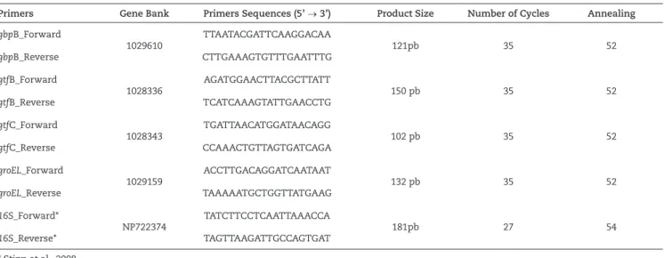
The PCR experiment was performed in triplicate for each cDNA. One microliter of cDNA (obtained from 24 ng of RNA) was used as a template in the PCR (25 μl) analysis, with 50 mM of MgCl2, 0.3 mM of each specific primer, 200 μM of dNTPs, 1.25 U of Taq polymerase and 10x PCR buffer, to generate amplicons ranging from 150-200 base pairs (Table 1). The amplicons were visualized on agarose gels (2% w/v) with ethidium bromide (0.3 µg/ml). Positive controls and a 100 bp DNA ladder were included in each gel. Digital images were captured under ultraviolet light using the Gel logic 100 Imaging System (Eastman Kodak Co.). The curves of the amplicons per number of thermal cycles (1-60 cycles), specific for each primer pair, were then generated to identify the middle of the exponential phase amplification or to establish the optimal number of cycles for each target gene. The thermal cycling parameters were as follows: 94°C for 45 s, annealing temperature (Table 1) for 60 s, and 72°C for 60 s.
All cDNA samples were tested with their respective primers for a specific target gene as well as with primers for the housekeeping gene 16S, a constitutively transcribed control gene whose expression is invariant under the used experimental conditions (Stipp et al., 2008). The intensities of the amplicons captured in the gels, using the same imaging system, were expressed as the relative arbitrary units of expression (UR). Controls for the RT-PCR included: reaction mixtures lacking template cDNA to effectively rule out (detect) the presence of contaminating DNA, and/or the formation of primer dimers. The positive controls included genomic DNA of the respective pathogenic bacterial strain.
Statistical analysis
Primers Gene Bank Primers Sequences (5’ → 3’) Product Size Number of Cycles Annealing
gbpB_Forward
1029610
TTAATACGATTCAAGGACAA
121pb 35 52
gbpB_Reverse CTTGAAAGTGTTTGAATTTG
gtfB_Forward
1028336
AGATGGAACTTACGCTTATT
150 pb 35 52
gtfB_Reverse TCATCAAAGTATTGAACCTG
gtfC_Forward
1028343
TGATTAACATGGATAACAGG
102 pb 35 52
gtfC_Reverse CCAAACTGTTAGTGATCAGA
groEL_Forward
1029159
ACCTTGACAGGATCAATAAT
132 pb 35 52
groEL_Reverse TAAAAATGCTGGTTATGAAG
16S_Forward*
NP722374
TATCTTCCTCAATTAAACCA
181pb 27 54
16S_Reverse* TAGTTAAGATTGCCAGTGAT * Stipp et al., 2008.
Table 1
Primer sequence, Gene Bank reference, amplicon size, and thermal conditions applied in the semi-quantitative reverse transcription polymerase chain reactions of known biofilm-associated genes and house-keeping gene 16S.
(after 30 min of exposure to the antimicrobial agent) and their respective controls (after 1 min of exposure) were applied an ANOVA test. The gene expression by planktonic cells submitted to 1% (v/v) DMSO vehicle for 30 min was also used as a control. The p values < 5% were considered statistically significant for all tests.
Results
Halistanol sulfate A (1), isolated from the sponge Petromica ciocalyptoides (Kossuga et al., 2007) and the modified diketopiperazines rodriguesines A (2) and B (3) isolated from the ascidian Didemnum sp. (Kossuga et al., 2009) (Fig. 1) inhibited the growth of Streptococcus mutans strains in low concentrations. Other bacteria growth were inhibited with higher concentrations of these antimicrobials agents (> 62.5 µg/ml).
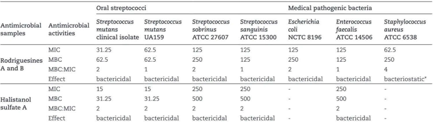
Table 2 shows the MIC and MBC values, as well as the MBC/MIC ratio, displayed by the halistanol sulfate A (1) and by rodriguesines A (2) and B (3). Rodriguesines A (2) and B (3) were inseparable under several HPLC conditions, as previously reported by Kossuga et al., (2009), and were tested in combination. Both samples presented a broad inhibitory spectrum against the tested bacterial strains. Rodriguesines A (2) and B (3) inhibited all of the tested bacteria, with MIC values between 15-125 µg/ml and MBC values between 31.2-250 µg/ml. These modified diketopiperazines had a bactericidal effect on all the bacterial strains except Staphylococcus aureus ATCC 6538. Halistanol sulfate A showed the lowest MIC and MBC values when tested on Streptococcus mutans. However, its inhibitory effect against other species was much less potent (MIC > 125 µg/ml) (Table 2). Therefore, it appears that halistanol sulfate A selectively inhibits the Streptococcus mutans pathogen at low concentrations, without modifying the ecological balance between more and less pathogenic microorganisms. The specificity of an oral antimicrobial agent at low concentrations
against Streptococcus mutans is important for reducing the selective pressure on other bacterial species, particularly initial colonizers, such as Streptococcus sanguinis. Therefore, both halistanol sulfate A and rodriguesines A and B were selected for additional studies of their antibiofilm activities.
The pathogenic Streptococcus mutans CI and Streptococcus mutans UA159 showed response to CV-stained biofilms greater than 2.0 at λmax OD575 nm in control conditions and were categorized as good biofilm formers (Loo et al., 2000). Fig. 1 shows the effect of rodriguesines A and B and halistanol sulfate A on planktonic cells and on biofilm formation, as well as the MIC values of biofilm formation by Streptococcus mutans strains. No significant differences in the inhibition of planktonic cells were observed in these MIC compared to the DMSO control (Student’s t test, p > 0.05). As for the MIC of biofilm formation, we observed reductions of up to 38% in the optical density of planktonic cells (at λmax OD550 nm) relative to the DMSO vehicle (Fig. 1). There were no significant differences in the viable cells counts of the planktonic cultures (Student’s t test, p > 0.05, data not shown). These results indicate that these compounds present modest selectivity in their inhibition of biofilm formation, despite their ability to inhibit the growth of planktonic cells.
Halistanol sulfate A (1) showed potent antibacterial activity against Streptococcus mutans CI and Streptococcus mutans UA159 strains at 3.0 µg/ml, inhibiting approximately 85.2 and 99.5% the biofilm formation by these species, respectively. The strain Streptococcus mutans UA159 was more susceptible to the antimicrobial activity of all tested substances in comparison with Streptococcus mutans CI. Biofilm formation by Streptococcus mutans UA159 was inhibited by 77.5 to 100%, while Streptococcus mutans CI biofilm formation was inhibited by 34.8 to 85.2% after antimicrobial treatment during 18 h.
Oral streptococci Medical pathogenic bacteria
Antimicrobial samples
Antimicrobial activities
Streptococcus mutans
clinical isolate
Streptococcus mutans
UA159
Streptococcus sobrinus
ATCC 27607
Streptococcus sanguinis
ATCC 15300
Escherichia coli
NCTC 8196
Enterococcus faecalis
ATCC 14506
Staphylococcus aureus
ATCC 6538
Rodriguesines A and B
MIC 31.25 62.5 125 125 125 125 62.5
MBC 62.5 62.5 250 125 250 125 250
MBC:MIC 2 1 2 1 2 1 4
Effect bactericidal bactericidal bactericidal bactericidal bactericidal bactericidal bacteriostatic*
Halistanol sulfate A
MIC 15 15 250 250 - 250
-MBC 31.25 31.25 500 500 - 500
-MBC:MIC 2 2 2 2 - 2
-Effect bactericidal bactericidal bactericidal bactericidal - bactericidal
-The antimicrobials were considered bactericidal when the MBC:MIC ratio was between 1:1 to 2:1 and bacteriostatic if this ratio was higher than 2:1 (Galvão et al., 2012). DMSO (1%) was used as a negative control. (*)The bacteriostatic effect of the tested samples could be bactericidal if bacterial inocula were used at approximately 1x10⁵ CFU/ml, as described by CLSI (2009).
Table 2
Antimicrobial activities (values of MIC, MBC and the MBC/MIC ratio in µg/ml, bactericidal or bacteriostatic effects) of halistanol sulfate A and rodriguesines A/B against oral streptococci and medical pathogenic bacteria.
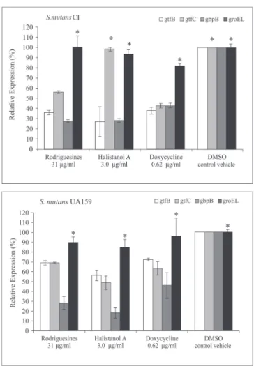
sulfate A (1) affected the expression of both S. mutans UA 159 and S. mutans CI genes involved in cell adhesion and biofilm formation, except the expression of groEL and 16S genes. The ANOVA test showed a statistically significant difference (p < 0.05) in the levels of gtfB, gtfC and gbpB gene transcription after the treatment of planktonic cells in the mid-log phase with the antimicrobial compounds. A 15 to 85% reduction of the gene transcription levels relatively to the respective controls (T1) was observed. The gene expression was normalized relatively to 16S gene transcription, which showed invariable expression in all tested conditions (data not shown), as well as relative to groEL, which codes for the chaperone GroEL, a class I heat shock protein of Streptococcus mutans.
Comparing DMSO 1% at T1 and at T30, we observed an increase of 12 to 55% (average 26.3 ± 15.3) in the transcription levels of biofilm-associated genes of S. mutans CI only, at T30 (data not shown). Fig. 2 shows the percentage of gtfB, gtfC, gbpB and groEL transcription levels after treatment with the compounds 1 - 3 after 30 min. Treatment with compounds 1 - 3 inhibited gene expression between 1.5 and 82% (51.56 ± 18.52) (Fig. 2). We observed a decrease in the gene expression levels of all tested genes, except gtfC transcription in S. mutans CI, which was not altered after treatment with halistanol sulfate A (ANOVA, p > 0.05). In addition, there were no alterations in the expression levels of the groEL and 16S genes after the antimicrobial treatments.
The gene gbpB was more severely affected after treatment for 30 min with the antimicrobial compounds, with an expression reduction of 53 to 82% for both pathogenic bacterial strains. The expression of gtfB and gtfC decreased by approximately 1.5 to 57%, depending on the tested strain and the antimicrobial compounds (Fig. 2). The gbpB gene of Streptococcus mutans UA159 strain was inhibited (average 72.5% ± 13.4) to a higher level than the gtf genes (average 35,8% ± 8,0). A higher inhibition of gtfB expression by Streptococcus mutans CI was observed relatively to Streptococcus mutans UA159.
Discussion
A series of marine natural products inhibit biofilm formation of Pseudomonas aeruginosa, Acinetobacter baumannii, Bordetella bronchiseptica, Staphylococcus aureus and Streptococcus mutans (Huigens et al., 2007, Rogers et al., 2008, Liu et al., 2011). In the present investigation we verified that halistanol sulfate A (1), isolated from the sponge Petromica ciocalyptoides (Kossuga et al., 2007), and the mixture of modified diketopiperazines rodriguesines A (2) and B (3) isolated from an ascidian of the genus Didemnum (Kossuga et al., 2009), displayed low MIC values against Streptococcus mutans. The rodriguesines A and B had broad antimicrobial spectra, inhibiting both oral streptococci as well as pathogenic bacteria with low MIC values.
Diketopiperazines are dipeptides that have also been isolated from Gram-positive bacteria, fungi and higher organisms (De Carvalho and Abraham, 2012). Diketopiperazines are reported to modulate the LuxR-mediated quorum-sensing systems of Gram-negative and Gram-positive bacteria and are considered to influence cell-cell bacterial signaling, offering alternative ways to control biofilms by interfering with
Figure 2 – The percentage of gtfB, gtfC and groELrelative expres-sion by planktonic cells of Clinical Isolated (CI) Streptococcus mu-tans and Streptococcus mutans UA159 after treatment for 30 min with rodriguesines A/B, halistanol sulfate A and doxycycline in the MIC of biofilm inhibition. Data were expressed relative to treatment with 1% DMSO for 30 min (control vehicle). An as-terisk represents that there is no significant difference relative to control (ANOVA p > 0.05). The mRNA expression levels were calibrated using 16S rRNA. Bars represent the means ± standard deviations of triplicate experiments.
microbial communication (Withers et al., 2001; Abraham, 2005; Ryan and Dow, 2008). Streptococcus mutans LuxS affects acid and oxidative-stress tolerance, bacteriocin production and biofilm formation (Merritt et al., 2003; 2005, Wen and Burne, 2004; Yoshida et al., 2005; Wen et al., 2011).
for the control of S. mutans because these systems regulate the transcription of several virulence target genes, some of which have been studied in detail (Li et al., 2001; Van Der Ploeg Jr, 2005). However, the lack of a detailed knowledge of the involved cell-cell communication mechanisms has hindered the development of effective therapeutic strategies to fight bacterial infections. The increased interest in studying the bacterial lifestyle in biofilm formation should lead to the discovery of new therapeutic strategies for caries treatment (Bayles, 2007).
Halistanol sulfate A inhibited biofilm formation by two Streptococcus mutans strains at low MIC, but it did not inhibit the initial colonizers represented by S. sanguinis. This activity profile is highly suitable because antibacterial agents that inhibit pathogenic bacteria and biofilms without affecting the healthy normal flora can be used in preventive treatments based on specifically targeted molecules. Agents with this mechanism tend to not disturb the ecological balance between pathogens and commensal residents in the oral cavity (Liu et al., 2011).
Halistanol sulfates A-E were first described as antifungal and antithrombin agents (Kanazawa et al., 1992). Previously it has been observed that halistanol sulfate A affects Staphylococcus aureus cells, possibly via a disruption membrane mechanism (Marinho et al., 2012). Sterol sulfates related to halistanol trisulfate A may act as detergents, inhibiting the growth of positive bacteria without affecting Gram-negative bacteria, possibly via a membranolytic mechanism and may also inhibit bacterial protein-tyrosine kinases (Slate, et al., 1994).
Streptococcus mutans CI showed higher resistance to the antibiofilm action of small-molecules than S. mutans UA159. The resistance mechanisms or intermediary resistance to antimicrobials agents are known to be variable among bacterial genotypes. The existence of a high diversity of S. mutans genotypes in human oral cavities (Kamiya et al., 2005) suggests that such antibacterial small-molecules should be tested against different strains with possible distinct resistance phenotype/genotypes to antimicrobial agents. The mechanisms controlling the expression of the gtfB, gtfC and gbpB virulence genes are strain-specific, illustrating the genetic diversity of S. mutans (Stipp et al., 2008). Such genotypic variation may have further implications in the selection of therapeutic targets to control this cariogenic bacterium.
After antimicrobial treatment with halistanol, rodriguesines or doxycycline, there was a significant reduction in the Streptococcus mutans biofilm formation, although there were no alterations in the groEL expression levels. The gene groEL codes for a chaperone, that has essential roles in cellular metabolism by assisting in the folding of newly synthesized or denatured proteins, as well as in the assembly, transport, and degradation of cellular proteins (Bukau and Horwich, 1998). In S. mutans, GroEL is classified as a class I heat shock protein as well is the chaperone DnaK. Lemos et al. (2007), showed that the down-regulation of dnaK and groEL reduced S. mutans biofilm formation and acid tolerance, suggesting that the regulatory response by S. mutans to environmental stresses for the development and maturation of a biofilm has a significant influence on the biofilm structure. Our data suggest that groEL
expression is not essential for the development of S. mutans biofilms because groEL is not a target gene of halistanol sulfate A and rodriguesines. These antimicrobial agents inhibited biofilm formation by the down regulation of gtfB / gtfC and gbpB, which codes for a water-insoluble glucan matrix and increases the interaction between S. mutans and glucans of the matrix.
The evaluation of natural products against different Streptococcus mutans genotypes is important for establishing the antibacterial effectiveness of these substances and for elucidating their effects on the selective pressure of possible S. mutans genotypes that are resistant to antimicrobial agents. The results presented herein illustrate the potential of marine secondary metabolites to act as antibacterial agents against S. mutans with distinct modes-of-action. Further studies are necessary to clarify the mechanisms-of-action of rodriguesines and halistanol sulfate A, their effects on the regulation of other S. mutans virulence genes and/or S. mutans quorum sensing signaling pathways, and their respective species-specific antibacterial activities in multi-species biofilm models, both in vitro and in vivo, associated with transcriptome and/or proteome analyses.
Conclusion
The modified diketopiperazines rodriguesines A and B as well as halistanol sulfate A displayed inhibitory effects on biofilm formation by Streptococcus mutans and down regulate the expression of the gtfB, gtfC and gbpB genes. The antimicrobial properties of halistanol sulfate A suggest that it may be useful in the development of new drug candidates for anti-Streptococcus mutans therapy because it selectively inhibits biofilm formation by S. mutans strains at low concentrations, not affecting initial buccal colonizing species such as S. sanguinis.
Authors’ contributions
semi-quantitative RT-PCR and performed statistical analysis. Contributed extensively to the preparation of the manuscript. All the authors have read the final manuscript and approved its submission.
Conflicts of interest
The authors declare no conflicts of interest.
Acknowledgments
This study was financially supported by Fundação de Amparo à Pesquisa de São Paulo (FAPESP, grants 2005/60175-2 and 2010/50190-2) and Fundação de Desenvolvimento da Unicamp (FUNCAMP, grant Faepex 68807). B.A.L. was supported by a FAPESP MSc scholarship (proc. 2007/01078-2), R.U.K. was supported by FAPESP (proc. 2005/58479-8 and 2006/60037-1), while S.P.L. and M.H.K. were supported by CAPES and CNPq scholarships.
R E F E R E N C E S
Abraham, W-R, 2005. Controlling gram-negative pathogenic bacteria by interfering with their biofilm formation. Drug. Des. Rev. 2, 13-33.
Bader, J.D., Shugars, D.A., Bonito. A.J., 2001. Systematic reviews of selected dental caries diagnostic and management methods. J. Dent. Educ. 65, 960-968.
Bayles, K.W., 2007. The biological role of death and lysis in biofilm development. Nat. Rev. Microbiol. 5, 721-726.
Blunt, J.W., Copp, B.R., Keyzers, R.A., Munro, M.H.G., Prinsep, M.R., 2014. Marine natural products. Nat. Prod. Rep. 31, 160-258. Bukau, B., Horwich, A.L., 1998. The Hsp70 and Hsp60 chaperone
machines. Cell. 92, 351-366.
Butler, M.S., Robertson, A.A.B., Cooper, M.A., 2014. Natural product and natural product derived drugs in clinical trials. Nat. Prod. Rep. 31, 1612-1661.
Chia, J.S., Lee, Y.Y., Huang, P.T., Chen, J.Y., 2001. Identification of stress-responsive genes in Streptococcus mutans by differential display reverse transcription-PCR. Infect. Immun. 69, 2493-2501.
CLSI, 2009. Methods for Dilution Antimicrobial Susceptibility Tests for Bacteria that Grow Aerobically, Approved Standard, 29, no. 2, Clinical And Laboratory Standards Institute document M07-A8, Wayne, Pa, USA, 8th ed.
Coenye, T., Honraet, K., Rigole, P., Nadal, J.P., Nelis, H.J., 2007. In vitro inhibition of Streptococcus mutans biofilm formation on hydroxyapatite by subinhibitory concentrations of anthraquinones. Antimicrob. Agents. Chemother. 51, 1541-1544.
De Carvalho, M.P., Abraham, W.R., 2012. Antimicrobial and biofilm inhibiting diketopiperazines. Curr. Med. Chem. 19, 3564-3577. Duque, C., Stipp, R.N., Wang, B., Smith, D.J., Höfling, J.F.,
Kuramitsu, H.K., Duncan, M.J., Mattos-Graner, R.O., 2011. Downregulation of GbpB, a component of the VicRK regulon, affects biofilm formation and cell surface characteristics of Streptococcus mutans. Infect. Immun. 79, 786-796.
Galvão, L.C., Furletti, V.F., Bersan, S.M., Da Cunha, M.G., Ruiz, A.L., De Carvalho, J.E., Sartoratto, A., Rehder, V.L.G., Figueira,
G.M., Duarte, M.C.T., Ikegaki, M., de Alencar, S. M., Rosalen, P.L., 2012. Antimicrobial activity of essential oils against Streptococcus mutans and their antiproliferative effects. Evid. Based Complement. Alternat. Med. 2012, http://dx.doi. org/10.1155/2012/751435
Huigens III, R.W., Richards, J.J., Parise, G., Ballard, T.E., Zeng, W., Deora, R., Melander, C., 2007. Inhibition of Pseudomonas aeruginosa biofilm formation with Bromoageliferin analogues. J. Am. Chem. Soc. 129, 6966-6967.
Kamiya, R.U., Höfling, J.F., Gonçalves, R.B., 2008. Frequency and expression of mutacin biosynthesis genes in isolates of Streptococcus mutans with different mutacin-producing phenotypes. J. Med. Microbiol. 57, 626-635.
Kamiya, R.U., Napimoga, M.H., Rosa, R.T., Höfling, J.F., Gonçalves, R.B., 2005. Mutacins production in Streptococcus mutans genotypes isolated from caries-affected and caries-free individuals. Oral. Microbiol. Immunol. 20, 20-24. Kanazawa, S., Fusetani, N., Matsunaga, S., 1992. Halistanol
sulfates A-E, new steroid sulfates, from a marine sponge, Epipolasis sp. Tetrahedron. 48, 5467-5472.
Klein, M.I., Flório, F.M., Pereira, A.C., Höfling, J.F., Gonçalves, R.B., 2004. Longitudinal study of transmission, diversity, and stability of Streptococcus mutans and Streptococcus sobrinus genotypes in Brazilian nursery children. J. Clin. Microbiol. 42, 4620-4626.
Koo, H., Gomes, B.P., Rosalen, P.L., Ambrosano, G.M., Park, Y.K., Cury, J.A., 2000. In vitro antimicrobial activity of propolis and Arnica montana against oral pathogens. Arch. Oral Biol. 45, 141-148.
Koo, H., Hayacibara, M.F., Schobel, B.D., Cury, J.A., Rosalen, P.L., Park, Y.K., Vacca-Smith, A.M., Bowen, W.H., 2003. Inhibition of Streptococcus mutans biofilm accumulation and polysaccharide production by apigenin and tt-farnesol. J. Antimicrob. Chemother. 52, 782-789.
Koo, H., Seils, J., Abranches, J., Burne, R.A., Bowen, W.H., Quivey, R.G. Jr., 2006. Influence of apigenin on gtf gene expression in Streptococcus mutans UA159. Antimicrob. Agents Chemother. 50, 542-546.
Kossuga, M.H., De Lira, S.P., McHugh, S., Torres, Y., Lima, B.A., Gonçalves, R.B., Veloso, K., Ferreira, A.G., Rocha, R.M., Berlinck, R.G.S., 2009. Antibacterial modified diketopiperazines from two ascidians of the genus Didemnum. J. Braz. Chem. Soc. 20, 704-711.
Kossuga, M.H., De Lira, S.P., Nascimento, A.M., Gambardella, M.T.P., Berlinck, R.G.S., Torres, Y.R., Nascimento, G.G.F., Pimenta, E.F., Silva, M., Thiemann O.H., Oliva, G., Tempone, A.G., Melhem, M.S.C., de Souza, A.O., Galetti, F.C.S., Silva, C.L., Cavalcanti, B., Pessoa, C.O., Moraes, M.O., Hajdu, E., Peixinho, S., Rocha, R.M., 2007. Isolamento e atividades biológicas de produtos naturais das esponjas Monanchora arbuscula, Aplysina sp., Petromica ciocalyptoides e Topsentia ophiraphidites, da ascídia Didemnum ligulum e do octocoral Carijoa riisei. Quim. Nova 30, 1194-1202.
Kossuga, M.H., Romminger, S., Xavier, C., Milanetto, M.C., Do Valle, M.Z., Pimenta,E.F., Morais, R.P., Carvalho, E., Mizuno, C.M., Coradello, L.F.C., Barroso, V. M., Vacondio, B., Javaroti, D.C.D., Seleghim, M.H.R., Cavalcanti, B.C., Pessoa, C., Moraes, M.O., Lima, B.A., Gonçalves, R.B., Bonugli-Santos, R.C., Sette, L.D., Berlinck, R.G.S., 2012. Evaluating methods for the isolation of marine-derived fungal strains and production of bioactive secondary metabolites. Rev. Bras. Farmacogn. 22, 257-267. Lemos, J.A., Luzardo, Y., Burne, R.A., 2007. Physiologic effects
Li, Y.H., Lau, P.C., Lee, J.H., Ellen, R.P., Cvitkovitch, D.G., 2001. Natural genetic transformation of Streptococcus mutans growing in biofilms. J. Bacteriol. 183, 897-908.
Liu, C., Worthington, R.J., Melander, C., Wu, H., 2011. A new small molecule specifically inhibits the cariogenic bacterium Streptococcus mutans in multispecies biofilms. Antimicrob. Agents Chemother. 55, 2679-2687.
Lonn-Stensrud, J., Petersen, F.C., Benneche, T., Scheie, A.A., 2007. Synthetic bromated furanone inhibits autoinducer-2-mediated communication and biofilm formation in oral streptococci. Oral Microbiol. Immunol. 22, 340-346. Loo, C.Y., Corliss, D.A., Ganeshkumar, N., 2000. Streptococcus
gordonii biofilm formation: identification of genes that code for biofilm phenotypes. J. Bacteriol. 182, 1374-1382.
Mai, J., Tian, X-L., Gallant, J.W., Merkley, N., Biswas, Z., Syvitski, R., Douglas, S.E., Ling, J., Li, Y.H., 2011. A novel target-specific, salt-resistant antimicrobial peptide against the cariogenic pathogen Streptococcus mutans. Antimicrob. Agents Chemother. 55, 5205-5213.
Marinho, P.R., Simas, N.K., Kuster, R.M., Duarte, R.S., Fracalanzza, S.E., Ferreira, D.F., Romanos, M.T., Muricy, G., Giambiagi-Demarval, M., Laport, M.S., 2012. Antibacterial activity and cytotoxicity analysis of halistanol trisulphate from marine sponge Petromica citrina. J. Antimicrob. Chemother. 67, 2396-2400.
Mattos-Graner, R.O., Jin, S., King, W.F., Chen, T., Smith, D.J., Duncan, M.J., 2001. Cloning of the Streptococcus mutans gene encoding glucan binding protein B and analysis of genetic diversity and protein production in clinical isolates. Infect. Immun. 69, 6931-6941.
Mayer, A.M., Rodríguez, A.D., Taglialatela-Scafati, O., Fusetani, N., 2013. Marine pharmacology in 2009-2011: marine compounds with antibacterial, antidiabetic, antifungal, anti-inflammatory, antiprotozoal, antituberculosis, and antiviral activities; affecting the immune and nervous systems, and other miscellaneous mechanisms of action. Mar. Drugs. 11, 2510-2573.
Merritt, J., Kreth, J., Shi, W., Qi, F., 2005. LuxS controls bacteriocin production in Streptococcus mutans through a novel regulatory component. Mol. Microbiol. 57, 960-969.
Merritt, J., Qi, F., Goodman, S.D., Anderson, M.H., Shi, W. 2003. Mutation of LuxS affects biofilm formation in Streptococcus mutans. Infect. Immun. 71, 1972-1979.
Murata, R.M., Almeida, L.S.B., Yatsuda, R., Dos Santos, M.H., Nagem, T.J., Rosalen, P.L., Koo, H., 2008. Inhibitory effects of 7-epiclusianone on glucan synthesis, acidogenicity and biofilm formation by Streptococcus mutans. FEMS Microbiol. Lett. 282, 174-181.
O’Toole, G.A., Kolter, R., 1998. Initiation of biofilm formation in Pseudomonas fluorescens WCS365 proceeds via multiple, convergent signalling pathways: a genetic analysis. Mol. Microbiol. 28, 449-461.
Oliveira, J.H., Seleghim, M.H., Timm, C., Grube, A., Kock, M., Nascimento, G.F.G., Martins, A.C.T., Silva, E.G.O., de Souza, A.O., Minarini, P.R.R., Galetti, F.C.S., Silva, C.L., Hajdu E., Berlinck, R.G.S., 2006. Antimicrobial and antimycobacterial activity of cyclostelletamine alkaloids from sponge Pachychalina sp. Mar. Drugs. 4, 1-8.
Pasquantonio, G., Greco, C., Prenna, M., Ripa, C., Vitali, L.A., Petrelli, D., Di Luca, M.C., Ripa, S., 2008. Antibacterial activity and anti-biofilm effect of chitosan against strains of Streptococcus mutans isolated in dental plaque. Int. J. Immunopathol. Pharmacol. 21, 993-997.
Rogers, S.A., Melander, C., 2008. Construction and screening of a 2-aminoimidazole library identifies a small molecule capable of inhibiting and dispersing bacterial biofilms across order, class, and phylum. Angew. Chem. Int. Ed. Engl. 47, 5229-5231. Ryan, R.P., Dow, J.M., 2008. Diffusible signals and interspecies
communication in bacteria. Microbiol. 154, 1845-1858. Seleghim, M.H.R., De Lira, S.P., Kossuga, M.H., Batista, T., Berlinck,
R.G.S., Hajdu, E., Muricy, G., da Rocha, R.M., do Nascimento, G.G.F., Silva, M., Pimenta, E.F., Thiemann, O.H.,Oliva, G., Cavalcanti, B.C., Pessoa, C., de Moraes, M.O., Galetti, F.C.S., Silva, C.L., de Souza, A.O., Peixinho, S., 2007. Antibiotic, cytotoxic and enzyme inhibitory activity of crude extracts from Brazilian marine invertebrates. Rev. Bras. Farmacogn. 17, 287-318.
Slate, D.L., Lee, R.H., Rodriguez, J., Crews, P., 1994. The marine natural product, halistanol trisulfate, inhibits pp60v-src protein tyrosine kinase activity. Biochem. Biophys. Res. Commun. 203, 260-264.
Smith, D.J., 2002. Dental caries vaccines: Prospects and concerns. Crit. Rev. Oral Biol. Med. 13, 335-349.
Stipp, R.N., Goncalves, R.B., Höfling, J.F., Smith, D.J., Mattos-Graner, R.O., 2008. Transcriptional analysis of gtfB, gtfC, and gbpB and their putative response regulators in several isolates of Streptococcus mutans. Oral Microbiol. Immunol. 23, 466-473. Van Der Ploeg Jr, 2005. Regulation of bacteriocin production in
Streptococcus mutans by the quorum-sensing system required for development of genetic competence. J. Bacteriol. 187, 3980-3989.
Wen, Z.T., Burne, R.A., 2004. LuxS-mediated signaling in Streptococcus mutans is involved in regulation of acid and oxidative stress tolerance and biofilm formation. J. Bacteriol. 186, 2682-2691.
Wen, Z.T., Nguyen, A.H., Bitoun, J.P., Abranches, J., Baker, H.V., Burne, R.A., 2011. Transcriptome analysis of LuxS-deficient Streptococcus mutans grown in biofilms. Mol. Oral Microbiol. 26, 2-18.
Whiley, R.A., Beighton, D., 1998. Current classification of the oral streptococci. Oral Microbiol. Immunol. 13, 195-216.
Withers, H., Swift, S., Williams, P., 2001. Quorum sensing as an integral component of gene regulatory networks in gram-negative bacteria. Curr. Opin. Microbiol. 4, 186-193. Yoshida, A., Ansai, T., Takehara, T., Kuramitsu, H.K., 2005.