Mariana Passos DE LUCA(a) Irlan Almeida FREIRES(b) Alfonso GALA-GARCÍA(c) Vagner Rodrigues SANTOS(d) Miriam Pimenta VALE(a)
Severino Matias de ALENCAR(e) Pedro Luiz ROSALEN(f)
(a) Universidade Federal de Minas Gerais –
UFMG, School of Dentistry, Department of Pediatric Dentistry and Orthodontics, Belo Horizonte, MG, Brazil.
(b)University of Florida, College of Dentistry,
Department of Oral Biology, Gainesville, FL, United States.
(c) Universidade Federal de Minas Gerais –
UFMG, School of Dentistry, Department of Restorative Dentistry, Belo Horizonte, MG, Brazil.
(d) Universidade Federal de Minas Gerais –
UFMG, School of Dentistry, Department of Oral Pathology and Oral Surgery, Belo Horizonte, MG, Brazil.
(e) Universidade de São Paulo – USP, “Luiz
de Queiroz” College of Agriculture, Department of Agri-Food industry, Piracicaba, SP, Brazil.
(f) Universidade de Campinas – UNICAMP,
Piracicaba Dental School, Department of Physiological Sciences, Piracicaba, SP, Brazil.
The anti-caries activity and toxicity of an
experimental propolis-containing varnish
Abstract: We investigated the anti-caries effects of an experimental
propolis varnish in vivo, and further tested its toxicity against
ibroblasts. Fifty-six SPF female Wistar rats were infected with
Streptococcus mutans UA159 (SM) and allocated into four groups
(n = 14/group): G1, propolis varnish (15%/PV); G2, chitosan varnish (CV/vehicle); G3, gold standard (GS/Duraphat®); and G4, untreated.
The animals received a single varnish application on their molars and
were submitted to a high cariogenic challenge (Diet-2000, 56% sucrose, and 5% sucrose-added water, ad libitum) for 4 weeks. Total cultivable
microbiota and SM were counted, and smooth-surface and sulcal caries were scored. PV, CV and GS cytotoxic effects were tested against ibroblasts. The data were analyzed using ANOVA with the Tukey-Kramer test (p ≤ 0.05). Total microbiota and SM counts did not differ among the treatments (p = 0.78), or in relation to the untreated group (p = 0.52). PV reduced development of smooth-surface enamel caries compared with the untreated group (p = 0.0018), with no signiicant difference from GS (p = 0.92); however, the PV effects were no longer observed when the dentin was affected. Neither PV nor GS prevented enamel sulcal lesion onset, but GSsigniicantly reduced the severity of dentinal sulcal lesions (p < 0.0001). No signiicant difference was observed in ibroblast viability between PV and GS (p < 0.0001). In conclusion, PV prevented smooth-surface enamel caries and showed low cell toxicity. Nevertheless, due to the high cariogenic challenge, its effects were not sustained throughout the experiment. Further studies are encouraged to establish a protocol to sustain the long-term anti-caries activity of PV in the oral cavity.
Keywords: Propolis; Streptococcus mutans; Dental Caries; Models, Animal.
Introduction
Dental caries remains a major public health issue worldwide with high prevalence and signiicant social impact.1 It results from a demineralization process in which acidogenic and acidophilic bacteria embedded in a mature, well-arranged bioilm degrade the tooth substance, ultimately leading to cavitation.2
Although several studies have related the participation of other bacteria in
the pathogenesis of dental caries,2,3,4,5 Streptococcus mutans plays a central role
in the development of cariogenic bioilms, mainly due to its acid-tolerant and Declaration of Interests: The authors
certify that they have no commercial or associative interest that represents a conflict of interest in connection with the manuscript.
Corresponding Author:
Mariana Passos De Luca
E-mail: delucamariana@gmail.com, rosalen@fop.unicamp.br
https://doi.org/10.1590/1807-3107BOR-2017.vol31.0045
Submitted: Aug 29, 2016
acidogenic characteristics. This microorganism uses dietary sucrose to synthetize extracellular polysaccharides (EPS), which are functional structures that mediate its adherence to the tooth surfaces. The carbohydrate fermentation process creates low-pH microenvironments that favor enamel and dentin demineralization.6
Natural products are major sources of bioactive
molecules and pharmaceutical leads, and have therefore
contributed signiicantly to drug development.7 Among these, propolis is one such product that stands out for
its biological properties, mainly as an antimicrobial, with direct application in dentistry.8,9 Its anti-caries
mechanism of action is associated with inhibition of glucosyltransferases and downregulation of speciic genes associated with stress survival and tolerance
in S. mutans.10 Moreover, some lavonoids, terpenoids,
isolavones and other phenolic acids contained in propolis were found to diminish S. mutans acid
production and tolerance.10,11,12
Overall, a large number of biological activities have been attributed to different types of propolis, including antimicrobial, antifungal, antitumor, and others.9 Propolis type 12, from southeastern Brazil,13 demonstrated strong antibacterial effects and
prevented caries development in rats.14 In addition,
topical application of the ethanolic extract of other
types of propolis was also found to effectively reduce
the incidence and severity of carious lesions in vivo.14,15,16
However, the biological effects of propolis may vary
according to the geographical origin and chemical
composition of the collected sample.16
Varnishes are materials widely used in dentistry for the prevention of dental caries.17 Some active
principles can be incorporated into these formulations
to promote and prolong their anti-caries effects,
including luoride, with remineralizing action,18 and
chlorhexidine, with antimicrobial action.19
An experimental propolis-containing varnish
was developed by our research group, as reported elsewhere.20 Our previous indings demonstrated that
the experimental varnish has in vitro antimicrobial
activity against cariogenic bacteria, and showed a
very satisfactory sustained release of propolis in vitro.20
These data have encouraged us to investigate the anti-caries activity of this experimental formulation, using an animal model under a high cariogenic
challenge. Herein, we investigated the anti-caries effects
of the experimental propolis varnish in a rat model,
and further tested its toxicity against ibroblasts, to
provide enough information to perform clinical trials
and eventually promote future clinical use.
Methodology
Animals
Fifty-six speciic pathogen-free (SPF) female Wistar rats were obtained from CEMIB (Multidisciplinary
Center for Biological Research, University of Campinas,
SP, Brazil) and maintained at the animal facility of Piracicaba Dental School (Piracicaba, SP, Brazil). All procedures were performed in accordance with
the ethical standards for the use and care of the
animals. This study had the prior approval of the
Ethics Committee on Animal Use (CEUA, University
of Campinas, SP, Brazil; protocol no. 3142-1).
Experimental caries model
The rats were initially screened for the presence of
indigenous S. mutans on mitis salivarius agar (Difco
Laboratories, Detroit, USA) and mitis salivarius agar plus bacitracin (MSB/Sigma Chemical Co., St Louis, USA.). This is a quality control of the SPF condition of the animals. The initial screening using MSA and MSB plates conirmed that all the animals were streptococci- and mutans streptococci-free. At the age of 21 days, the rats were infected with an actively growing overnight culture of S. mutans UA159 using
a cotton swab, and then fed pellet chow, Diet2000 with 56% sucrose,21 plus 5% sucrose in drinking
water ad libitum.22 At the age of 25 days, the animals
were screened for the establishment of oral infection, by plating oral samples onto MSB agar plates. On the following day, the rats were randomly assigned to four groups (n = 14), anesthetized with chloral hydrate (440 mg/kg) and submitted to the following treatments: G1 – topical application of propolis varnish (PV) (type 12 propolis from southeastern Brazil13 - propolis ethanolic extract, 15%, w/v); G2 –
(negative control). The varnishes were applied on the occlusal surfaces of the molars using one microbrush per hemiarch. After topical application, the animals fasted for 2 hours and were kept in individual cages for four weeks. Diet2000, containing 56% of sucrose, and 5% sucrose-added sterile distilled water were provided
ad libitum. The animals were weighed weekly, and
their behavior and physical appearance were noted daily. At the end of the experiment, the animals were euthanized and submitted to microbiological analysis.
Microbiological analysis
The lower left jaw was aseptically dissected, suspended in 5.0 mL of sterile saline solution (0.9% NaCl, w/v), and sonicated using three 10-second pulses at 30W with 5-second intervals (Vibracell, Sonics & Material Inc.), in order to obtain the maximum recoverable viable counts. A spiral plater (Whitley Automatic Spiral Plater, DW Scientiic®) was used to streak the suspensions onto blood agar (5% sheep blood) and mitis salivarius agar containing 100 µg/mL streptomycin sulfate (MSB, Sigma®), to determine the number of CFU/mL of total cultivable microorganisms
and S. mutans, respectively.23 The counting of total
cultivable microbiota was performed (i) to determine the proportion (%) of S. mutans in the oral cavity of the
animals in relation to the other microorganisms, which
is directly related to strain implantation on the tooth
surfaces; (ii) to ensure that the animals from all the groups had a similar microbial load at the endpoint; (iii) to identify whether treatment signiicantly affects the indigenous total microbiota of the animals and could potentially cause a microbiological disturbance.
Smooth-surface and sulcal caries and their severity (E, enamel
lesion; Ds, slight dentinal caries; Dm, moderate dentinal caries – 3/4 of the dentin affected; Dx, extensive dentinal caries – all dentin affected) were scored according to Larson’s modiication of Keyes’ system.24 Determining of
the caries score was blinded by codiication of the jaws and performed by one calibrated examiner (Intraclass Correlation Coeficient: 0.93, which indicates excellent intra-rater agreement).
Cytotoxic effects against 3T3-L1 fibroblasts Fibroblasts 3T3-L1 (ATCC® CL-173™) were grown in DMEM-F12, supplemented by 10%
synovial fibroblasts and 1% antibiotic-antimycotic solution (10,000 units of penicillin, 10 mg of streptomycin and 25 µg/mL of Amphotericin B in 0.9% sodium chloride; Sigma, St. Louis, MO) in humidified air – 5% CO2 atmosphere at 37ºC.
The materials tested were: a. culture medium + cells (control), b. culture medium, c. PV (type 12 propolis from southeastern Brazil – propolis ethanolic extract, 15%, w/v), d. CV (varnish base, vehicle control), e. gold-standard (GS) varnish (Duraphat,
fluoride 2.26%, w/v, positive control), and f. acetic acid (1%, v/v).20
One millimeter of each product was placed on round glass coverslips (9-mm diameter) and dried under vacuum for 24 h. Then the ibroblasts were plated into 48-well microtiter plates (4 x 10⁵ cells/well). After
4 h, the cells received conditioned media respective
to each experimental group. Cell conditioning was carried out for 1 h, at 37°C, in a humid atmosphere with 5% CO2. Then the conditioned media of all the groups
were replaced by fresh media. Cell mitochondrial activity was analyzed using the MTT-based method 24 h and 48 h after conditioning.25 Cell growth
curves were plotted, and the absorbance data were transformed into percentages of viable cells.
Statistical analysis
Smooth-surface and sulcal caries scores were expressed as proportions of their maximum possible values (124 and 56, respectively).24 The data were
submitted to one-way analysis of variance (ANOVA) followed by Tukey-Kramer HSD (Honest Standard Deviation) pairwise comparison test on JMP version 3.1 software, with a 5% signiicance level.
Results
All the rats gained weight and remained apparently healthy and active throughout the experiment. The average weight gains among the individual groups of rats were not signiicantly different (P>0.05,
data not shown).
animals are expressed in Table 1. At the endpoint, the total microbiota and SM counts did not differ signiicantly among the treatments (p = 0.78), or in relation to the untreated group (p = 0.52), showing that microbial viability was not affected.
As seen in Table 2, PV reduced the development of smooth-surface enamel caries, as compared with the untreated group (p = 0.0018), with no signiicant difference from the GS (p = 0.92). Nevertheless,
the anti-caries effects of PV that were no longer observed as smooth-surface carious lesions became more severe, that is, when the dentin was affected. There was no signiicant difference between PV and the untreated group (p = 0.34, 0.99 and 0.48, respectively), either for slight, moderate or extensive dentinal caries.
Neither the experimental nor the standard varnishes were able to prevent the onset of sulcal lesions in the enamel (Table 3). However, the GS signiicantly reduced the severity of the sulcal lesions, affecting dentin, as compared with the untreated group (p < 0.0001).
No macroscopic tissue changes or abnormalities were observed in the oral cavity of the animals throughout the whole experiment.
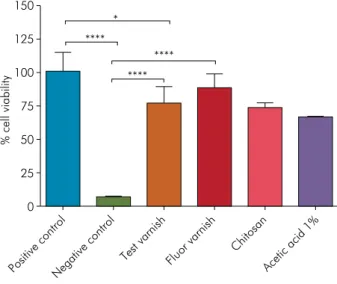
Toxicity assays were carried out in vitro to provide
preliminary evidence on whether PV would pose a risk against host cells. As seen in Figures 1 and 2, no signiicant difference was observed in ibroblast viability between PV and the GS after 24-h or 48-h exposure, respectively (p < 0.0001).
Table 1. Effects of the treatment with experimental and control varnishes on total oral microbiota and Streptococcus mutans counts in rats submitted to a high cariogenic challenge for four weeks. The values are expressed as mean (standard deviation).
Treatment
Total microbiota (CFU/mL) (M ± SD)
S. mutans (CFU/mL) (M ± SD)
% S. mutans (M ± SD)
Propolis varnish 75.4 (7.8)a 37.6 (4.2)a 40.7 (11.3)a
Gold standard* 43.5 (6.0)a 24.4 (3.7)a 46.2 (15.5)a
Chitosan Varnish** 74.3 (8.1)a 38.7 (4.7)a 47.1 (7.8)a
Untreated control*** 48.4 (10.1)a 33.1 (8.0)a 40.4 (11.3)a
*Duraphat®; **vehicle control; ***untreated group. Different superscript
letters in the same column indicate significant differences among
treatments (One-way ANOVA with Tukey-Kramer HSD, p ≤ 0.05).
Table 2. Effects of the treatment with the experimental propolis varnish on the development and severity of smooth-surface caries in rats. The values of the of the caries scores are given as mean ± standard deviation using the Keyes’ classification modified by Larson.
Treatment Smooth-surface caries
E (M ± SD) Ds (M ± SD) Dm (M ± SD) Dx (M ± SD)
Propolis varnish 19.8 (1.5)b 13.4 (5.2)b,c 1.4 (1.4)a 0.1 (0.3)a
Gold standard* 16.5 (1.6)b 10.3 (4.6)c 0.1 (0.4)b 0.0 a
Chitosan Varnish** 33.5 (1.6)a 19.8 (5.9)a 1.2 (1.4)a,b 0.1 (0.3)a
Untreated control*** 28.3 (1.5)a 16.5 (4.5)a,b 1.1 (1.0)a 0.3 (0.6)a
E: enamel caries; Ds: slight dentinal caries; Dm: moderate dentinal caries; Dx: extensive dentinal caries; *Duraphat®; **vehicle control,
***untreated group. Different superscript letters in the same column indicate significant differences among treatments (One-way ANOVA with
Tukey-Kramer HSD, p ≤ 0.05).
Table 3. Effects of the treatment with the experimental propolis varnish on the development and severity of sulcal caries in rats. The values of the of the caries scores are given as mean ± standard deviation using the Keyes’ classification modified by Larson.
Treatment Sulcal caries
E (M ± SD) Ds (M ± SD) Dm (M ± SD) Dx (M ± SD)
Propolis varnish 43.2 (6.5)a 34.6 (6.7)a,b 14.6 (4.6)b 6.4 (1.6)a
Gold standard* 35.6 (6.0)a 21.0 (5.7)c 4.9 (3.2)c 2.8 (1.9)b
Chitosan Varnish** 46.5 (4.0)a 40.0 (5.9)a 20.1 (4.2)a 7.5 (2.5)a
Untreated control*** 41.6 (5.3)a 34.1 (6.4)b 15.4 (4.2)a,b 6.0 (2.4)a
E: enamel caries; Ds: slight dentinal caries; Dm: moderate dentinal caries; Dx: extensive dentinal caries; *Duraphat®; **vehicle control,
***untreated group. Different superscript letters in the same column indicate significant differences among treatments (One-way ANOVA with
Discussion
Propolis contains bioactive molecules in its composition, which may affect several targets in the human body, including those related to the onset of oral diseases. Greater scientiic knowledge of the
chemical composition of various types of propolis has helped envision their potential use and application
in dental practice.8,9,26,27
Previous studies have reported the strong antimicrobial activity of propolis against several bacteria of the oral microbiome, including
S. mutans.1,2,28,29,30,31 As conirmed in our study, propolis
and some of its isolated compounds, such as apigenin
and tt-farnesol,11 have been found to display a major
effect on microbial virulence (e.g., polysaccharide
production and acid tolerance), rather than affecting unspeciic or speciic microbiome viability, considered a highly desirable quality.
In our st udy, the experimental
propolis-containing varnish was able to reduce the onset
of smooth-surface caries in the enamel, similarly to
the GS (Duraphat®). However, it was not effective
in diminishing the severity of carious lesions
affecting dentin, either on smooth-surface or sulcal
areas. While this result may mean that the propolis varnish had no satisfactory antimicrobial activity
to prevent advanced smooth-surface and sulcal
caries, one should take into consideration that such an effect was evaluated only at one endpoint, when the material was probably no longer adhered to the tooth structure. Hence, it seems pertinent to note that the experimental propolis varnish was found to be more effective against early smooth-surface lesions under a high cariogenic challenge. The lack of a residual effect conirms that propolis activity occurs while it is in direct contact with the teeth. Hence, repeated and prolonged applications of this new product could reduce the virulence of cariogenic bacteria, and thus achieve effective anti-caries activity. Further studies should also
focus on the incorporation of isolated anti-caries
compounds (e.g.: apigenin, tt-farnesol) into the
varnish matrix.
Other studies have investigated the anti-caries
effects of propolis extracts using this same animal
model.10,15,16,23,32 In these studies, ethanolic extracts
of propolis, or its bioactive fractions, were applied
Figure 2. MTT-based viability assay of fibroblast culture after 48 hours in contact with the following materials: positive control (culture medium+cells); negative control (culture medium); test varnish (propolis varnish); fluoride-containing varnish (gold standard, Duraphat®); vehicle control (chitosan varnish);
and 1% acetic acid. Four asterisks (****) indicate significant differences among the groups at p < 0.0001 (One-way ANOVA with Tukey-Kramer HSD).
% cell viability
150
125
75
50
25
0
Positive controlNegative control Test varnish Fluor varnish Acetic acid 1%
100
Chitosan
****
****
**** *
Figure 1. MTT-based viability assay of fibroblast culture after 24 hours in contact with the following materials: positive control (culture medium + cells); negative control (culture medium); test varnish (propolis varnish); fluoride-containing varnish (gold standard, Duraphat®); vehicle control (chitosan varnish);
and 1% acetic acid. Four asterisks (****) indicate significant differences among the groups at p < 0.0001 (One-way ANOVA with Tukey-Kramer HSD).
% cell viability
150
125
75
50
25
0
Positive controlNegative control Test varnis h
Fluor varnish
Acetic acid 1%
100
Chitosan
****
topically to the molars of rats twice a day for a period of 4 to 5 weeks. Overall, the authors observed anti-caries activity similar to that of Duraphat®.
This result suggests that the continuing presence
of propolis in the oral cavity enabled more effective antimicrobial activity, leading to a signiicant decrease in the severity of carious lesions.
The experimental propolis varnish was not cytotoxic against fibroblasts or osteoblasts, as previously
reported,20 and showed no difference in comparison
with the GS Duraphat®. This suggests that application of propolis varnish in intimate contact with the oral tissues is not potentially harmful to host cells.
Chitosan is a biocompatible and biodegradable
p o l y m e r i c m a t e r i a l33 w it h non-tox ic a nd
antimicrobial properties.34 Chitosan of medium
molecular weight was used as a vehicle during preparation of the experimental varn ish. Our results showed that the chitosan varnish (no active principle included) became inert as the severity of dental caries increased. This may be due to the molecular weight of chitosan used in the formulation, which is different from that generally used in other studies (using low molecular weight) to test the antimicrobial activity of dental products.7,10,14,33,35,36,37,38,39,40 Changes in the molecular
weight of the chitosan used may have precluded polymeric film formation, which is necessary to mediate product adhesion to the teeth.
Fluoride, considered the GS in caries prevention, has little antimicrobial effects on the synthesis of
glucans and acid tolerance in S. mutans, and its
mechanism of action is based on the physicochemical dynamics of ions in order to keep the integrity of the enamel hydroxyapatite mineral content.6 Fluoride
prevents the onset of dental caries by reducing demineralization and enhancing remineralization of incipient lesions.15 Duraphat® is a luoride-containing varnish, and was used as positive control in this
experiment due to its proven effects in controlling
dental caries. This varnish did not interfere with the total microbiota and SM counts – as expected, because of luoride mechanism of action – but it signiicantly reduced the severity of carious lesions.
Concerning smooth-surface enamel caries, there
was no difference in the preventive effects of the experimental varnish versus the GS.
Further studies are now needed to establish an eficacious protocol to sustain long-term antimicrobial
activity of the propolis varnish, particularly to prevent advanced dentinal carious lesions under a
high cariogenic challenge.
Acknowledgments
The authors are grateful to the National Council for Scientific and Technological Development (CNPq) for the scholarship, and wish to thank André Augusto Gomes Faraco and Juçara Ribeiro Franca for manufacturing the propolis varnish, and Eliane Franco and José Carlos for their technical support, as well as Bruna Benso, Carina Denny, Laila Facin, Lívia Galvão, Camila Batista, Luiz Eduardo Ferreira, and Cleiton P. Santos for their valuable help in the experiments.
1. Petersen PE, Bourgeois D, Ogawa H, Estupinan-Day S, Ndiaye C. The global burden of oral diseases and risks to oral health. Bull World Health Organ. 2005;83(9):661-9. https:/doi.org/10.1590/S0042-96862005000900011 2. Wolff D, Frese C, Maier-Kraus T, Krueger T, Wolff B. Bacterial
biofilm composition in caries and caries-free subjects. Caries Res. 2013;47(1):69-77. https://doi.org/10.1159/000344022 3. Beighton D. The complex oral microflora of high-risk
individuals and groups and its role in the caries process. Community Dent Oral Epidemiol. 2005;33(4):248-55. https://doi.org/10.1111/j.1600-0528.2005.00232.x
4. Choi EJ, Lee SH, Kim YJ. Quantitative real-time polymerase chain reaction for Streptococcus mutans and Streptococcus sobrinus in dental plaque samples and its association with early childhood caries. Int J Paediatr Dent. 2009;19(2):141-7. https://doi.org/10.1111/j.1365-263X.2008.00942.x 5. Head DA, Marsh PD, Devine DA. Non-lethal
control of the cariogenic potential of an agent-based model for dental plaque. PLoS One. 2014;9(8):e105012.
https://doi.org/10.1371/journal.pone.0105012
6. Jeon JG, Rosalen PL, Falsetta ML, Koo H. Natural products in caries research: current (limited) knowledge, challenges and future perspective. Caries Res. 2011;45(3):243-63. https://doi.org/10.1159/000327250
7. Mishra BB, Tiwari VK. Natural products: an evolving role in future drug discovery. Eur J Med Chem. 2011 Oct;46(10):4769-807. https://doi.org/10.1016/j.ejmech.2011.07.057
8. Libério SA, Pereira AL, Araújo MJ, Dutra RP, Nascimento FR, Monteiro-Neto V et al. The potential use of propolis as a cariostatic agent and its actions on mutans group streptococci. J Ethnopharmacol. 2009;125(1):1-9. https://doi.org/10.1016/j.jep.2009.04.047
9. Freires IA, Alencar SM, Rosalen PL. A pharmacological perspective on the use of Brazilian Red Propolis and its isolated compounds against human diseases. Eur J Med Chem. 2016;110:267-79. https://doi.org/10.1016/j.ejmech.2016.01.033
10. Bueno-Silva B, Alencar SM, Koo H, Ikegaki M, Silva GV, Napimoga MH et al. Anti-Inflammatory and antimicrobial evaluation of neovestitol and vestitol isolated from Brazilian red propolis. J Agric Food Chem. 2013;61(19):4546-50. https://doi.org/10.1021/jf305468f
11. Koo H, Hayacibara MF, Schobel BD, Cury JA, Rosalen PL, Park YK et al. Inhibition of Streptococcus mutans biofilm accumulation and polysaccharide production by apigenin and tt-farnesol. J Antimicrob Chemother. 2003;52(5):782-9. https://doi.org/10.1093/jac/dkg449
12. Castro ML, Nascimento AM, Ikegaki M, Costa-Neto CM, Alencar SM, Rosalen PL. Identification of a bioactive compound isolated from Brazilian propolis type 6. Bioorg Med Chem. 2009;17(14):5332-5. https://doi.org/10.1016/j.bmc.2009.04.066 13. Park YK, Alencar SM, Scamparine ARP, Aguiar CL.
[Propolis produced in South Brazil, Argentine and Uruguay: phytochemical evidence for the plant origin]. Cienc Rural. 2002 2;32(6):997-1003. Portuguese. https://doi.org/10.1590/S0103-84782002000600013 14. Ikeno K, Ikeno T, Miyazawa C. Effects of propolis on
dental caries in rats. Caries Res. 1991;25(5):347-51. https://doi.org/10.1159/000261390
15. Duarte S, Rosalen PL, Hayacibara MF, Cury JA, Bowen WH, Marquis RE et al. The influence of a novel propolis on mutans streptococci biofilms and caries development in rats. Arch Oral Biol. 2006;51(1):15-22. https://doi.org/10.1016/j.archoralbio.2005.06.002 16. Koo H, Rosalen PL, Cury JA, Park YK, Ikegaki M, Sattler A.
Effect of Apis mellifera propolis from two Brazilian regions on caries development in desalivated rats. Caries Res. 1999;33(5):393-400. https://doi.org/10.1159/000016539 17. Petersson LG, Twetman S, Dahlgren H, Norlund A, Holm
AK, Nordenram G et al.. Professional fluoride varnish treatment for caries control: a systematic review of clinical trials. Acta Odontol Scand. 2004;62(3):170-6. https://doi.org/10.1080/00016350410006392
18. Marinho VC, Worthington HV, Walsh T, Clarkson JE. Fluoride varnishes for preventing dental caries in children and adolescents. Cochrane Database Syst Rev. 2013;(7):CD002279. https://doi.org/10.1002/14651858.CD002279.pub2
19. Jayabal J, Mahesh R. Current state of topical antimicrobial therapy in management of early childhood caries. ISRN Dent. 2014;2014:ID762458. https://doi.org/10.1155/2014/762458 20. De Luca MP, Franca JR, Macedo FA, Grenho L, Cortes
ME, Faraco AA, Moreira AN, Santos VR. Propolis varnish: antimicrobial properties against cariogenic bacteria, cytotoxicity, and sustained-release profile. Biomed Res Int. 2014;2014:ID348647. https://doi.org/10.1155/2014/348647 21. Keyes PH. Dental caries in the Syrian bamster. VIII.
The induction of rampant caries activity in albino and golden animals. J Dent Res. 1959;38(3):525-33. https://doi.org/10.1177/00220345590380031401 22. Murata RM, Branco-de-Almeida LS, Franco EM, Yatsuda R,
Santos MH, Alencar SM et al. Inhibition of Streptococcus mutans biofilm accumulation and development of dental caries in vivo by 7-epiclusianone and fluoride. Biofouling. 2010;26(7):865-72. https://doi.org/10.1080/08927014.2010.527435
23. Hayacibara MF, Koo H, Rosalen PL, Duarte S, Franco EM, Bowen WH et al. In vitro and in vivo effects of isolated fractions of Brazilian propolis on caries development. J Ethnopharmacol. 2005;101(1-3):110-5. https://doi.org/10.1016/j.jep.2005.04.001
24. Larson RM. Merits and modifications of scoring rat dental caries by Keyes’ method. In: Tanzer JM, editor. Animal models in cariology. Proceedings of a Symposium and Workshop on Animal Models in Cariology, April 21-23, 1980, Sturbridge. Washington, DC: IRL; 1981. p. 195-203.
25. Gala-García A, Carneiro MB, Silva GA, Ferreira LS, Vieira LQ, Marques MM et al. In vitro and in vivo evaluation of the biocompatibility of a calcium phosphate/poly(lactic-co-glycolic acid) composite. J Mater Sci Mater Med. 2012;23(7):1785-96. https://doi.org/10.1007/s10856-012-4657-8
26. Vagish Kumar LS. Propolis in dentistry and oral cancer management. N Am J Med Sci. 2014;6(6):250-9. https://doi.org/10.4103/1947-2714.134369
27. Więckiewicz W, Miernik M, Więckiewicz M, Morawiec T. Does propolis help to maintain oral health? Evid Based Complement Alternat Med. 2013;2013:ID351062. https://doi.org/10.1155/2013/351062
28. Anauate Netto C, Marcucci MC, Paulino N, Anido-Anido A, Amore R, Mendonça S et al. Effects of typified propolis on mutans streptococci and lactobacilli: a randomized clinical trial. Braz Dent Sci. 2013;16(2):31-6. https://doi.org/10.14295/bds.2013.v16i2.879
30. Tulsani SG, Chikkanarasaiah N, Siddaiah SB, Krishnamurthy NH. The effect of Propolis and Xylitol chewing gums on salivary Streptococcus mutans count: A clinical trial. Indian J Dent Res. 2014;25(6):737-41. https://doi.org/10.4103/0970-9290.152182 31. Mohsin S, Manohar B, Rajesh S, Asif Y.
The effects of a dentifrice containing propolis on Mutans Streptococci: a clinico-microbiological study. Ethiop J Health Sci. 2015;25(1):9-16. https://doi.org/10.4314/ejhs.v25i1.3
32. Koo H, Pearson SK, Scott-Anne K, Abranches J, Cury JA, Rosalen PL et al. Effects of apigenin and tt-farnesol on glucosyltransferase activity, biofilm viability and caries development in rats. Oral Microbiol Immunol. 2002;17(6):337-43. https://doi.org/10.1034/j.1399-302X.2002.170602.x 33. Uysal T, Akkurt MD, Amasyali M, Ozcan S, Yagci A,
Basak F et al. Does a chitosan-containing dentifrice prevent demineralization around orthodontic brackets? Angle Orthod. 2011;81(2):319-25. https://doi.org/10.2319/062910-359.1
34. Busscher HJ, Engels E, Dijkstra RJ, van der Mei HC. Influence of a chitosan on oral bacterial adhesion and growth in vitro. Eur J Oral Sci. 2008;116(5):493-5. https://doi.org/10.1111/j.1600-0722.2008.00568.x
35. No HK, Park NY, Lee SH, Meyers SP. Antibacterial activity of chitosans and chitosan oligomers with different molecular weights. Int J Food Microbiol. 2002;74(1-2):65-72. https://doi.org/10.1016/S0168-1605(01)00717-6 36. Decker EM, von Ohle C, Weiger R, Wiech I, Brecx M.
A synergistic chlorhexidine/chitosan combination for improved antiplaque strategies. J Periodontal Res. 2005;40(5):373-7. https://doi.org/10.1111/j.1600-0765.2005.00817.x 37. Salamat-Miller N, Chittchang M, Johnston TP.
The use of mucoadhesive polymers in buccal drug delivery. Adv Drug Deliv Rev. 2005;57(11):1666-91. https://doi.org/10.1016/j.addr.2005.07.003
38. Chen CY, Chung YC. Antibacterial effect of water-soluble chitosan on representative dental pathogens Streptococcus mutans and Lactobacilli brevis. J Appl Oral Sci. 2012;20(6):620-7. https://doi.org/10.1590/S1678-77572012000600006 39. Costa EM, Silva S, Tavaria FK, Pintado MM. Study of the
effects of chitosan upon Streptococcus mutans adherence and biofilm formation. Anaerobe. 2013;20:27-31. https://doi.org/10.1016/j.anaerobe.2013.02.002 40. Costa EM, Silva S, Madureira AR, Cardelle-Cobas A,