Trans-Synaptic Signaling by
Activity-Dependent Cleavage
of Neuroligin-1
Rui Peixoto
Oeiras, Abril de 2011
i
Table of contents
Table of contents
iAcknowledgements
iiiSummary (English)
vSumário (Português)
ixIntroduction
1Chapter 1
Acute regulation of synaptic Neuroligin-1 by Matrix-
Metalloprotease mediated cleavage
-Introduction 11
-Results 17
-Discussion 31
-Methods 37
-Supplementary Figures 43
ii
Chapter 2
Signaling pathways Regulating Neuroligin-1 cleavage
-Introduction 47
-Results 53
-Discussion 67
-Methods 75
-Acknowledgements 79
Chapter 3
Acute Cleavage of Neuroligin-1 Destabilizes Presynaptic
Neurexin-1β and Reduces Neurotransmitter Release
-Introduction 81
-Results 87
-Discussion 101
-Methods 109
-Acknowledgements 113
Final Discussion
115iii
Acknowledgements
This project was undoubtedly the biggest endeavor I ever embarked on and it is a pleasure to thank all those who helped me along the way. First and foremost I want to thank my parents and my sister for their unconditional love and support. It was their examples of courage, dedication and perseverance that gave me the strength to carry through all the frustration and frequent disappointment that lie behind the work presented here. As important to me was my wife Susana. Thank you for your everlasting patience and for being the best friend, partner and colleague one can wish for.
This thesis would have never been possible without the guidance and continued support from my advisor Michael Ehlers whose professionalism and rigor were truly inspiring and will remain a beacon for all my future pursuits. A big thank you goes as well to all the members of the lab, past and present, for their company, friendship, and all the precious tips and tricks that helped me get my experiments working. I am particularly grateful to Ian Davison for going out of his way on many occasions and for often being the voice of reason in more troubled times, and to Cyril Hanus for always speaking his mind and sparking many insightful discussions.
iv
to thank my committee members at Duke, Anne West, Ryohei Yasuda and Richard Weinberg for all the helpful comments and suggestions.
Regarding my previous advisors, I want to give a special thanks to Sukalyan Chatterjee for the opportunities and trust he gave me and for broadening my horizons at an important time in my life. I am also in debt to my professor Carlos Duarte for always being the most generous colleague and showing me early on that there is more to science than memorizing endless amounts of information.
I would also like to thank everyone involved in the Gulbenkian PhD program and particularly to Miguel Seabra (and Sukalyan) for letting me take part in it. The PGDB was a truly life changing experience and I will be forever grateful for this remarkable opportunity.
v
Summary
Throughout the brain, patterns of activity in postsynaptic neurons influence the properties of synaptic inputs. Such feedback regulation is central to neural network stability that underlies proper information processing and feature representation in the central nervous system. At the cellular level, tight coupling of presynaptic and postsynaptic function is fundamental to neural computation and synaptic plasticity. The cohort of protein complexes at the pre and postsynaptic membrane allows for tight synapse-specific segregation and integration of diverse molecular and electrical signals. Bridging these scaffolding complexes are trans-synaptic adhesion molecules that organize and stabilize synaptic specializations. Indeed, adhesive contact between pre and post synaptic neurons initiates synapse formation during brain development and provides a natural means of trans-synaptic signaling. Numerous adhesion molecules and their role during synapse development have been described in detail, however, once established, the mechanisms of adhesive disassembly and its function in acute regulation of synaptic transmission remain uncertain.
vi
complexes involved in synaptic transmission. During development, total NLG levels are correlated with the overall number of synapses generated reflecting the strong synaptogenic potential of this protein family. Furthermore, NLGs regulate synaptic transmission by modulating postsynaptic function and exocytosis of synaptic vesicles at the nerve terminals and have been implicated in synaptic plasticity. Moreover, NLG function is itself dependent of NMDA glutamate receptors (NMDAR) and Ca2+/calmodulin-dependent protein kinase (CaMK) activity indicating that these molecules are themselves regulated by synaptic activity. However, despite the numerous studies addressing NLG function, the basic molecular and cellular mechanisms regulating NLG levels at synapses remain mostly unknown.
vii
To characterize the molecular mechanisms regulating NLG1 cleavage I developed a novel biochemical method based on surface protein labeling with a biotin conjugate that allows the enrichment and isolation of cleaved protein fragments from the culture media. Using this technique I found that activity dependent NLG1 cleavage requires NMDA receptor activation and Ca2+/calmodulin-dependent protein kinase signaling. Moreover, using pharmacological manipulation of protease activity I determined that NLG1 cleavage occurs in its juxtamembrane extracellular region and is mediated by Matrix Metalloprotease-9 (MMP9). Interestingly, MMP9-dependent cleavage of NLG1 is also upregulated in vivo, in the hippocampus, during pilocarpine induced epileptic seizures, unveiling a potential link between NLG function and epileptogenesis.
Due to the redundant and promiscuous nature of MMP activity regulation it has been difficult to address how acute cleavage of adhesion molecules affects synaptic function. To overcome this limitation I developed a new in vitro system based on the application of an exogenous protease that allows the specific and temporally controlled cleavage of transmembrane proteins. Combining this method with real-time microscopy imaging analysis, I found that acute NLG1 shedding at the plasma membrane causes rapid destabilization of its presynaptic
viii
showed that NLG1 cleavage rapidly depresses synaptic transmission by abruptly reducing presynaptic release probability.
ix
Sumário
Os padrões de actividade neuronal influenciam as propriedades funcionais dos aferentes pré-sinápticos ao nível do sistema nervoso central. Este mecanismo regulador de feedback é essencial para a estabilização de redes neuronais e para o processamento de informação no cérebro. Ao nível celular, a correcta justaposição das especializações pré- e pós-sinápticas é essencial para a integração e transmissão de informação entre neurónios. Múltiplas famílias de proteínas de adesão trans-sinápticas estão envolvidas na organização e estabilização de sinapses. De facto, a formação de sinapses entre neurónios durante o desenvolvimento é iniciada pela interacção entre moléculas de adesão, o que desencadeia cascatas de sinalização que levam à eventual agregação e estabilização de proteínas sinápticas. Têm sido descrito em detalhe o papel de diversas proteínas de adesão existentes no sistema nervoso, assim como a sua função durante o desenvolvimento. No entanto, existe pouca informação acerca dos mecanismos responsáveis pela regulação destas moléculas no contexto de plasticidade sináptica.
x
adesão pré-sinápticas, as NLGs induzem a justaposição e a organização funcional de múltiplas proteínas nas sinapses. Durante o desenvolvimento, os níveis globais de NLG estão correlacionados com o número total de sinapses formadas, o que revela o elevado potencial sinaptogénico destas moléculas. Além disso, estudos recentes indicaram que as NLGs estão envolvidas na regulação de propriedades pós-sinápticas, na modulação de exocitose de vesículas sinápticas e em processos de plasticidade sináptica. Outros estudos demonstraram ainda que a função das NLGs depende da activação de receptores do glutamato do tipo NMDA (NMDAR) e da proteína cinase dependente de cálcio e calmodulina (CaMK), indicando que estas moléculas são elas próprias reguladas pela actividade sináptica. No entanto, apesar do elevado número de estudos centrados na função de NLGs, os mecanismos celulares e moleculares responsáveis pela regulação dos níveis de NLG nas sinapses permanecem desconhecidos.
xi
De forma a caracterizar os mecanismos moleculares responsáveis pela regulação da clivagem de NLG1 foi desenvolvida uma nova técnica baseada na marcação de proteínas de superfície por biotina que permite o isolamento de fragmentos proteicos clivados presentes no meio de cultura. Usando este método identificou-se que a clivagem de NLG1 induzida por actividade neuronal depende da activação de receptores NMDA e da proteína cinase CaMK. Além disso, através de manipulações farmacológicas demonstrou-se que a clivagem de NLG1 é mediada pela actividade proteolítica da metaloproteinase 9 (MMP9) e ocorre na região proximal extracelular da proteína. A clivagem de NLG1 através da acção de MMP9 acontece também in vivo no hipocampo e é potenciada num modelo animal de epilepsia induzida farmacologicamente, o que revela uma potencial associação entre este mecanismo e processos epileptogénicos.
A elevada redundância e reduzida especificidade da acção das MMPs tem dificultado o estudo dos efeitos específicos mediados pela clivagem de proteínas de adesão sinápticas. De forma a ultrapassar esta limitação, desenvolveu-se uma nova metodologia in vitro baseada na aplicação de uma protease exógena que permite a clivagem específica e controlada de proteínas de membrana. Combinando esta técnica com métodos de microscopia em tempo real, demonstrou-se que a proteólise aguda de NLG1 induz a desestabilização rápida da sua parceira de interacção situada ao nível pré-sináptico, Neurexina-1β
xii
propriedades electrofisiológicas, demonstrou-se que a clivagem de NLG1 origina uma depressão da transmissão sináptica por redução aguda da probabilidade de exocitose de vesículas ao nível pré-sináptico.
1
Introduction
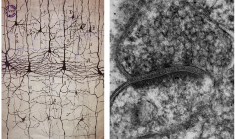
The vertebrate brain is arguably the most complex multicellular system in all Eukarya. In humans, the average brain contains approximately 100 billion neurons each participating in thousands of precise inter-cellular connections resulting in a network of staggering complexity (Kandel and Schwartz, 1985). Until the early 1900’s the brain was conceived as a continuous reticular network of interlacing nerve fibers where electricity could flow freely to and from different regions of the body. This “reticular” theory found a notable opponent in Ramon y Cajal, that by using a novel staining method was able to demonstrate that the brain was instead formed by an ordered array of multiple individual cells (Figure 1)(Cowan and Stevens, 2001).
Figure 1. Neuronal synapses are specialized adhesion junctions (Left)
Ramon y Cajal drawing based on Golgi staining of neurons in cortical sections
2
highlighting the electron dense material in the synaptic cleft (courtesy of Cam
Robinson).
_______________________________________________________________
3
observation long suggested the presence of strong trans-synaptic adhesive elements that would maintain the juxtaposition of both synaptic sides. Indeed, ultrastructural analysis of neuronal synapses revealed that the synaptic cleft is not empty, but is instead filled with electron dense material (Gray, 1959), which is now recognized to be composed mainly by highly glycosylated membrane proteins and synaptic cell adhesion molecules (CAMs).
Cell adhesion molecules are transmembrane proteins that span across the synaptic cleft and undergo homophilic and heterophilic interactions (Gerrow and El-Husseini, 2006). Importantly, the formation of a mature synapse, or synaptogenesis, is a multi-step process that gets initiated with the transient adhesive contact between two neurons. This initial contact is followed by the establishment of stable sites of cell-cell contact and subsequent recruitment of scaffolding proteins, which in turn contributes for the stabilization of neurotransmitter receptors, voltage-gated ion channels, and various second-messenger signaling molecules (Garner et al., 2002). Hence, CAMs are key structural players in synapses that provide anchoring points to intracellular cytoskeletal and scaffolding elements, and extracellular trans-synaptic adhesive interactions.
4
days, even in complete absence of ATP (Gray and Whittaker, 1962). Furthermore, the association between pre and postsynaptic sites resists the presence of high salt concentrations and even urea (Cotman and Taylor, 1972). Together, this indicates that the trans-synaptic adhesive complex is extremely strong and thermodynamically stable. Yet, neuronal synapses distinguish themselves from other cellular adhesive specializations for their capacity to undergo extremely fast functional and structural changes in response to brief stimuli (Citri and Malenka, 2008; Trachtenberg et al., 2002). The activity-dependent modification of synaptic properties, or synaptic plasticity, underlies the capacity of neuronal networks to adapt to changes in the environment and effectively process and transmit information. However, this highly dynamic nature of synapse remodeling is in stark contrast with the structural stability of the synaptic adhesive complex observed in synaptosomes. This suggests that the remodeling or disassembly of synaptic structures requires the involvement of active processes. However, the cellular and molecular basis for adhesive disassembly and its potential function in regulating synaptic transmission is still unknown and many questions remain about how nanomolar high-avidity adhesive interactions are uncoupled across the synaptic cleft in contexts of acute synaptic plasticity or elimination.
5
plasticity, long-term potentiation (LTP) and long-term depression (LTD), are thought to represent the cellular correlate of learning and memory, and can have different expression mechanisms depending on the neuronal circuits in which they operate (Citri and Malenka, 2008; Malenka and Bear, 2004). Other forms of plasticity act on a much broader regulatory scale. For example, homeostatic synaptic plasticity serves as a negative feedback mechanism in response to global changes in neuronal network activity, resulting in a compensatory and uniform scaling of all synaptic strengths (Pozo and Goda, 2010; Turrigiano, 1999, 2008).
6
7
were initially thought to play an important role during synaptogenesis (Chih et al., 2005; Graf et al., 2004; Scheiffele et al., 2000), have recently been implicated as critical regulators of synaptic function (Chubykin et al., 2007; Futai et al., 2007; Varoqueaux et al., 2006) and shown to be important for LTP expression in the amygdala (Jung et al., 2010; Kim et al., 2008). Together, these studies clearly demonstrate that cell adhesion molecules play important roles in the regulation of mature synapses. Hence the elucidation of the molecular mechanisms capable of acutely regulating CAMs at synapses is an important step to understand how these structural elements are regulated in the context of rapid synaptic remodeling.
8
adhesion molecules expressed at the plasma membrane. Moreover, given its acute and irreversible effects, proteolytic cleavage is a plausible mechanism for CAM regulation at synapses during synaptic plasticity. A recent study has employed the application of tissue inhibitor of metalloproteinase-1 (TIMP-1), an ADAM10 inhibitor, and a cell-permeable peptide capable of interfering with ADAM10 synaptic localization and activity to study the effects of N-Cadherin shedding at synapses. ADAM10 inhibition using these methods decreased N-cadherin cleavage, induced a significant increase in size of dendritic spines and potentiated AMPAR currents (Malinverno et al., 2010). However, ADAM10 is capable of targeting several proteins (Janes et al., 2005; Reiss et al., 2005) and TIMP-1 blocks multiple proteases including for example, MMP9 (Ethell and Ethell, 2007). Hence these results, despite showing that ADAM10 activity causes changes in synaptic properties, are inconclusive when it comes to providing information about the specific effects of N-Cadherin shedding at synapses. In fact, this highly redundant nature of ADAM and MMP activity has undermined the study of adhesion molecule shedding and its consequences in synapse maturation and function. Although manipulations of adhesion molecule levels and binding properties can alter synaptic transmission and influence synaptic plasticity, there is virtually no information on the consequences of acute shedding of trans-synaptic adhesion in response to neuronal activation.
9
a direct reduction of synaptic transmission by decreasing presynaptic function. The first chapter describes the initial observation that cleavage of NLG1 occurs in response to neuronal depolarization and is the major form of NLG1 regulation under these conditions. Moreover, I also present data showing that NLG1 cleavage occurs over development and is upregulated in the visual cortex in response to sensory experience during early development. Chapter 2 is centered on the characterization of the signaling mechanisms regulating NLG1 cleavage using a newly developed biochemical method based on surface biotinylation. Using this technique I found that NLG1 cleavage is bi-directionally regulated by activity and is mediated by NMDAR and CaMK signaling. Moreover, I also identified the region where NLG1 is cleaved and that the protease involved in activity dependent NLG1 cleavage in vitro and in vivo is the Matrix Metalloprotease-9 (MMP9). To finalize, in Chapter 3, I describe the development of a new technique that allows the cleavage of any transmembrane protein in a specific and temporally controlled manner. Using this novel approach in combination with real time microscopy I show that acute cleavage of NLG1 causes
rapid destabilization of its presynaptic partner Neurexin-1β
11
Chapter 1
Acute regulation of synaptic Neuroligin-1 by Matrix-
Metalloprotease mediated ectodomain shedding
Introduction
In the mammalian brain, neuronal synapses are highly specialized adhesion junctions maintained by a complex network of adhesion molecules that span the synaptic cleft and juxtapose the presynaptic active zone of neurotransmitter release and the postsynaptic density (Dalva et al., 2007; Shapiro et al., 2007; Yamagata et al., 2003). Among these, Neuroligins (NLGs) and Neurexins (NRXs) have emerged as critical regulators of proper circuit development and function (Sudhof, 2008).
12
Interestingly, each NLG isoform exhibits a specific pattern of expression and subcellular distribution. In particular, NLG1 and NLG2 are exclusively localized to excitatory and inhibitory synapses, respectively (Graf et al., 2004; Song et al., 1999; Varoqueaux et al., 2004), whereas NLG3 can be present in both (Budreck and Scheiffele, 2007).
Neurexins were identified as receptors for α-latrotoxin (Ushkaryov et al., 1992), a toxin present in the venom of the black widow spider that induces massive release of neurotransmitters (Ushkaryov et al., 2008). The mammalian genome contains 3 NRX genes each encoding a long α-protein
and a shorter β-protein from independent promoters (Tabuchi and Sudhof, 2002). Moreover, NRXs are highly polymorphic and through extensive alternative splicing at 5 canonical sites have the potential to generate over 3000 possible isoforms (Ullrich et al., 1995). Interestingly, different NRX splice variants are differentially expressed in specific neuronal types (Ullrich et al., 1995) and have different binding affinities to different NLG isoforms (Chih et al., 2006) (Comoletti et al., 2006) (Ushkaryov and Sudhof, 1993) indicating that NRXs may provide a structural adhesive code at synapses.
13
of postsynaptic components (Graf et al., 2004; Heine et al., 2008; Nam and Chen, 2005) and regulate NMDA receptor function trans-synaptically (Kattenstroth et al., 2004). This synaptogenic potential of NLGs and NRXs is due in part to the fact that both these proteins contain intracellular domains that interact with important synaptic scaffold proteins such as PSD95 and CASK (Hata et al., 1996; Irie et al., 1997). Adhesion between NLGs and NRXs thus provides a direct structural bridge between pre- and postsynaptic scaffolding machinery.
14
Interestingly, regardless of no apparent change in the number of total synapses generated, NLG1-3 triple KO neurons present reduced evoked excitatory postsynaptic currents (eEPSCs) and decreased miniature EPSCs (mEPSCs) frequency, which is consistent with impaired presynaptic function (Varoqueaux et al., 2006). Moreover, overexpression of NLG1 in hippocampal slices and cultured neurons results in increased release probability through a NRX-dependent mechanism (Futai et al., 2007; Ko et al., 2009b; Stan et al., 2010), suggesting that NLG1 modulates presynaptic function trans-synaptically. Consistent with this hypothesis, neurons lacking αNRX1-3 exhibit deficits in synaptic transmission due to severe impairment of N-type Ca2+ channel function (Missler et al., 2003), while disruption of endogenous NLG-NRX interactions with soluble Fc-NRX fragments decreases mEPSC frequency and release probability (Levinson et al., 2005). Taken together, these results suggested a new model by which NLGs and NRXs validate and stabilize synapses during development by modulating synaptic transmission at the presynaptic level.
15
occur during early development (Hensch, 2004, 2005b). As such, the elucidation of the molecular and cellular pathways regulating NLGs may provide new insights regarding the function of these proteins in the etiology of ASDs and in broader terms, the role of adhesion molecules during the functional maturation of neuronal circuits. Despite the numerous studies addressing how NLG affects synaptic properties, little is known about how endogenous NLGs are themselves regulated at synapses. It is also still unclear if NLGs are stable structural elements in synapses or if they can be regulated by changes in neuronal activity.
17
Results
To determine the effect of neuronal activity on synaptic NLGs, we treated dissociated hippocampal cultures at 21 days in vitro
(DIV21) with 30 mM KCl for 2 h, a paradigm that elicits robust depolarization and is widely used as a model for activity-dependent neuronal signaling (Kim et al., 2010; Murase et al., 2002; Sheng et al., 1990).
Figure 1. Neuronal activity triggers loss of Neuroligins from synapses. (A)
Hippocampal neurons (DIV21) were incubated in Neurobasal media (Control) or
Neurobasal media supplemented with 30 mM KCl (KCl) for 2 h, fixed, and
immunolabeled for endogenous PSD-95 and pan-NLG (NLG1-4). Neurons were
transfected with mCherry as a cell fill. Solid arrows show decreased NLG1-4
labeling at PSD-95 positive synapses following KCl incubation. Open arrows
depict synaptic NLG1-4 labeling under control conditions. Scale bar, 5 µm. (B)
Data indicate means ± SEM of NLG1-4 fluorescence intensity in PSD-95
positive dendritic spines (synaptic NLG1-4) or (C) over the entire neuron (total
NLG1-4) normalized to controls. Control, n = 435 spines from 8 neurons; KCl, n
18
19
Figure 2. Neuronal activity triggers loss of NLG1 from excitatory
synapses. (A) Immunoblot analysis of NLG1 and β-Tubulin in membrane fractions isolated from control or KCl-treated cortical neurons (DIV21). EXT,
whole cell extract; SPM, synaptic plasma membrane; PSDI,II, III; sequential
postsynaptic density fractions. Note that 4-fold less protein by mass was loaded
in PSD fraction lanes. See Experimental Procedures for details. (B) Data
indicate means ± SEM of NLG1 proteins levels in indicated fractions relative to
the total extract control. n = 3, *p < 0.05, **p<0.005, ***p<0.001.
20
these results indicate that the neuronal levels of NLG1 are extensively reduced following 2h of neuronal depolarization.
Several synaptic membrane proteins and receptors are degraded in response to changes in neuronal activity through the lysosomal pathway upon regulated endocytosis (Ehlers, 2000). To address whether the decrease in NLG1 levels is due to increased internalization and lysosomal degradation, we measured internalization rates of NLG1 in dissociated cortical cultures during KCl depolarization using a biochemical strategy based on surface biotinylation of proteins (Ehlers, 2000) (Figure 3A). In these experiments, neurons were pre-incubated with leupeptin for 1 h to inhibit lysosomal proteolysis.
Figure 3. Neuronal depolarization does not increase NLG1 internalization.
(A) Surface biotinylation based assay of endocytosis performed on cultured
cortical neurons. Surface proteins are covalently labeled with 1mg/ml
21
(Ctrl) or in media supplemented with 30 mM KCl (KCl), and subsequently
treated with 50mM Glutathione pH8 for 30min to remove surface biotin label (B)
Immunoblot of GluA1 and NLG1 indicate that GluA1 internalization was
increased by KCl while NLG1 was not. (C) Quantitative analysis of GluA1 and
(D) NLG1 internalization over time. Immunoblot values were compared to a
calibration standard of total surface protein at time zero (Surf) to quantify
percent internalization.
_________________________________________________________
Under basal conditions 5.3 ± 1.2% of surface NLG1 was internalized over 2h (Figures 3B and 3D). This low internalization rate was unaltered by KCl incubation (5.7 ± 0.8% of surface NLG1) indicating that KCl-induced NLG1 loss is not due to increased internalization. The GluA1 AMPA receptor was used as a positive control and exhibited a marked increase in internalization under KCl stimulation (Figure 3B and 3C, 7.4 ± 2.4% of total surface protein internalized in control conditions; 25 ± 4.7% in KCl), similar to previous reports (Ehlers, 2000). These results suggested that KCl induced NLG1 loss is not caused by protein internalization and lysosomal degradation.
22
Figure 4. Activity dependent loss of NLG1 is mediated by
Metalloproteases. (A) Immunoblot analysis of NLG1 in total lysates from
cortical cultures following 2 h incubation in neurobasal medium (Ctrl) or medium
supplemented with 30 mM KCl alone (KCl) or with MG132 (10 µM), leupeptin
(200 μM), leupeptin plus MG132 (Leup/MG) or GM6001 (10 µM). Note that GM6001 prevents KCl-induced loss of total NLG1. (B) Data indicate means ±
SEM of total NLG1 levels under the indicated conditions. n = 3, *p < 0.05.
As previously shown (Figure 2) depolarization induced by KCl incubation resulted in a 48 ± 3% reduction in total NLG1 levels. Interestingly, this effect was unaffected by proteasome
inhibition (50 μM MG132), blockade of lysosomal degradation (200 µM leupeptin), or both together (46 ± 7% decrease with MG132, 52 ± 6% with leupeptin, 47 ± 4% with both; Figures 4A and 4B). These results indicated that NLG1 degradation was occurring through a different degradation pathway and prompted us to test an alternative hypothesis. Several membrane proteins are targeted and degraded by Matrix Metalloproteases, which are a large family of secreted proteases. Indeed, incubation with the broad-spectrum Matrix Metalloprotease (MMP) inhibitor GM6001
23
proteolysis is the major regulatory mechanism mediating the rapid and extensive degradation of NLG1 in response to KCl depolarization.
To determine if NLG1 is also cleaved in vivo we analyzed soluble fractions of cortical, hippocampal, and cerebellar tissue from adult P60 mice (Figure 5A).
Figure 5. NLG1 cleavage fragments are ubiquitous throughout the brain.
(A) Immunoblot analysis of NLG1 in whole homogenates (5μg, Total) and soluble (50μg, Sol) fractions obtained from cortical, hippocampal and cerebellar tissue from P60 WT mice reveals soluble ~90 kDa NLG1-NTFs. Na,K-ATPase
was used to assess purity of soluble fraction. (B) Schematic representation of
NLG-1 cleavage fragments (C) Immunoblot analysis of NLG1 C-terminal
fragments in whole homogenates (Total), soluble (Sol) and membrane enriched
(Memb) fractions obtained from cortical tissue from P60 WT mice reveals ~20
kDa NLG1-CTFs. (D) Immunoblot analysis of NLG1 in whole brain
homogenates (Total) and soluble fractions (Sol) from WT and NLG1-KO mice
indicating the absence of NLG1-NTFs in NLG1-KO tissue. (E) Immunoblot
analysis of NLG1 C-terminal in whole brain homogenates from WT and
24
Interestingly, several bands of approximately 90 kDa were recognized by the N-terminal NLG1 antibody in the soluble fractions of extracts of different brain regions, suggesting that multiple NLG1 extracellular cleavage fragments are generated in vivo. A logical outcome of the ectodomain shedding of NLG1 is the generation of corresponding intracellular C-terminal fragments of approximately 20kDa (Figure 5B). Analysis of mouse cortical fractions using an antibody targeted against the C-terminal domain of NLG1 (NLG1-C) revealed in fact, multiple membrane bound bands of approximately 20KDa, a size consistent with the predicted mass based on the size of the N-terminal cleavage fragments (Figures 5B and 5C). To further confirm these findings we expressed a dual labeled version of NLG1 with GFP tagged to the N-terminus and HA tagged to the C-terminus (GFP-NLG1-HA) in COS7 cells (Figure S2). Immunoblot analysis of cell extracts using an anti-HA antibody revealed the presence of similar ~20 kDa bands that were absent in extracts of COS7 cells transfected with GFP-NLG1.
25
Several 90 kDa NLG1 species have been widely observed throughout the literature and have generally been considered to be immature unglycosylated forms of NLG1 (Ichtchenko et al., 1995; Ko et al., 2009b). This is due to the fact that NLG1 has an amino-acid mass of approximately 90KDa, but due to extensive N- and O-glycosylation, it migrates electrophoretically with an apparent weight of 110kDa. To test whether the 90 kDa NLG polypeptide(s) detected in our western blots correspond to immature or incompletely glycosylated isoforms of NLG1, we enzymatically deglycosylated all N- and O-linked glycans from P5 mouse cortical extracts and respective soluble fraction and measured changes in electrophoretic mobility.
Figure 6. NLG1-NTFs originate from fully glycosylated NLG1. (A)
Schematic representation of the electrophoretic mobility shift induced by
proteolytic cleavage of the N-terminal domain and subsequent deglycosylation
of N- and O-linked glycans of full form NLG1. (B) Deglycosylation (Deglyco) of
N- and O-linked residues in whole homogenates and soluble fractions of P5 WT
mouse cortex results in a 20 kDa apparent mass shift of NLG1-NTFs.
26
glycosylated forms of NLG1 (Figures 6A and 6B). Importantly, whereas deglycosylation of total cortical extracts collapsed NLG1 to both a 90 kDa and 70 kDa species corresponding to full-length and N-terminal cleaved NLG1, respectively, deglycosylation of soluble fractions produced only a 70 kDa species (Figures 6B), confirming that all soluble NLG1-NTFs correspond to cleavage fragments of mature, fully glycosylated NLG1.
27
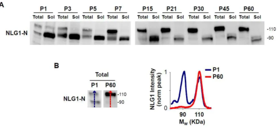
Figure 7. NLG1-NTFs are highly abundant during early development and
decrease with age. (A) Developmental profile of NLG1-NTFs from WT mice
cortical extracts. Postnatal days (P) are shown at the top. Total, 10 μg whole cortical homogenates; Sol, 50 µg soluble fractions from mouse cortex. (B)
Analysis of the relative abundance of full form NLG1 and NLG1-NTFs present in
whole cortical extracts from P1 and P60 mice. Note that NLG1 cleavage
products are more abundant during early developmental stages. The graph
plots signal intensity along the lines shown on the blots on the left.
28
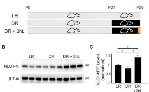
compared with control animals (LR) reared in normal light cycle (DR group - 0.71 ± 0.04 NLG1-NTFs normalized to LR group) (Figures 8B and 8C). Notably, 2 hours of re- exposure to light after dark rearing caused a robust increase in NLG1 cleavage (1.51 ± 0.14 compared with LR group).
Figure 8. NLG1 cleavage is regulated during the critical period of visual
cortex development. (A) Schematic representation of the experimental
paradigm. Mice are reared in normal 12h day/night cycle (LR) or submitted to
dark rearing for 5 days from P21-P26 (DR). DR+2hL indicates experimental
group submitted to a brief 2h light exposure following light deprivation. (B)
immunoblot of P26 mouse primary visual cortex of animals reared under
different conditions. Note that 2h of light exposure after dark rearing induces
increase in NLG1-NTF levels. (C) Data indicate means ± SEM of NLG1-NTFs
levels under the indicated conditions normalized to LR animals. n = 12 animals
per condition, *p < 0.005.
29
31
Discussion
Although highly studied for its role in synapse maturation and stabilization, it has been unclear whether the NLG-NRX trans-synaptic complex undergoes dynamic regulation and dissociation. Here we have shown that acute depolarization leads to a robust decrease in synaptic NLG1 levels. MMP inhibitors but not lysosome or proteasome inhibitors block this effect, indicating that under these conditions NLG1 is predominantly regulated by proteolytic cleavage. The extent of NLG1 loss observed using biochemical methods (Figures 2 and 4) is somewhat greater than that observed by immunostaining (Figure 1A). This disparity may be attributed to the broad specificity of the pan-NLG antibody used for immunolabeling or to the fact that it targets the C-terminal domain of NLGs, an epitope that may be differently regulated after ectodomain shedding. In addition, the presence of a residual C-terminal fragment (CTF) of cleaved NLG-1 (Figure 5C) raises the possibility that further intracellular processing of NLG1 may trigger downstream signaling cascades similar to what has been described for other membrane proteins such as Notch and E-Cadherin (Marambaud et al., 2002; Mumm et al., 2000). Moreover, these results suggest that caution is warranted in interpreting changes in Neuroligin synaptic abundance measured exclusively by an N-terminal or C-terminal antibody.
32
33
34
shown that tissue plasminogen activator (tPA), a potent activator of proteolytic cascades, is involved in the stabilization and pruning of synapses during early development (Mataga et al., 2004; Mataga et al., 2002). In addition, visual experience during the critical period affects dendritic spine morphology in visual cortex and leads to an increase in the fraction of thin spines and filopodia (Tropea et al., 2010). Together, this raises the interesting possibility that NLG cleavage may contribute for the maturation of synapses during cortical development.
35
37
Methods
Reagents and Antibodies
Dissociated primary neuronal cultures were prepared from hippocampi or cortex of embryonic day 18 or 19 Wistar rats. Tissue was dissociated by enzymatic papain digestion followed by brief mechanical trituration. Cultures were grown in Neurobasal media supplemented with B27 and Glutamax. For biochemical experiments 700k cortical cells were plated onto 60mm petri dishes. For immunocytochemistry, 100k hippocampal cells were plated onto 12 well plates containing Poly-L-Lysine coated 18mm glass coverslips. Plasmid transfection was done using lipid mediated gene transfer using Lipofectamine 2000 (Invitrogen). Briefly, for each coverslip, 1μg total DNA was mixed
with 1μl Lipofectamine in 100 μl of Neurobasal media for 15min.
Following incubation time 500 μl of conditioned media was mixed to the reaction and added to cell cultures for 30min. After this period, lipofectamine containing media was removed and replaced with a 1:1 mixture of conditioned media + fresh growth media. COS7 cells were grown in DMEM (Invitrogen) supplemented with 10% fetal bovine serum. Plasmid tranfections
were performed at approximately 70% confluency using 3 μg of
DNA and 3 μl of Lipofectamine per 60mm petri dish and let express for 2 days.
38
(mouse, Chemicon), Na+,K+-ATPase (rabbit, Cell Signaling), GFP (mouse, Millipore), HA.11 (mouse, Covance). Rabbit anti-panNLG antibody used for immunocytochemistry was a gift from Peter Scheiffele (Biozentrum).
Imaging
39
Biochemical Analysis of Brain and PSD Fractions
Mouse brains or brain regions were homogenized in
homogenization buffer (4 mM HEPES, 0.32 M sucrose pH 7.4)
with protease and phosphatase inhibitors (Roche). Crude
membrane fractionation was performed by centrifugation at
150,000 x g for 30 min. High density cortical cultures were
incubated in Neurobasal media or media supplemented with 30
mM KCl for 2 h. Whole cell, synaptic plasma membrane, and
PSD fractions from cultured cortical neurons were prepared as
described previously (Ehlers, 2003) and immunoblotted for
proteins of interest. For PSD fractions, 5 μg of protein was
loaded per lane, while for remaining fractions 20 μg protein was
loaded. Deglycosylation was performed using an enzymatic
deglycosylation kit according to the manufacturer’s instructions
(Calbiochem). All protein concentrations were measured with Dc
protein assay (Bio-Rad).
Biotinylation-Based Internalization Assay
High-density cortical neuron cultures were incubated with 100
μg/ml of the lysosomal protease inhibitor leupeptin beginning 1 hr
40
linkage with glutathione cleavage buffer (50 mM glutathione in 75 mM NaCl and 10 mM EDTA containing 1% BSA and 0.075 N NaOH) two times for 15 min each at 4ºC. Whole cell extracts were prepared, and biotinylated proteins were precipitated essentially as described (Ehlers, 2000). Biotinylated receptors were detected by immunoblot (ECL Plus, Amersham), and quantification was performed on an LAS-3000 gel reader (Fujifilm), using Multigauge 3.0 software. The percent receptor internalized was determined by measuring the band intensity after 37ºC incubation, subtracting the nonspecific band intensity obtained after 4ºC incubation (always <5%), and comparing to the total surface receptor calibration curve.
Dark rearing experiments
Male and female mice were reared in a normal light dark cycle (12 h light/12 h light) from birth until P26 (LR group) or were transferred to a dark room in complete darkness at P21 (DR group). Handling of animals in the dark room was done with the aid of night vision goggles and infra-red light. Mice were all sacrificed at P26 at the peak of the critical period of visual cortex development (Gordon and Stryker, 1996). One experimental group (DR+2hL) was re-exposed to light for 2 hours before being euthanized. Animals were sacrificed after 2 hours of light exposure and primary visual cortex was extracted, homogenized in homogenization buffer (4 mM HEPES, 0.32 M sucrose pH 7.4) containing protease and phosphatase inhibitors (Roche). Soluble
fraction was obtained by centrifugation at 150,000 x g for 30 min.
41
(Bio-Rad) and 50μg of protein were loaded on western blot and
probed for NLG1-NTFs using a NLG1 specific antibody targeted
43
Supplementary Figures
Figure S1
Figure S1. Biochemical Fractionation of Cultured Cortical Neurons.
Shown are immunoblots of biochemical fractions from cortical neurons cultures.
EXT, whole cell extract; S1, supernatant; SPM, synaptic plasma membranes;
S3, synaptic vesicle fraction; PSDI, washed SPM pellet; PSDII,
Triton-washed PSDI pellet; PSDIII, Sarcosyl-Triton-washed PSDI pellet. Note that 4-fold less
protein by mass was loaded in PSD fraction lanes. See Chapter1 Experimental
Procedures for details.
Figure S2
Figure S2. Generation of NLG1-CTFs in COS7 cells.
Lysates from COS7 cells expressing GFP-NLG1 or GFP-NLG1-HA were
analysed by immunoblot using anti-GFP and anti-HA antibodies. Anti-HA
immunoblot revealed a fragment of approximately 20 kDa corresponding to a
45
Acknowledgements
47
Chapter 2
Signaling Pathways Regulating Neuroligin-1 Cleavage
Introduction
Neuroligins and Neurexins undergo high affinity molecular interactions and subsequently activate multiple trans-synaptic signaling mechanisms resulting in stabilization and maturation of synapses during development (Dean et al., 2003; Graf et al., 2004; Heine et al., 2008; Ichtchenko et al., 1995; Nam and Chen, 2005; Prange et al., 2004; Scheiffele et al., 2000; Song et al., 1999; Wittenmayer et al., 2009). Furthermore, several studies indicate that manipulation of NLG levels in vitro and in vivo alters synaptic function (Chubykin et al., 2007; Futai et al., 2007; Varoqueaux et al., 2006). However, the detailed molecular and cellular mechanisms regulating endogenous NLGs in neuronal synapses remain mostly unknown.
48
surface receptors and cell adhesion molecules (Hubschmann et al., 2005; Maretzky et al., 2005; Monea et al., 2006; Sternlicht and Werb, 2001). Hence, MMPs can regulate a large array of biological processes in diverse tissues from cell migration and tissue morphogenesis to wound repair and inflammation (Ethell and Ethell, 2007; Sternlicht and Werb, 2001). Moreover, the expression profile of different MMPs can be tuned in response to specific stimuli or different cellular contexts. Given the potential deleterious effects caused by unrestrained proteolytic activity, MMPs are tightly regulated at the level of transcription, compartmentalization and enzymatic activity. Importantly, MMPs are secreted as inactive zymogens containing an auto-inhibitory N-terminal prodomain with a cysteine residue that blocks Zn2+ in the active catalytic site. Removal of this prodomain via proteolysis or S-nitrosylation exposes the catalytic site and activates the protein (Gu et al., 2002; Morgunova et al., 1999; Van Wart and Birkedal-Hansen, 1990).
49
in response to activity (Sbai et al., 2010; Wilczynski et al., 2008). This is further supported by recent work based on high resolution in situ zymography showing that gelatinase activity is increased in glutamatergic synapses of hippocampal neurons 2h after kainite (KA) induced seizures (Gawlak et al., 2009). Epileptogenesis, the process that leads to recurrent seizures after the initial epileptic episode, involves extensive circuit remodeling and fiber sprouting in the hippocampus (Pitkanen and Sutula, 2002). Thus, the robust upregulation of MMP9 levels and activity after seizures long suggested a potential involvement of this protease in synaptic remodeling. This hypothesis has been recently confirmed in a study showing that MMP9-KO mice do not exhibit synapse remodeling following seizures (Wilczynski et al., 2008). This significant result indicates that cleavage of MMP9 targets constitutes an important regulatory step mediating synapse stability in the hippocampus after status epilepticus.
Besides being involved in the molecular signaling cascades induced by epileptogenic activity, MMP9 is also upregulated in CA1 after LTP induction in acute hippocampal slices (Nagy et al., 2006) and in vivo (Bozdagi et al., 2007). Moreover, MMP-9 blockade impairs LTP under these conditions whereas recombinant MMP9 is sufficient to induce LTP (Wang et al., 2008). The substrates of MMP-9 associated with hippocampal LTP remain however, unknown. Interestingly,
zinc-50
mediated TrkB activation (Hwang et al., 2005). In fact, zinc has
been shown to be released from presynaptic sites during
high-frequency stimulation, suggesting that this may be an additional
regulatory mechanism of MMP activity at synapses (Vogt et al.,
2000). In addition, NMDAR antagonists also block the MMP9
effect in LTP suggesting that MMP9 dependent proteolysis can
be regulated by synaptic activity (Nagy et al., 2006).
51
(Thyagarajan and Ting, 2010). Interestingly, this effect is also blocked by NMDAR antagonist APV suggesting that NMDAR can regulate NLG1 trafficking. Hence, several lines of evidence suggest that NLG levels at synapses can be modulated in response to changes in neuronal activity via NMDAR dependent processes.
53
Results
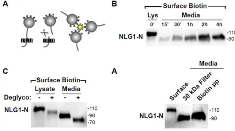
To dissect the signaling mechanisms regulating NLG1 cleavage, we developed a novel in vitro assay based on surface biotinylation that allows the purification of protein fragments cleaved and released to the culture media (Figure 1A).
Figure 1. Surface biotinylation based assay reveals NLG1-NTFs in
neuronal cultures. (A) Schematic diagram indicating the biotinylation-based
cleavage assay. Media from cultured neurons previously subjected to surface
biotinylation is collected and soluble biotin conjugates isolated by streptavidin
precipitation. (B) Precipitation of soluble biotinylated proteins released to the
media followed by immunoblot detection reveals time-dependent release of
soluble ~90 kDa NLG1 N-terminal fragment (NLG-NTF). The ~110 kDa full
form NLG1 in a total lysate (Lys, 10% of precipitate) is shown. Molecular mass
markers in kDa are shown on the right. (C) Deglycosylation (Deglyco) of N- and
O-linked glycans induces an equivalent mass shift of both full-length NLG1
(lysate) (D) Collection and 30 kDa mass cutoff filtration of media collected from
cultured cortical neurons (DIV21) reveals the presence of ~90 kDa NLG1-NTFs
54
(Biotin pp). The slight increase in molecular mass of the streptavidin-isolated
NLG1-NTF relative to filter-isolated NLG1-NTF is likely due to biotinylation.
_______________________________________________________________
Briefly, neuronal cultures are covalently labeled with cell impermeable biotin (Sulfo-LC-Biotin-NHS, 1 mg/ml) for 10 min to exclusively label surface proteins. Following incubation at 37ºC, culture media was collected, centrifuged, and incubated overnight with streptavidin beads to isolate released soluble biotin-conjugates prior to immunoblot analysis. Compared with traditional filtration and protein precipitation techniques this method specifically isolates protein fragments that originate from proteins expressed at the plasma membrane at the time of labeling and excludes peptides released via the secretory pathway. Moreover, the labeling of surface proteins allows the use of conditioned media during experiments given that previous accumulation of NTFs in the media does not confound the results.
55
30 kDa cutoff filter. Similar ~90 kDa bands were detected in concentrated filtered fractions (Figure 1D), indicating that NLG1 N-terminal fragments (NLG1-NTFs) are not a by-product of biotin labeling.
Neuroligin-based signaling is regulated by activity (Chubykin et al., 2007) although the underlying mechanisms remain obscure. Using our biotinylation based assay we investigated whether activity regulates NLG1 cleavage in vitro. For this, we first bidirectionally manipulated network activity in cortical neuron cultures using a pharmacological approach.
Figure 2. NLG1 cleavage is bidirectionally regulated by activity. (A)
Biotinylation-based isolation and detection of NLG1-NTFs from cortical neurons
incubated under control conditions or in the presence of TTX (2 µM) or
bicuculline (50 µM) plus 4AP (25 μM, Bic/4AP) to decrease or increase network activity, respectively. Total surface-labeled NLG1 corresponding to 10% of
precipitated protein is shown in the left lane (Surface). Molecular mass markers
in kDa are shown on the right. (B) Data represent means ± SEMs of
NLG1-NTFs produced under the indicated conditions normalized to control. n = 3, *p
< 0.05 relative to control.
56
resulted in decreased generation of NLG1-NTFs (0.65 ± 0.09 of control), increasing network activity by simultaneously blocking inhibitory synaptic transmission with the GABAA receptor
antagonist bicuculline (50 μM) and K+ channels with 4-aminopyridine (4AP, 25 μM) significantly increased NLG1 cleavage (Bic/4AP, 1.5 ± 0.1 of control) (Figures 2A and 2B). Besides confirming our previous data showing that NLG1 cleavage is increased by neuronal depolarization these results also show that this process is bidirectionally regulated and can be reduced in response to decreased neuronal activity.
To mimic conditions that lead to robust loss of synaptic NLG1 (See Chapter 1, Figures 1 and 2), we depolarized cortical neurons by incubation with KCl (30 mM, 2 h).
Figure 3. Activity Induces NLG1 Cleavage Through NMDARs and CaMK.
(A) Biotinylation-based isolation and detection of NLG1-NTFs from cortical
neurons incubated under control conditions or in the presence of KCl (30 mM, 2
h) with or without indicated pharmacological agents. Note the robust increase in
NLG1-NTFs induced by KCl depolarization that is blocked by the NMDA
receptor antagonist APV and CaMK inhibitors KN93 and KN62. (B) Data
represent means ± SEMs of NLG1-NTFs produced under the indicated
57
As previously described (Chapter 1, Figures 1 and 2) KCl mediated depolarization led to a dramatic increase in NLG1-NTFs (4.4 ± 0.5 fold) compared to control conditions (Figures 3A and 3B). Interestingly, this depolarization-induced cleavage of NLG1 was abolished by the NMDA receptor antagonist APV (50
μM). Activation of NMDA receptors elicits Ca2+ influx and activation of Ca2+/calmodulin-dependent kinases (CaMK) including CaMKII (Fink and Meyer, 2002). Accordingly, inhibition
of CaMK using either KN93 (5 μM) or KN62 (10 μM), but not the
inactive isomer KN92 (5 μM), significantly reduced
activity-induced cleavage of NLG1 (KN93, 1.5 ± 0.6; KN62, 1.5 ± 0.5; KN92, 5.0 ± 1.1 fold increase in NLG1-NTFs relative to control). These results show that activity-induced cleavage of NLG1 is triggered by NMDA receptor activation and requires Ca2+/calmodulin-dependent kinase activity.
58
4B). In order to determine the specific Matrix Metalloprotease involved in NLG1 ectodomain shedding we followed a candidate approach based on pharmacological inhibition of MMP activity. MMP-2, -3 and -9 are the most abundant MMPs in the brain and have been implicated in synaptic maturation and multiple forms of synaptic plasticity (Ethell and Ethell, 2007; Yong, 2005). This prompted us to address how these specific proteases affected KCl-induced cleavage of NLG1.
Figure 4. MMP9 Mediates Activity-Dependent Cleavage of NLG1. (A)
Isolation and detection of ~90 kDa NLG1-NTFs following biotinylation of cortical
neurons incubated under control conditions or in the presence of KCl with or
without MMP inhibitors. Molecular mass markers in kDa are shown on the right.
(B) Data represent means ± SEMs of NLG1-NTFs produced under the indicated
59
Incubation with the selective MMP9 inhibitor I (0.5 μM) or
MMP3 inhibitor III (50 μM) significantly reduced KCl-induced cleavage (0.68 ± 0.02 and 1.27 ± 0.27 fold increase in NLG1-NTFs relatively to control, respectively), with MMP9 inhibitor I eliciting a more robust reduction. Conversely, the MMP1 inhibitor
GM1489 (5 nM), MMP2 inhibitor III (50 μM) and MMP8 inhibitor I
(10 μM) had no significant effect on activity-induced NLG1 cleavage (2.82 ± 0.14, 2.40 ± 0.19 and 1.92 ± 0.37 relatively to control, respectively).
60
Figure 5. MMP9 Mediates Activity-Dependent Cleavage of NLG1. (A)
Following surface biotinylation, cortical neurons were treated with the
nonselective MMP activator APMA (0.5 mM, 15 min) with or without MMP
inhibitors. Lysates (Lys) and soluble media (Med) were subjected to streptavidin
precipitation prior to immunoblot analysis for NLG1 using an N-terminal
antibody (4C12). Note that MMP9 inhibitors block APMA-induced cleavage. (B)
Data represent means ± SEMs of NLG1-NTFs produced under the indicated
conditions normalized to control. n = 3, *p < 0.05 relative to APMA alone.
Indeed, brief incubation of cortical neurons with 0.5 mM APMA for 15 min induced robust generation of NLG1-NTFs (Figures 5A and 5B). Co-incubation with MMP9 inhibitor I almost completely blocked APMA-induced cleavage while inhibition of MMP3 or MMP2 had no effect. Based on these results we conclude that MMP9 is most likely the terminal effector protease responsible for cleavage of NLG1 while MMP3 might be an upstream component of the signaling cascade activating MMP9.
61
mutants with sequential deletions and amino acid replacements in its juxtamembrane domain (Figure 6A).
Figure 6. Extracellular Cleavage of NLG1 Occurs Near Its Transmembrane
Domain. (A) Model illustrating the location of the NLG1-ΔSD substitution mutants in relation to NLG1 domains: AEHD, acetylcholinesterase homology
domain; DD, dimerization domain; SD, stalk domain; TM, transmembrane
domain; CT, C-terminal domain. Numbers indicate amino acids. (B)
Biotinylation-based cleavage assay of COS7 cells expressing the indicated
NLG1 mutants. Total lysates (Lys) or media (Med) were collected and subjected
to immunoblot analysis following treatment with the MMP activator APMA with
or without the broad spectrum MMP inhibitor GM6001 (GM). Both
NLG1-ΔSDfull and NLG-ΔSD3 were resistant to APMA-induced cleavage indicating that MMP cleavage sites lie between residues 672 and 695. Molecular mass
markers are shown at the right.
62
cleavage is MMP-dependent under these conditions (Figure 6B). Substituting 60 amino acids (aa) of the NLG1 stalk domain (aa 636-695) with the polylinker GAAAAA resulted in a mutant
(NLG1-ΔSDfull) that is resistant to APMA-induced cleavage (Figures 6A and 6B). Within this 60 residue stretch, deletion of
amino acids 672-695 (NLG1-ΔSD3) and replacement with the polylinker GAAAAA likewise abolished APMA-induced cleavage whereas mutation of more membrane-distal sequences did not
(aa 636-660, NLG-ΔSD1; aa 654-677, NLG-ΔSD2). This indicates that NLG1 cleavage occurs in it juxtramembrane region within the 24 aminoacid sequence upstream of the transmembrane domain. We attempted to further resolve the precise cleavage site, but shorter deletions or different single site substitution mutants were all cleaved upon APMA treatment (data not shown), suggesting the presence of multiple MMP target sequences within this 24 aa domain. However, these results indicate that cleavage of NLG1 occurs in the region corresponding to amino acids 672-695 of NLG1.
63
Figure 7. Differential regulation of NLG2 cleavage. (A) Immunoblot analysis
of NLG1, NLG2 and NLG3 in total lysates from cortical cultures following 2 h
incubation in neurobasal medium (Ctrl) or medium supplemented with 30 mM
KCl alone (KCl) or with MG132 (10 µM), leupeptin (200 μM), leupeptin plus MG132 (Leup/MG) or GM6001 (10 µM). Note that GM6001 prevents
KCl-induced loss of total NLG1 and NLG3 but not NLG2. (B) Immunoblot analysis of
NLG1, NLG2 and NLG3 in total lysates from cortical cultures following 15min
incubation in neurobasal medium (Ctrl) or medium supplemented with 0.5 mM
APMA (APMA). Note that NLG2 levels are not reduced by APMA (C)
Biotinylation-based cleavage assay of COS7 cells expressing the indicated NLG
isoform HA tagged mutants. Total lysates (Lys) or media (Med) were collected
and subjected to immunoblot analysis following treatment with the MMP
activator APMA. Note the presence of residual soluble NLG2-HA bands in the
media.
64
15 min resulted in a dramatic reduction of NLG1 and NLG3 levels, but not NLG2 (Figure 7B). Again, this is consistent with a differential regulation of NLG2 compared with NLG1 and NLG3. Unfortunately, due to the lack of NLG2 specific antibodies that target the N-terminal domain of the protein we could not address NLG2 cleavage based on surface biotinylation. However, to address if NLG2 also undergoes ectodomain shedding we expressed HA-tagged forms of NLG2 and NLG3 in COS7 cells and measured the release of NLG-NTFs after APMA treatment using surface biotinylation (Figure 7C). While surface NLG3-HA was completely cleaved and released to the extracellular media in response to APMA stimulation, NLG2-HA remained mostly associated with the membrane fraction. This result is consistent with lack of NLG2 degradation following APMA incubation in neuronal cultures (Figure 7B). However, residual amounts of soluble NLG2-HA fragments could still be detected in the media of APMA treated cultures (Figure 7C, top panel) indicating that NLG2 can also undergo ectodomain shedding. Nevertheless, despite being cleaved this data indicates that NLG2 is differentially regulated when compared with other NLG isoforms, potentially by a different set of proteases that are not blocked by GM6001 and not activated by APMA.
65
Figure 8. NLG1 is cleaved in response to epileptic seizures in the
hippocampus by MMP-9.(A) Schematic of the experimental conditions (B) WT
and MMP9 KO mice were injected with pilocarpine (315 mg/kg) to induce status
epilepticus (Pilocarpine) or saline (Control). After 2 h, hippocampi were isolated
and homogenized and either total extracts (5 μg) or soluble fractions (Sol, 50
μg) were immunoblotted for NLG1. Note the increased NLG1-NTFs after pilocarpine-induced seizures in WT and the abrogation of this response in
MMP9-KO animals. Pilocarpine upregulates Arc in both wildtype and MMP9 KO
animals. (C) Data represent means ± SEMs of NLG1-NTFs produced under the
indicated conditions normalized to control. n = 9, *p < 0.05.
Intraperitoneal administration of pilocarpine in P60 mice induced robust epileptic seizures and resulted in a 3.12 ± 0.47 fold increase of soluble NLG1-NTFs in the hippocampus after 2h of PSE induction (Figures 8B and 8C). These findings indicate that increased neuronal activity also upregulates NLG1 cleavage
66
MMP9-KO hippocampus under basal conditions still exhibit normal levels of NLG1-NTFs indicating that NLG1 cleavage still occurs in the absence of MMP9. However, 2h of PSE characterized by robust seizures failed to elevate soluble NLG1-NTFs in hippocampus when compared to saline controls (Figures 8B and 8C). As a control for seizure activity we measured levels of the activity-regulated cytoskeleton-associated protein (Arc) and both wild-type and MMP9 KO mice exhibited significant Arc upregulation after pilocarpine administration (Figure 8B). These findings indicate that activity triggers NLG1 cleavage in vivo
67
Discussion
In this section, we have characterized the molecular and signaling pathways regulating activity-dependent NLG1 cleavage. Using a newly developed assay based on biotinylation of surface proteins we found that cleavage of NLG1 at the plasma membrane is bidirectionally regulated by neuronal activity, requires NMDA receptor signaling, Ca2+/calmodulin-dependent kinase activity and is mediated by the activity-regulated and brain-expressed Matrix Metalloprotease-9.
Figure 9. Schematic model for activity-induced MMP9 cleavage of NLG1.
Increased neuronal activity activates a proteolytic cascade of MMPs via
NMDAR and CaMK signaling. Ultimately, MMP9 activation cleaves NLG1
releasing NLG1-NTFs to the media. NMDAR, N-methyl-D-aspartate receptor;
68
Moreover, these results define a novel link between NLG-NRX adhesion and a NMDAR-CaMK-MMP9 signaling cascade. In addition, we found that cleavage of NLG1 is highly upregulated in the hippocampus during epileptic seizures. Previous data described in Chapter 1 already has shown that NLG1 cleavage can be regulated by changes in neuronal activity in vivo. Besides providing additional evidence that NLG1 shedding is dynamically regulated by activity in vivo, the increase of NLG1-NTFs levels during epileptic seizures indicates that this process can also occur in adult brains outside developmental periods of circuit maturation. Indeed, recent work has implicated NLG1 in the expression of LTP in the amygdala suggesting that NLG1 is not only involved in the maturation and stabilization of synapses but is also required for synaptic function in later stages of development (Jung et al., 2010; Kim et al., 2008). Moreover, the increase in NLG1-NTFs following pilocarpine injection was absent in MMP9 KO mice indicating that MMP9 is required for seizure-induced cleavage of NLG1 in the hippocampus. This result is consistent with our findings in dissociated cultures where MMP9 inhibition abrogated the increase in NLG1-NTFs induced by KCl stimulation. Thus, MMP9 is likely an important protease regulating NLG1 cleavage in contexts of increased neuronal activity.
69
following seizure induction (Szklarczyk et al., 2002; Wilczynski et al., 2008). This important finding suggests that the molecular targets of MMP9 in hippocampal neurons may be critical regulators of synapse maintenance and stability during epileptogenesis. Indeed, MMP7, an upstream activator of MMP-9, induces morphological changes in dendritic spines converting them to elongated filopodia-like structures (Bilousova et al., 2006). Conversely, MMP9 inhibition accelerates spine maturation in neuronal cultures and increases the total fraction of mushroom spines (Bilousova et al., 2009). Together these results suggest that MMP9 activity may indeed be an important regulator of synapse maintenance.
Besides interacting with NLGs, presynaptic NRXs also
bind to other postsynaptic molecules such as β-dystroglycan
70
response to visual experience during critical period, a paradigm that also elicits subsequent synapse remodeling. Hence, cleavage of NLG1 emerges as a potential general mechanism associated with synaptic remodeling during development and disease.
71
al., 2009), whereas transgenic overexpression of NLG2 leads to increased mIPSC frequency in prefrontal cortical regions (Hines et al., 2008). This interesting hypothesis will certainly be the focus of future studies.
Excitotoxicity driven by unrestrained glutamate release contributes to neuronal degeneration in many acute CNS diseases, including ischemia, trauma, and epilepsy (Arundine and Tymianski, 2003). In particular, excessive NMDAR dependent Ca2+ influx has been shown to be prone to induce deleterious effects in neurons (Arundine and Tymianski, 2004). Interestingly, NLG1 levels in dissociated neurons are directly correlated with NMDAR EPSC amplitudes (Chubykin et al.,
2007). Moreover, αNRX deletion induces loss of NMDAR function in cortical slices (Kattenstroth et al., 2004). Hence, an acute loss of NLG1 in response to increased activity could mediate decreased NMDAR dependent Ca2+ influx, protecting neurons from glutamate excitotoxicity. Consistent with this hypothesis, it has been shown that MMP9 activity increases synaptic NMDAR mobility which potentially decreases NMDAR synaptic currents (Michaluk et al., 2009). This effect has been
attributed to Integrin-β1 signaling however integrin function was assessed by overnight antibody cross linking which may cause indirect secondary effects. Thus, other mechanisms may be involved in the destabilization of synaptic NMDAR by MMP9.
72
indicates the existence of parallel proteolytic mechanisms targeting NLG1 under basal conditions. Consistent with this idea, none of the pharmacological MMP inhibitors tested in our in vitro
assays was able to fully block NLG1 cleavage. Moreover, multiple soluble NLG1-NTF species can be detected throughout the brain (Chapter 1, Figure 5), indicating that NLG1 probably contains multiple cleavage sites. This observation provides a plausible explanation for why single amino acid point mutations in the stalk region of NLG1 failed to generate mutants that would resist APMA induced MMP-dependent cleavage, which instead required substitution of a longer 24 amino acid segment (Figures 6A and 6B). Hence, these results suggest that the regulation of NLG1 cleavage is probably under control of multiple proteolytic mechanisms that may be differently regulated depending of cellular context. It will be important for future studies to define in detail the activity-independent and MMP9-independent mechanisms responsible for NLG1 cleavage and their potential role in synapse development and plasticity.
73
contexts (Nagy et al., 2006). Recent work has further shown that in response to brief depolarization, NLG1 internalization is halted via NMDAR signaling (Thyagarajan and Ting, 2010). Taken together, this data suggests that NLG1 is tightly regulated by NMDAR and CaMK activity at multiple levels and that the regulation of proteolysis and trafficking of NLGs may occur in tandem though similar signaling mechanisms.