RESUMO: Este trabalho teve por objetivo avaliar, in vitro e in vivo, a eficácia de produtos naturais no controle da antracnose do mamão. Os experimentos in vitro foram instalados em delineamento intei-ramente casualizado, sendo para a avaliação de crescimento micelial considerado o esquema fatorial 10 × 4 (tratamentos x períodos de avaliação) com oito repetições, com a esporulação avaliada ao final do experimento. Os tratamentos utilizados foram: extrato aquoso nas concentrações de 5 e 15% de Syzygium aromaticum (L.) Merr. & Perry (cravo-da-índia), Cinnamomum zeylanicum Breym (canela) e Zingiber officinalis Rox (gengibre), quitosana 1 e 3%, fungicida Procloraz a 100 μg.mL-1 e testemunha (sem tratamento). Para a ava-liação da germinação de conídios, foram considerados seis tratamen-tos com cinco repetições. Foram utilizados os mesmos tratamentratamen-tos, porém, nas concentrações de 7,5% de cada extrato (cravo-da-índia, canela e gengibre), 1,5% de quitosana, e 50 μg.mL-1 de Procloraz. Para o experimento in vivo, foram utilizados mamões “Formosa” “Tainung 1”, em delineamento inteiramente casualizado, sendo seis tratamentos com quatro repetições para avaliação de severidade de antracnose cau-sada por Colletotrichum gloeosporioides. Os frutos foram tratados por imersão durante 5 minutos com extratos de cravo-da-índia, canela e gengibre a 15%, quitosana a 8% e testemunha com água destilada, e imersão por 2 minutos em fungicida Procloraz (33,75 g i.a./100 L). Os tratamentos com extrato de cravo-da-índia a 15% e quitosana a 8% foram eficazes em todas as avaliações, sendo uma alternativa viável ao fungicida Procloraz. Os tratamentos com extrato de gengibre foram menos eficientes, com controle intermediário para o extrato de canela.
PALAVRAS-CHAVE:Carica papaya; doenças pós-colheita; extratos vegetais; quitosana.
ABSTRACT: We evaluated the efficacy of natural products in the control of papaya anthracnose, in vitro and in vivo. The in vitro experiments for evaluation of mycelial growth used a completely randomized 10 × 4 factorial design (treatments × evaluation periods) with eight replicates, with sporulation evaluated at the end of the experiment. The treatments involved the use of aqueous extract at concentrations of 5 and 15% for Syzygium aromaticum (L.) Merr. & Perry (clove), Cinnamomum zeylanicum Breym (cinnamon), and Zingiber officinalis Rox. (ginger); 1 and 3% chitosan; the fungicide Prochloraz at 100 μg.mL-1; and a control (no treatment). For evaluating conidia germination, we used six treatments with five replicates. The treatments included 7.5% of each extract (clove, cinnamon, and ginger), 1.5% chitosan, and 50 μg.mL-1 of Prochloraz. For the in vivo experiment, “Formosa” papaya “Tainung 1” was used in a completely randomized design, with six treatments and four replicates to evaluate the severity of anthracnose caused by Colletotrichum gloeosporioides. The fruits were treated by immersion for 5 min with 15% clove, cinnamon, and ginger extracts, 8% chitosan, and control with distilled water, and immersion for 2 min in Prochloraz (33.75 g a.i./100 L). The treatments with 15% clove extract and 8% chitosan were effective in all evaluations, resulting in a viable alternative to the fungicide Prochloraz. The treatments with ginger extract were less effective and those with cinnamon offered intermediate control.
KEYWORDS: Carica papaya; post-harvest diseases; plant extracts; chitosan.
In vitro
antimicrobial activity and
alternative control of anthracnose in papaya*
Atividade antimicrobiana
in vitro
e controle alternativo da antracnose em mamão
Héber Ferreira dos Reis1** , Lilian Maria Arruda Bacchi1,
Silvana de Paula Quintão Scalon1, Jasna Karoliny Pereira Flores1
INTRODUCTION
Anthracnose of papaya, caused by Colletotrichum gloeosporioides Penz., is very important economically — the fruits affected by this disease become unsuitable for commercialization and consumption. Although the fruits do not show symptoms of the disease at harvest, it manifests itself during the packaging, transportation, ripening, and commercialization phases, causing substantial losses (SIQUEIRA JUNIOR et al., 2011).
Post-harvest disease control in papaya is usually done by immersing the fruits in water at 48–49ºC for 20 min, follo-wed by another immersion at 14ºC for 20 min. Subsequently, the application of waxes and fungicides is recommended to guarantee longer fruit survival (CARNELOSSI et al., 2009). In addition, the use of refrigeration or controlled atmosphere is fundamental to enhance control efficacy (CIA et al., 2010).
Although the use of fungicides is an important control strategy, their residues in fruits have generated cause for con-cern owing to their negative effects on human health and the environment. For example, in 2016, ANVISA prohibi-ted the use of the active ingredient Prochloraz in pesticides, based on the results of its toxicological reassessment per the newly adopted guidelines and toxicological evaluation requi-rements (ANVISA, 2016).
As an alternative to fungicides, and for the post--harvest conservation of several fruits, the use of bio-films such as chitosan (a natural, edible polysaccharide extracted from the exoskeleton of crustaceans) has been investigated. It can form a semipermeable coating, pro-longing post-harvest life by minimizing respiration rate and reducing fruit water loss (HERNÁNDEZ et al., 2011). Chitosan also has antifungal and antibacterial activity, showing in vitro its potential for inhibition of mycelial growth, as obtained for Rhizopus stolonifer
(CIA et al., 2010).
Research with plant extracts or essential oils, obtained from medicinal plants of native Brazilian flora, such as clove (LUCAS, 2012; FONTANA et al., 2017), ginger (BRITO; NASCIMENTO, 2015; DIEMER, 2016), and cinnamon (ANDRADE; VIEIRA, 2016; BONILLA; SOBRAL, 2017), has indicated their potential to control phytopathogens. Essential oils not only exhibit a direct fungitoxic action, inhibiting mycelial growth and spore germination, but also affect the induction of phytoalexins, indicating the presence of compounds with elicitor characteristics (SCHWAN-ESTRADA, 2009).
The objective of this study was to evaluate, in vitro and in vivo, the control of C. gloeosporioides, a causal agent of papaya anthracnose in the post-harvest period, using chitosan and extracts of cloves, cinnamon, and ginger.
MATERIAL AND METHODS
Research site
The in vitro experiments were conducted at the Laboratory of Agricultural Microbiology and Phytopathology of the Federal University of Grande Dourados (UFGD), Dourados, Mato Grosso do Sul, from September to December 2013.
Mycelial sensitivity and sporulation
of
C. gloeosporioides
in response
to natural products and a fungicide
The fungus C. gloeosporioides was isolated from disease lesions in “Formosa” papaya fruits and cultivated in potato-dextrose--agar (PDA) medium for six days at 25ºC, with a 12h pho-toperiod. After fungal colony growth, consecutive repetitions were made until pure cultures were obtained. The confirma-tion of fungus identificaconfirma-tion was based on its morphological characteristics, per TARNOWSKI (2009) and TORRES-CALZADA et al. (2012), through slide preparation and microscopic observations.
We used a completely randomized design, with ten treatments and eight replications. For evaluation of mycelial growth, we implemented a 10 × 4 factorial scheme, with ten treatments and four incubation periods (2, 4, 6, and 8 days). The treatments were aqueous extract at concentrations of 5 and 15% of clove, cinnamon and ginger, 1 and 3% chitosan, the fungicide Prochloraz at 100 μg.mL-1, and a control (no
treatment). The concentrations established for the extracts were based on the results of previous tests.
Ginger rhizome, cinnamon bark, and clove bud were used to prepare the aqueous extracts. Thirty grams of plant material were ground in 120 mL of distilled water for 1 min. After grinding, the material was filtered through a sieve, gauze, and cotton, conditioned in an Erlenmeyer flask, and kept in a water bath at 65ºC for 60 min to decontaminate. From this solution, the necessary volume was subsequently removed to obtain a concentration of 5 and 15% of the plant extract in the PDA culture medium.
One and three percent chitosan solutions were prepa-red and autoclaved at 15 psi for 15 min, per HERNÁNDEZ et al. (2011).
The evaluations were performed every two days for eight days; we measured mycelial diameter growth in two direc-tions, perpendicularly, with a graduated ruler graduated in millimeters (mm).
At the end of the evaluation of mycelial growth, sporula-tion of C. gloeosporioides was also evaluated. For each repeti-tion, a suspension of conidia was prepared by placing 10 mL of sterile distilled water plus Tween 20 (0.1 mL.mL-1) on the
surface of the Petri dish with fungal mycelium, smoothly scraping it with Drigalsky’s handle. The suspension was filte-red through a double layer of sterile gauze to count conidia in a hemocytometer.
The mycelial growth and sporulation data were submitted to analysis of variance. The average values, when significant, were compared by Tukey’s test and the average of the evalua-tion periods for mycelial growth was adjusted by the regres-sion analysis, at 5% level of significance.
Sensitivity of
C. gloeosporioides
conidia
to natural products and a fungicide
For the evaluation of inhibition of spore germination, a com-pletely randomized design was used, with six treatments and five replications. The treatments were 7.5% aqueous extract of clove, cinnamon and ginger, 1.5% chitosan, the fungicide Prochloraz at 50 μg.mL-1, and control (sterilized distilled water).
After 12 days of propagation, the suspension of coni-dia in the culture was adjusted to 1.4 × 10⁵ conidia.mL-1,
using a hemocytometer. The spore germination test followed the methodology described by TAVARES; SOUZA (2005).
The conidia germination data were submitted to analy-sis of variance, and, when significance was found, the avera-ges were compared by Tukey’s test at 5% level of significance.
Effect of natural products and
fungicide on the control of
C. gloeosporioides
during post-harvest
of “Formosa” papaya “Tainung 1”
“Formosa” papaya “Tainung 1” from a traditional growing area, in the municipality of Ivinhema, Mato Grosso do Sul, were harvested in May 2014. We selected fruits at stage 2 of ripeness (up to 25% yellow surface), per COSTA et al. (2010).
After being harvested, the fruits were wrapped in newspa-per and packed in plastic boxes, and then transported to the Laboratory of Agricultural Microbiology and Phytopathology at UFGD, in Dourados, Mato Grosso do Sul, where they were classified. We discarded those with lesions or inadequate coloration, to standardize maturation stage and fruit quality. The fruits were washed with water and detergent, and kept at room temperature until reaching 50% yellow surface; then, they were submitted to the treatments.
The fruits were immersed in sterilized distilled water (trol), aqueous extracts (clove, cinnamon, and ginger) at 15% con-centration and 8% chitosan for 5 min, while immersion in the fungicide Prochloraz, at a concentration of 33.75 g a.i./100 L, was carried out for 2 min. In both cases, drying occurred naturally.
The plant extract solutions were prepared using the methodology described in the in vitro experiment for myce-lial sensitivity of C. gloeosporioides. The 8% chitosan solution was prepared by dissolving 800 g of chitosan in 10,000 mL of distilled water, then heating at 100ºC with constant stir-ring for 5h. Dustir-ring the preparation, the solution was adjusted and maintained at pH 5.5 by the addition of acetic acid P.A. After the treatments, the fruits were packed in polystyrene cups with the stem region facing upwards, inoculated with
C. gloeosporioides, per NERY-SILVA et al. (2007), and stored in a temperature-controlled chamber at 25ºC with a 12h photoperiod. The evaluations were performed after 2, 4, and 6 days of inoculation, and scores were given based on the severity of stem-end-rot symptoms, as suggested by NERY-SILVA et al. (2007):
1. no symptoms of stem-end-rot;
2. presence of small superficial aqueous punctures (up to 3 mm) in the stem region;
3. presence of aqueous or mummified lesions, not coales-cing, covering a larger area around the stem;
4. presence of coalescing lesions, with translucent or mum-mified appearance, limited to the stem region; and 5. lesions similar to those described in scale 4, including
grea-ter extension in the fruit pulp, reaching the seed cavity, the tissue of this region may present a loss in consistency.
A completely randomized design was used, in a 6 × 3 fac-torial design, with six treatments and three evaluation periods (2, 4, and 6 days), with four replicates per treatment. The data were submitted to analysis of variance and, when significance was found, the averages between the treatments were compa-red by Tukey’s test at 5% level of significance.
RESULTS AND DISCUSSION
Mycelial sensitivity and sporulation
of
C. gloeosporioide
s in response to
natural products and a fungicide
the two extracts resulted in lower mycelial growth. The effect of 15% ginger on mycelial growth was significantly greater than the treatment with 15% cinnamon from the first evaluation. Inhibition of mycelial growth by ginger extract was also confirmed by BRITO; NASCIMENTO (2015): they found that the 5% concentration of ginger extract provided 70% inhi-bition of mycelial growth of Curvularia eragrostidis. According to DIEMER (2016), it was not possible to relate the antimi-crobial activity of ginger only to the amounts found for the
major constituents (α-zingiberene, geranial, β-bisabolene, neral, and β-sesquiphellandrene). Therefore, these may be related to the synergistic effect between the constituents.
Inhibition of mycelial growth by cinnamon extract was also observed by VENTUROSO et al. (2011). They found higher antifungal activity in the highest concentrations of cinnamon aqueous extract (10 and 20%), in vitro, without total inhibition on the phytopathogenic fungi studied, including Colletotrichum
spp. The antimicrobial action of cinnamon extract is related to cinnamaldehyde, its main component (BERALDO et al., 2013). The results obtained with the Prochloraz treatment con-cur with those obtained by TAVARES; SOUZA (2005). These authors also verified, in vitro, 100% inhibition of myce-lial growth of C. gloeosporioides, with 10 μg.mL-1 of Prochloraz,
Propiconazole, and Tebuconazole.
The effectiveness of in vitro mycelial growth inhibition by clove extract and chitosan resembled the fungicide, demonstra-ting them to be promising alternatives for the control of phyto-pathogenic fungi. According to SILVA et al. (2009), the effective antimicrobial action of clove extract is related to the presence of eugenol (83.6%), its main constituent. However, regarding chi-tosan, according to MAIA et al. (2010) and FREDDO (2012), the control efficacy is not only a function of concentration and pathogen but also of factors such as degree of deacetylation, molecular weight, pH, and temperature.
Although sporulation did not occur in treatments with 5 and 15% clove, 1 and 3% chitosan, and the fungicide Prochloraz, it did not differ from the 15% cinnamon and control treatments (Table 1).
The occurrence of higher production of conidia with the highest concentration of ginger was also found by RODRIGUES et al. (2006). The in vitro study conducted by this author sho-wed that under an aqueous extract of ginger there was greater
Helminthosporium spp. conidia production at concentrations of 20 and 40%, compared to those of 1, 5, and 10%.
Greater inhibition of sporulation with 15% cinnamon, compared to 5%, suggests the possibility of complete inhi-bition with a higher concentration. However, according to ARAUJO et al. (2009), the control effectiveness of an extract does not only vary by its concentration but also by the spe-cies of fungus to be controlled. This was observed with the
Figure 1. Mycelial growth (cm) of Colletotrichum gloeosporioides
under the effect of treatments: control (Cont.); 5% ginger (Ging. 5); 15% ginger (Ging. 15); 5% cinnamon (Cin. 5); 15% cinnamon (Cin. 15); 5% clove (Clo. 5); 15% clove (Clo. 15); 1% Chitosan (Ch. 1); 3% Chitosan (Ch. 3) and fungicide Prochloraz (FgP). Dourados, Mato Grosso do Sul, UFGD, 2014.
Y = 0.006 + 1.091 X – 0.029 X²; R² = 0.999 Y = 0.231 + 0.851 X; R² = 0.999
Y = 0.621 + 0.135 X; R² = 0.999 Y = 0.237 + 0.790 X; R² = 0.998 Y = 0.190 + 0.892 X; R² = 1.000 Y = ns
Y = ns Y = ns Y = ns Y = ns Cont.
Ging. 5 Ging. 15 Cin. 5 Cin. 15 Clo. 5 Clo. 15 Ch. 1 Ch. 3 FgP
Days
a
a
a
ab
ab
ab
c
c
c
c
d
d
d
d e e e
b
b
b
b
Colony Diameter (cm)
8,0
6,0
4,0
2,0
0,0
0 2 4 6 8
Treatments
Cont. Ging. 5 Ging. 15 Cin. 5 Cin. 15 Clo. 5 Clo. 15 Ch. 1 Ch. 3 FgP
26.8b* 69.1ab 185.2a 195.0a 17.5b 0.0b 0.0b 0.0b 0.0b 0.0b
CV%: 98.319
Table 1. Sporulation, expressed by the number of C. gloeosporioides conidia (× 104 conidia.mL‑1) under different treatments: control
(Cont.); 5% ginger (Ging. 5); 15% ginger (Ging.15); 5% cinnamon (Cin. 5); 15% cinnamon (Cin. 15); 5% clove (Clo. 5); 15% clove (Clo. 15); 1% chitosan (Ch. 1); 3% chitosan (Ch. 3), and fungicide Prochloraz at 100 μg.mL‑1 (FgP). Dourados, Mato Grosso do Sul,
UFGD, 2014.
aqueous extract of 10% cinnamon, which inhibited the spo-rulation of Penicillium roqueforti, but had no effect on the production of Aspergillus ochraceus spores.
The results obtained with the clove extract are equivalent to those obtained with the fungicide, showing it to be a pro-mising alternative in the control of phytopathogenic fungi. LORENZETTI et al. (2011), when evaluating the effect of clove essential oil on Botrytis cinerea, found that, although this pathogen had presented mycelial growth, it did not pro-duce spores during the evaluation. This result supports disease control, due to increasing the time required for reproduction. These authors attributed the fungistatic and fungicidal effect to the presence of eugenol, which is the major component of clove.
The inhibition of sporulation by chitosan was also observed by BARRETO (2016). It was verified that the incorporation of chitosan at 4 mg.mL-1 in culture media with Aspergillus niger and Rhizopus stolonifer inhibited sporulation of both to less than 50%, which differed from the control. However, the association of chitosan and oregano oil (Origanum vulgare L.) provided 100% inhibition of sporulation. Similar results were also obtained for spore germination and mycelial growth. SILVA et al. (2015) found that the in vivo use of 2% chitosan reduced the spore concentration and the production of Aspergillus parasiticus in peanuts, and promoted, in vitro, morphological changes in the spores, such as swelling, larger diameter (20 μm), and absence of spicules. According to COQUEIRO; DI PIERO (2011), the exact mechanism of action on the pathogen is not yet fully understood. However, it is believed that its cationic nature con-tributes to the performance of chitosan, since its positive char-ges can interact with the negatively charged residues of macro-molecules exposed on the surface of the pathogen cell, altering cell membrane permeability.
Sensitivity of
C. gloeosporioides
conidia
to natural products and a fungicide
The germination of C. gloeosporioides spores presented high sensitivity to treatments with 1.5% chitosan, 7.5% clove, and the fungicide Prochloraz; these treatments presented signifi-cantly lower average values than the other treatments. The ger-mination of conidia exposed to 7.5% cinnamon extract was significantly lower than in the control (Table 2).
The result obtained with 1.5% chitosan was also found by MAIA et al. (2010). They found that treatment with 160 mg.L-1
of chitosan reduced the germination of spores of Elsinoe ampelina by 98%, 12 and 24h after incubation. Inhibition of spore germ tubes by chitosan is related to the modification of plasma membrane permeability, making it difficult for nutrients to enter the cells (DI PIERO; GARDA, 2008).
The sensitivity observed with the 7.5% clove treatment was also shown by LUCAS (2012), who observed complete inhibition of Alternaria solani conidia germination, under the effect of clove essential oil in vitro. CELOTO et al. (2008)
found that aqueous extracts provided a greater percentage of inhibition of C. gloeosporioides spore germination, in relation to hydroethanolic extracts.
The inhibition verified with 7.5% aqueous cinnamon extract was also found by MAMPRIM et al. (2013) with M. anisopliae. When spraying aqueous extract of 10% cinnamon on the fun-gus that was previously inoculated in PDA culture medium, they verified that conidia germination was reduced by 24%. LIMA et al. (2012) observed that with the increase in cinna-mon aqueous extract concentration (10, 25, 50, and 100%), there was a reduction in Aspergillus flavus spore germination.
The germination of spores shown with 7.5% gin-ger extract was also observed by CELOTO et al. (2008), who found that the 20% aqueous extract of ginger did not inhibit C. gloeosporioides germination in vitro.
The efficacy observed in the fungicide treatment was also obtained by TAVARES; SOUZA (2005); their study showed that the fungicide Prochloraz, in the concentration of 100 μg. mL-1, provided 100% inhibition of the C. gloeosporioides
spore germination.
Effect of natural products and
fungicide on the control of
C. gloeosporioides
in post-harvest
“Formosa” papaya “Tainung 1”
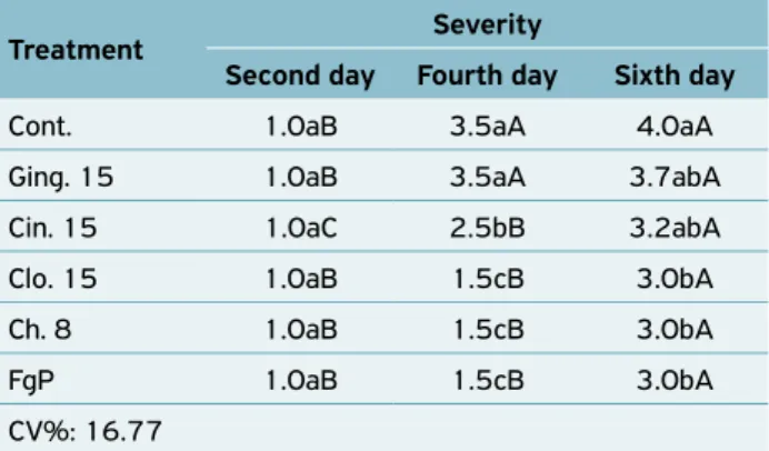
In vivo evaluations showed no significant differences between treatments on the second day of evaluation. However, anthrac-nose control was verified by treatments with 15% clove, 8% chi-tosan, and Prochloraz on the fourth and sixth day. The treatment with 15% cinnamon presented intermediate control, differing from the control on the fourth day. The 15% ginger treatment did not differ from the control group on the fourth and sixth day of evaluation, showing the same pattern of lesion growth
Treatment* Germination (%)
Cont. 15.3a
Ging. 7.5 12.0ab
Cin. 7.5 7.9b
Clo. 7.5 0.5c
Ch. 1.5 0.0c
FgP 0.0c
CV%: 30.796
Table 2. Spore germination (%), in vitro, of C. gloeosporioides
under different treatments: control (Cont.); 7.5% ginger (Ging 7.5); 7.5% cinnamon (Cin. 7.5); 7.5% clove (Clo. 7.5); 1.5% chitosan (Ch. 1.5), and the fungicide Prochloraz at 50 μg.mL‑1
(FgP). Dourados, Mato Grosso do Sul, UFGD, 2014.
REFERENCES
ANDRADE, W.P.; VIEIRA, G.H.C. Efeito dos óleos essenciais sobre
a antracnose in vitro e em frutos de mamoeiro. Revista Brasileira
de Plantas Medicinais, Campinas, v.18, n.1, suppl.1, p.367‑
372, 2016. Available from: <http://www.scielo.br/pdf/rbpm/ v18n1s1/1516‑0572‑rbpm‑18‑1‑s1‑0367.pdf>. Access on: 18 May 2016. http://dx.doi.org/10.1590/1983‑084X/15_089
AGÊNCIA NACIONAL DE VIGILÂNCIA SANITÁRIA (ANVISA). Resolução RDC n.60, de 3 de fevereiro de 2016. Dispõe sobre a
proibição da utilização do ingrediente ativo procloraz em produtos
agrotóxicos, em decorrência de sua reav. toxicológica. Diário
Oficial da União, Brasília, 4 fev., 2016. Available from: <http:// pesquisa.in.gov.br/imprensa/jsp/visualiza/index.jsp?jornal=1& pagina=48&data=04/02/2016>. Access on: 18 May 2016.
ARAUJO, R.C.Z.; CHALFOUN, S.M.; ANGÉLICO, C.L.; ARAUJO, J.B.S.; PEREIRA, M.C. Avaliação in vitro da atividade
fungitóxica de extratos de condimentos na inibição de fungos isolados de pães artesanais. Ciência e Agrotecnologia,
Lavras, v.33, n.2, p. 545‑551, 2009. Available from: <http://www.scielo.br/pdf/cagro/v33n2/v33n2a29.pdf>. Access on: 1 Dec. 2017.
throughout the evaluations; and the severity of the disease increa-sed between the second and fourth day, without differing bet-ween the fourth and sixth day. However, in treatments where there was greater control of the disease, the severity increased significantly after the second evaluation (Table 3).
The lower efficacy of the treatment with 15% ginger extract indicates the control indexes shown in the in vitro
experiments, in relation to the other treatments. However, STANGARLIN et al. (1999), verifying the elicitor effect of phytoalexins in bioassays with sorghum and soybean, found that the raw aqueous extract of ginger had the capacity to activate defense mechanisms in these plants.
The intermediate control obtained with the 15% cinna-mon extract indicates the possibility of obtaining a better result with a higher concentration. In the literature, no studies eva-luating the phytopathogenicity of cinnamon aqueous extract were found in vivo, but only in vitro. However, there are in vivo studies with cinnamon essential oil that have demons-trated its antimicrobial action. ANDRADE; VIEIRA (2016) observed that the low efficacy of the cinnamon essential oil in the post-harvest control of C. gloeosporioides in “Golden” papaya was due to low dosage (100 μL), since there was gra-dual inhibition of this fungus with increased concentrations.
The inhibition of the growth of C. gloeosporioides lesions found in the 8% chitosan treatment is related, according to MAIA et al. (2010), to the fungistatic effect of chitosan and its potential to activate enzymes and phenolic compounds related to the plant defense mechanism.
Although our study demonstrated anthracnose control with the 15% clove treatment, there are no studies that demons-trate the in vivo behavior of clove in the control of phytopa-thogens. However, in vitro studies by COSTA et al. (2011) and LUCAS (2012) show that eugenol and β-caryophyllene act on the degradation of the cell wall, with morphological changes in fungal mycelium, disorganization of cellular con-tents, intense fragmentation, and less turgidity of the hyphae.
CONCLUSION
The aqueous extracts of cinnamon and ginger showed potential inhi-bition of C. gloeosporioides development in vitro. However, in vivo, the anthracnose control effectiveness of the 15% doses was low.
Chitosan and aqueous clove extract are effective in inhibiting mycelial growth, and production and germination of C. gloeosporioides
conidia in vitro, as well as in reducing the severity of anthracnose
in vivo. They present a viable alternative to Prochloraz fungicide for post-harvest protection of “Formosa” papaya.
Treatment Severity
Second day Fourth day Sixth day
Cont. 1.0aB 3.5aA 4.0aA
Ging. 15 1.0aB 3.5aA 3.7abA Cin. 15 1.0aC 2.5bB 3.2abA
Clo. 15 1.0aB 1.5cB 3.0bA
Ch. 8 1.0aB 1.5cB 3.0bA
FgP 1.0aB 1.5cB 3.0bA
CV%: 16.77
Table 3. Severity of anthracnose in “Formosa” papaya “Tainung
1”, under different treatments: control (Cont.); 15% ginger (Ging. 15); 15% cinnamon (Cin. 15); 15% clove (Clo. 15); 8% chitosan (Ch. 8), and the fungicide Prochloraz 33.75 g i.a./100 L (FgP). Dourados, Mato Grosso do Sul, UFGD, 2014.
Averages followed by the same letter, lowercase in the column and upper case in the row, do not differ statistically from each other by the F‑test and Tukey’s test at 5% level of significance; CV: coefficient of variation. Level of disease severity by a score scale of 0 to 5: (1) absence of symptoms of stem‑end‑rot; (2) presence of small superficial aqueous punctures (up to 3 mm) in the peduncle region; (3) presence of aqueous or mummified lesions, not coalescing, covering a larger area around the stem; (4) presence of coalescing lesions, with translucent or mummified appearance, limited to the stem region; and (5) lesions similar to those described in scale 4, including greater extension in
the fruit pulp, reaching the seed cavity, the tissue of this region may
BARRETO, T.A Efeitos da aplicação de revestimento de quitosana e óleo essencial de orégano no controle da qualidade pós-colheita em tomates
cereja. 2016. 88f. Dissertação (Mestrado em Ciência e Tecnologia
de Alimentos) – Universidade Federal da Paraíba, João Pessoa, 2016. Available from: <https://repositorio.ufpb.br/jspui/bitstream/ tede/9438/2/arquivototal.pdf>. Access on: 27 Nov. 2017.
BERALDO, C.; DANELUZZI, N.S.; SCANAVACCA, J.; DOYAMA, J.T.; FERNANDES JÚNIOR, A.; MORITZ, C.M.F. Eficiência de óleos essenciais de canela e cravo‑da‑índia como sanitizantes na indústria de alimentos. Pesquisa Agropecuária Tropical, Goiânia,
v.43, n.4, p.436‑440, 2013. Available from: <http://www. scielo.br/pdf/pat/v43n4/06.pdf>. Access on: 27 Nov. 2017.
BONILLA, J.; SOBRAL, P.J.A. Antioxidant and antimicrobial
properties of ethanolic extracts of guarana, boldo, rosemary
and cinnamon. Brazilian Journal of Food Technology, Campinas,
v.20, e2016024, 2017. Available from: <http://www.scielo.br/ pdf/bjft/v20/1981‑6723‑bjft‑20‑e2016024.pdf>. Access on: 27 Nov. 2017. http://dx.doi.org/10.1590/1981‑6723.2416
BRITO, N.M.; NASCIMENTO, L.C. Potencial fungitóxico de extratos
vegetais sobre Curvularia eragrostidis (P. Henn.) Meyer in vitro.
Revista Brasileira de Plantas Medicinais, Paulínia, v.17, n.2, p.230‑
238, 2015. Available from: <http://www.scielo.br/pdf/rbpm/ v17n2/1516‑0572‑rbpm‑17‑2‑0230.pdf>. Access on:27 Nov. 2017. http://dx.doi.org/10.1590/1983‑084X/10_057
CARNELOSSI, P.R.; SCHWAN‑ESTRADA, K.R.F.; CRUZ, M.E.S.; ITAKO, A.T.; MESQUINI, R.M. Óleos essenciais no controle pós‑
colheita de Colletotrichum gloeosporioides em mamão. Revista
Brasileira de Plantas Medicinais, Botucatu, v.11, n.4, p.399‑
406, 2009. Available from: <http://www.scielo.br/pdf/rbpm/ v11n4/a07v11n4.pdf>. Access on: 27 Nov. 2017. http://dx.doi. org/10.1590/S1516‑05722009000400007
CELOTO, M.I.B.; PAPA, M.F.S.; SACRAMENTO, L.V.S.; CELOTO, F.J. Atividade antifúngica de extratos de plantas a Colletotrichum
gloeosporioides. Acta Scientiarum Agronomy, Maringá, v.30, n.1,
p.1‑5, 2008. Available from: <http://www.scielo.br/pdf/asagr/ v30n1/v30n1a01.pdf>. Access on: 27 Nov. 2017. http:// dx.doi.org/10.4025/actasciagron.v30i1.1104
CIA, P.; BENATO, E.A.; PASCHOLATI, S.F.; GARCIA, E.O. Quitosana no controle pós‑colheita da podridão mole em caqui ‘Rama Forte’. Bragantia, Campinas, v.69, n.3, p.745‑752, 2010. Available from: <http://www.scielo.br/pdf/brag/v69n3/28. pdf>. Access on: 27 Nov. 2017. http://dx.doi.org/10.1590/ S0006‑87052010000300028
COQUEIRO, D.S.O.; DI PIERO, R.M. Atividade de quitosanas com
diferentes pesos moleculares sobre alternaria solani. Arquivos
do Instituto Biológico, v.78, p.459‑463, 2011. Available from:
<http://www.biologico.sp.gov.br/uploads/docs/arq/v78_3/ coqueiro.pdf>. Access on: 27 Nov. 2017.
COSTA, A.R.T.; AMARAL, M.F.Z.J.; MARTINS, P.M.; PAULA, J.A.M.; FIUZA, T.S.; TRESVENZOL, L.M.F.; PAULA, J.R.; BARA, M.T.F. Ação do óleo essencial de Syzygium aromaticum (L.) Merr. & L. M. Perry sobre as hifas de alguns fungos fitopatogênicos. Revista
Brasileira de Plantas Medicinais, Paulínia, v.13, n.2, p.240‑245,
2011. Available from: <http://www.scielo.br/pdf/rbpm/v13n2/ v13n2a18.pdf>. Access on: 27 Nov. 2017. http://dx.doi. org/10.1590/S1516‑05722011000200018
COSTA, F.B.; MENEZES, J.B.; ALVES, R.E.; NUNES, G.H.S.; MARACAJÁ, PB. Armazenamento refrigerado do mamão Havaí “Golden” produzido na chapada do Apodi – RN – Brasil. Revista
Verde, Mossoró, v.5, n.4, p.37‑54, 2010. Available from: <https://
www.alice.cnptia.embrapa.br/alice/bitstream/doc/876244/1/ PG10006.pdf>. Access on: 27 Nov. 2017.
DIEMER, AW. Ação antimicrobiana de Rosmarinusofficinalis e
Zingiber officinale frente à Escherichia coli e Staphylococcus
aureus em carne mecanicamente separada de frango. 2016. 69f.
Dissertação (Mestrado em Biotecnologia) – Centro Universitário
UNIVATES, Lajeado, 2016. Available from: <https://www.univates. br/bdu/bitstream/10737/1075/1/2016AndreaWolfDiemer. pdf>. Access on: 27 Nov. 2017.
DI PIERO, R.M.; GARDA, M.V. Quitosana reduz a severidade da antracnose e aumenta a atividade de glucanase em feijoeiro‑ comum. Pesquisa Agropecuária, Brasília, v.43, n.9, p.1121‑ 1128, 2008. Available from: <http://www.scielo.br/pdf/pab/ v43n9/04.pdf>. Access on: 27 Nov. 2017.
FONTANA, D.C.; KULCZYNSKI, S.M.; TREVISAN, R.; SCHMIDT, D.; CARON, B.O.; PINHEIRO, M.V.M.; PRETTO, M.M.; DIEL, M.I. Uso de extratos vegetais no controle alternativo da podridão parda do pessegueiro.
Revista Cultivando o Saber, Cascavel, v.10, n.2, p.148‑165, 2017.
Available from: <https://www.fag.edu.br/upload/revista/cultivando_o_ saber/59a5b4143085c.pdf>. Access on: 27 Nov. 2017.
FREDDO, A.R. Quitosana in vitro e no tratamento de sementes de eucalipto e acácia-negra no controle de Rhizoctoniasolani e no
desenvolvimento inicial das plântulas. 2012. 78f. Dissertação
(Mestrado em Agronomia) – Universidade Tecnológica Federal do Paraná, Pato Branco, 2012. Available from: <http://repositorio. utfpr.edu.br/jspui/handle/1/276>. Access on: 27 Nov. 2017.
HERNÁNDEZ, A.M.A.; NECHA, L.L.B.; LAUZARDO, A.N.H.; DEL VALLE, M.G.V. Actividad antifúngica del quitosano y aceites
esenciales sobre Rhizopus stolonifer (Ehrenb.:Fr.) Vuill., agente causal de la pudrición blanda del tomate. Revista Colombiana
de Biotecnologia, Bogotá, v.13, n.2, p.127‑134, 2011. Available
from: <http://www.scielo.org.co/pdf/biote/v13n2/v13n2a11. pdf>. Access on: 27 Nov. 2017.
LIMA, C.Q.; ALMEIDA, F.A.C.; ARAÚJO, E.; SILVA, J.F.; MORAES, A.M.; MEDEIROS, D.S. Bioatividade de extratos e óleos vegetais no
controle in vitro de Aspergillus flavus em sementes de amendoim.
Tecnologia & Ciência Agropecuária, João Pessoa, v.6, n.1, p.13‑
18, 2012. Available from: <http://revistatca.pb.gov.br/edicoes/ volume‑06‑2012/volume‑6‑numero‑1‑marco‑2012/tca6103. pdf>. Access on: 27 Nov. 2017.
LORENZETTI, E.R.; MONTEIRO, F.P.; SOUZA, P.E.; SOUZA, R.J.; SCALICE, H.K.; DIOGO JUNIOR, R.; PIRES, M.S.O. Bioatividade de óleos essenciais no controle de Botrytiscinerea isolado de morangueiro.
Revista Brasileira de Plantas Medicinais, Botucatu, v.13, n.especial,
p.619‑627, 2011. Available from: <http://www.scielo.br/pdf/ rbpm/v13nspe/a19v13nspe.pdf>. Access on: 27 Nov. 2017.
© 2018 Instituto Biológico This is an open access article distributed under the terms of the Creative Commons license.
MAIA, A.J.; BOTELHO, R.V.; FARIA, C.M.D.R.; LEITE, C.D. Ação de quitosana sobre o desenvolvimento
de Plasmoporaviticola e Elsioneampelina, in vitro e em videiras
cv. ‘Isabel’. Summa Phytopathologica, Botucatu, v.36, n.3, p.203‑209, 2010. Available from: <http://www.scielo.br/pdf/ sp/v36n3/v36n3a03.pdf>. Access on: 27 Nov. 2017. doi: 10.1590/S0100‑54052010000300003
MAMPRIM, A.P.; ALVES, L.F.A.; PINTO, F.G.S.; FORMENTINI, M.A.; MARTINS, C.C.; BONINI, A.K. Efeito de defensivos agrícolas naturais e extratos vegetais sobre parâmetros biológicos de
Metarhizium anisopliae (Metsch.) Sorok. Semina, Londrina, v.34,
n.4, p.1451‑1466, 2013. Available from: <http://www.redalyc. org/html/4457/445744122002/>. Access on: 27 Nov. 2017. DOI: 10.5433/1679‑0359.2013v34n4p1451
NERY‑SILVA, F.A.; MACHADO, J.C.; RESENDE, M.L.V.; LIMA, L.C.O.
Metodologia de inoculação de fungos causadores da Podridão
peduncular em mamão. Ciência e Agrotecnologia, Lavras, v.31, n.5, p.1374‑1379, 2007. Available from: <http://www.scielo. br/pdf/cagro/v31n5/15.pdf>. Access on: 27 Nov. 2017.
RODRIGUES, E.; SCHWAN‑ESTRADA, K.R.F.; STANGARLIN, J.R.; CRUZ, M.E.S; TUTIDA‑FIORI, A.C.G. Avaliação da atividade
antifúngica de extratos de gengibre e eucalipto in vitro e em fibras
de bananeira infectadas com Helminthosporium sp. Acta Scientiarum
Agronomy, Maringá, v.28, n.1, p.123‑127, 2006. Available
from: <http://www.redalyc.org/pdf/3030/303026568006. pdf>. Access on: 27 Nov. 2017.
SCHWAN‑ESTRADA, K.R.F. Extratos vegetais e de cogumelos no controle de doenças de plantas. Horticultura Brasileira, Brasília,
v.27, n.2, S4038‑S4045, 2009. Available from: <http://www. abhorticultura.com.br/eventosx/trabalhos/ev_3/MR_4_Artigo_ Katia_Regina_Estrada.pdf>. Access on: 27 Nov. 2017.
SILVA, A.C.; SALES, N.L.P.; ARAÚJO, A.V.; CALDEIRA JUNIOR, C.F. Efeito in vitro de compostos de plantas sobre o fungo
Colletotrichum gloeosporioides penz. Isolado do maracujazeiro.
Ciência e Agrotecnologia, Lavras, v.33, Edição Especial, p.1853‑
1860, 2009. Available from: <http://www.scielo.br/pdf/cagro/ v33nspe/26.pdf>. Access on: 27 Nov. 2017.
SILVA, J.F.M.; PRADO, G.; MADEIRA, J.E.G.C.; OLIVEIRA, M.S.; FARACO, A.A.G.; MALTA, C.M.; NICOLI, J.R.; PIMENTA, R.S. Utilização de filme de quitosana para o controle de aflatoxinas em
amendoim. Bragantia, Campinas, v.74, n.4, p.467‑475, 2015. Available from: <http://www.scielo.br/pdf/brag/v74n4/0006‑ 8705‑brag‑1678‑44990120.pdf>. Access on:27 Nov. 2017. http://dx.doi.org/10.1590/1678‑4499.0120
SIQUEIRA JUNIOR, C.L.; MORAES, T.C.; MARTINS, J.A.B.; FREIRE, M.G.M. Controle de antracnose em mamão por extratos vegetais.
Biológicas & Saúde, v.1, n.1, p.99‑105, 2011. Available from:
<http://www.seer.perspectivasonline.com.br/index.php/ biologicas_e_saude/article/viewFile/517/429>. Access on: 27 Nov. 2017. DOI: https://doi.org/10.25242/8868112011517
STANGARLIN, J.R.; SCHWAN‑ESTRADA, K.R.F.; CRUZ, M.E.S.; NOZAKI, M.H. Plantas medicinais e o controle alternativo de doenças de plantas. Biotecnologia Ciência & Desenvolvimento,
Brasília, v.11, p.16‑21, 1999.
TARNOWSKI, T.L.B. Using molecular analysis to investigate
phylogenetic relationships in two tropical pathosystems: witches’ broom of cacao, caused by Moniliophthora perniciosa, and mango anthracnose, caused by Colletotrichum spp. 2009. 236 p. Thesis (Doctorate in Philosophy) University of Florida, Gainesville, 2009. Available from: <http://etd.fcla.edu/UF/UFE0041003/ tarnowski_t.pdf>. Access on:26 Apr. 2018.
TAVARES, G.M.; SOUZA, P.E. Efeito de fungicidas no controle in vitro
de Colletotrichum gloeosporioides, agente etiológico da antracnose
do mamoeiro (Caricapapaya L.). Ciência e Agrotecnologia, Lavras, v.29, n.1, p.52‑59, 2005. Available from: <http://www.scielo. br/pdf/cagro/v29n1/a06.pdf>. Access on: 27 Nov. 2017. http://dx.doi.org/10.1590/S1413‑70542005000100006
TORRES‑CALZADA, C.; TAPIA‑TUSSELL, R.; HIGUERA‑CIAPARA, I.; PEREZ‑BRITO, D. Morphological, pathological and genetic
diversity of Colletotrichum species responsible for anthracnose in papaya (Carica papaya L). European Journal of Plant Pathology,
Sochi, v.135, n.1, p.67‑79, 2012. Available from: <https:// www.researchgate.net/publication/257558376>. Access on: 26 Apr. 2018. DOI: 10.1007/s10658‑012‑0065‑7
VENTUROSO, L.R.; BACCHI, L.M.A.; GAVASSONI, W.L.; CONUS, L.A.; PONTIM, B.C.A.; SOUZA, F.R. Inibição do crescimento in vitro de