Clinical and Epidemiologic
Studies
of Chagas’ Disease in Rural Communities
of Oaxaca, Mexico, and an Eight-year
Followup:
II. Chilal
R. S.
GOLDSMITH,~
R. J.Z~~RATE,~
L. G.ZARATE,~
G. MORALES,~I.
KAGAN,~
R.DRICKEY,~ &
L. B.JACOBSON~
444
A seroepidemiologic survey conducted in 1971 in the rural Pacific coastal community of
Ckila in the Mexican state of Oaxaca showed an unusually high prevalence of antibody against the Ckagas’ disease agent Trypanosoma cruzi. Further studies were undertaken in 1973 and 1981 to (1) determine the pathologic impact of T. cruzi infecfion in humans, (2) investigate the natural kistoy of the disease, (3) confirm that serologically positive persons were parasitologically positive, and (4) evaluate whether T. cruzi transmission continued into the next decade. This article reports results derived from those studies.
A
serosurvey in the community of Chila, situated on the south coast of the Mexican state of Oaxaca, revealed an unusually high chagasic antibody prevalence. Electrocardiographic eval- uation demonstrated significant abnor- malities in seropositive as compared to‘This article will also be published in Spanish in the Bolefin de la Oficina Sanitaria Panamerzcana, Vol. 113, 1992.
‘Professor of Tropical Medicine and Epidemiology, Department of Epidemiology and Biostatistics, University of California, San Francisco, California 94143, U.S.A.
3Researcher, Centro de Investigaciones Ecologicas de1 Sureste, San Cristobal de las Casas, Chiapas, Mexico.
4Director Emeritus, Parasitology Division, United States Centers for Disease Control, Atlanta, Geor- gia, U.S.A.
5Assistant Professor, Department of Family and Community Medicine, University of California, San Francisco, California, U.S.A.
6Attending Cardiologist, Department of Medicine, Pacific Presbyterian Medical Center, San Francisco, California, U.S.A.; and Clinical Professor, Depart- ment of Medicine, University of California, San Francisco.
seronegative people, and some of these abnormalities progressed during an eight-year followup period.
Until fairly recently, Chagas’ disease was not thought to occur commonly in Mexico or to cause significant cardiac pa- thology. Antibody prevalences to Trypa- nosoma cruzi reported from several com- munities have ranged from 7% to 13% (1-6). Salazar Schettino et al. reported an overall seroprevalence of 6% for serum samples from about 6,600 persons that were collected between 1949 and 1985 in several states of southern and central Mexico (3). In 1986, Tay et al. reported that 88% of 52 schoolchildren tested, all from the town of Miahuatlan, Oaxaca, were seropositive (7).
Our 1971 seroepidemiologic surveys in 60 rural communities on the Pacific side of Oaxaca State found unusually high T. cruzi infection rates; in particular, three communities (Cerro de1 Aire, Tataltepec, and Chila) exhibited prevalences among adults ranging from 51% to 76% (8, 9).
This article, which supplements our previous report from Cerro de1 Aire (ZO),
presents clinical, electrocardiographic, serologic, and parasitologic findings de- rived from a cross-sectional study and an eight-year longitudinal study conducted in Chila. The objectives of these two studies were (1) to determine the path- ologic impact of T. cruzi infection in hu- mans from this area, (2) to investigate the natural history of the disease, (3) to con- firm that serologically positive persons were parasitologically positive, and (4) to find out whether T. cruzi transmission was continuing. Within this context, it should be noted that although many community-based cross-sectional studies of Chagas’ disease have been reported (IO-13), relatively few longitudinal com- munity-based studies have been per- formed (13-28); such longitudinal stud- ies are needed, particularly to obtain
information about the natural history of the disease (20, 29).
METHODS
Study Area
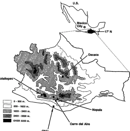
Oaxaca, one of Mexico’s southernmost states, is around 17” north latitude and 96” west longitude (Figure 1). The town of Chila is a rural settlement located on the state’s Pacific Coast. Chila has an annual rainfall of about 150 cm, most of which occurs between May and October.
Between 1971 and 1973 Chila had ap- proximately 2,600 inhabitants. Thus, our sample of 238 people represented about 9% of the total population in that period
Figure 1. A map of Mexico’s Oaxaca State showing the location of Chila.
1600 - 2400 m. 2400 - 3200 In. OVER 3200 in.
(9). By 1981, however, the population had grown to approximately 3,100 people.
Homes in Chila were made from a va- riety of materials. Some had concrete walls, concrete or dirt floors, and tin or thatch roofs (the latter being made of palm fronds or straw); while others had wood slat and adobe walls, dirt floors, and thatched roofs.
cross-sectional study; because of time limitations, histories and physical exam- inations were obtained from only 58 of these 135 persons. This resulted in med- ical history and physical examination data being obtained from a total of 177 people and ECGs being administered to 254. Longitudinal Clinical Study
Field Trips to Chila
In 1981 repeat ECGs, clinical evalua- tions, and serologic tests were obtained Field trips to Chila, which took place from 57 people who had been tested in in 1971, 1973, and 1981, involved the fol- 1973. The participants were those who lowing activities and objectives: continued to reside in the community and agreed to participate.
Serologic Surveys Xenodiagnosis
Our 1971 serologic survey included 238 people of all ages. The methods used in this survey have previously been de- scribed (9). In brief, Chila households were selected at random and, for the most part, all residents of the selected households were included in the survey sample.
In 1981, primarily to evaluate contin- ued transmission of T. cruzi infections to humans, blood specimens obtained from 144 children under the age of 10 were tested for T. cruzi antibody. The subjects were “self-selected” by their parents or guardians in response to an invitation is- sued through the town mayor asking mothers to have their children tested. Cross-sectional Clinical Study
In 1973, we evaluated the cardiac sta- tus of 119 people from the original 1971 cohort of 238. These 119 were self- selected in response to an invitation ask- ing cohort members to participate. The subjects’ cardiac status was evaluated by reviewing their history and by means of a physical examination, electrocardio- gram (ECG), and repeat serology.
In 1981, a group of 135 newly seen self- selected adults was added to the ECG
Xenodiagnosis was performed on 40 seropositive people in 1981.
Methods of Blood Collection,
Serology, and Xenodiagnosis
The methods used for the collection and storage of blood (9), T. cruzi antigen preparation (8, 30), conduct of the indi- rect hemagglutination (IHA) serologic test (positive titer ~128) (S), and xenodiag- nosis (10) have already been described.
Electrocardiograms
ECGs, recorded with either a Cam- bridge or Burdick portable ECG machine, included the six standard limb leads and either three or six standard precordial leads. The ECGs were evaluated by a car- diologist (LBJ) for rhythm, rate, PQRSTU contours, PR interval, and QRS axis, du- ration, and configuration in accordance with standard criteria but without knowl- edge of the patient’s serologic status.
Statistical Analysis
The statistical association between two
dichotomous outcomes was evaluated
using the chi-square test or, when the expected number for a cell was less than 5, Fisher’s exact test.
RESULTS
Serologic Surveys and
Seroconversion
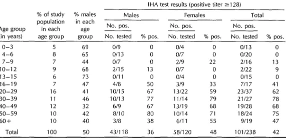
Results of the 1971 serologic survey, in which sera from 238 people were tested by IHA, are shown in Table 1, which groups the subjects by age and sex. Of the 86 children (subjects under age 16) who were tested, four (5%) were found to be seropositive. However, for all sub- sequent age groups from ages 16 through 39, the percentage of seropositives in- creased progressively-from 41% in the 16-19 year age group to 78% in the 30- 39 group. Thereafter, in the older age groups, the rates either remained about the same or declined. The difference in seropositivity among females (48%) ver- sus males (36%) was not statistically sig- nificant. Overall, of the 238 people tested, 42% were seropositive; but of the 135
people over 19 years old who were tested, 67% were seropositive.
IHA results from the 1981 serologic survey, which tested 144 children under age 10 and 135 adults over age 19, are shown in Table 2. Of the 144 children, only one was positive. Of the 135 adults, 43 (32%) were positive.
Regarding seroconversion, of 61 sub- jects tested in 1971 and 1973 and retested in 1981, one of 21 seronegatives con- verted to positive and four of 39 sero- positives converted to negative.
Of the 238 persons seen in 1971, four had died by 1981, three of unknown causes.
Cross-sectional
Study of 254
Subjects
Comparison of Clinical Findings among Seropositive and Seronegative Subjects
Medical histories were provided by 177 subjects. As indicated in Table 3, differ- ences in the frequency of signs and symptoms reported by the 83 seroposi-
Table 1. Age- and sex-specific indirect hemagglutination test (IHA) results for 238 people tested in the Chila serologic survey of 1971.
IHA test results (positive titer 2128) % of study % males Males Females Total population in each
Age group in each age ___ No. pos. No. pos. No. pos.
(in years) age group group No. tested % pos. No. tested % pos. No. tested % pos.
o-3 5
4-6 8
7-9 7
IO-12 9 13-15 6 16-19 7 20-29 16 30-39 11 40-49 12 50-59 10 60+ 10
Total 100
69 o/9 0 o/4 65 o/13 0 o/7 44 o/7 0 219 68 2115 13 Of7 73 0111 0 o/4 47 4/a 50 319 41 10115 67 13122 46 10113 77 11114 32 619 67 13/19 42 a/10 80 10114 40 3/a 38 6fll
0 o/13 0 0 O/20 0
22 2f16 13 0 2/22 9 0 o/15 0 33 7117 41 59 23137 62 79 21127 78 68 19f28 68 71 la/24 75 55 9/l 9 47 50 431118 36 58/120 48 101/238 42
Table 2. IHA test results for 144 children and 135 adults tested in the 1981 survey.
Indirect hemagglutination test resul& Age group”
(in years)
No. pos. No. tested
% positive Children
o-3 4-6 7-9 Total Adults
20-29 30-39 40-49 50-59 60+ Total
o/41 0139
1 I64 l/l44
4136 11
20146 43 7125 28 8/l 7 47 4/l 1 36 43/l 35 31.9
0 0 2 0.7
aNo people in the 1 O-l 9 age range were tested. bPositwe titer 2128.
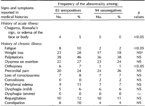
tives and the 94 seronegatives were sta- tistically significant (p < 0.05) in the fol- lowing instances: (1) initial bite lesions (Romana’s sign [unilateral bipalpebral
edema] or chagoma [furuncle-like skin le- sion]) or edema of the face or body was recalled by four (4.8%) seropositives but no seronegatives; (2) fatigue affected eight (9.6%) of the seropositives but only two (2.1%) seronegatives; and (3) orthopnea was reported by six (7.2%) seropositives but only one seronegative.
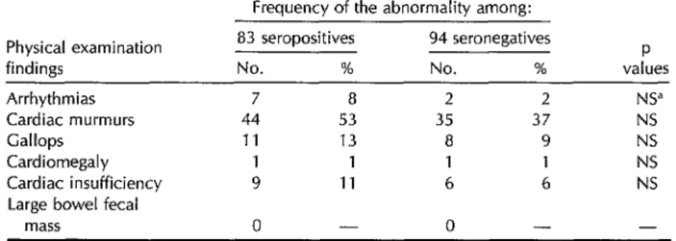
No statistically significant differences were found in the frequencies of physical examination abnormalities among the 83 seropositives and 94 seronegatives (Table 4).
ECG Findings among Seropositives and Seronegatives
Of the 254 people tested by ECG, 115 were seropositive and 139 seronegative (Table 5); the age distribution of these subjects is shown in Table 6.
As Table 5 indicates, a statistically sig- nificant difference (p < 0.005) was ob- served between the frequency of com-
Table 3. Frequency of abnormalities in medical histories from 83 seropositive and 94 seronegative subjects.
Signs and symptoms reported in medical histories
Frequency of the abnormality among: 83 seropositives 94 seronegatives
P
No. % No. % values History of acute illness:
Chagoma, Romana’s sign, or edema of the face or body Hfstory of chronic illness:
Fatigue Weight loss Palpitations Dyspnea on exertion Orthopnea
Precordial pain Loss of consciousness Convulsions
Peripheral edema Dysphagia (mild) Dysphagia (severe) Regurgitation Constipation
dNS = not significant.
4 5 0 0 co.05
8 10 2 2 co.05 23 28 17 18 NS” 38 46 36 38 NS 22 27 23 24 NS 6 7 1 1 co.05 20 24 24 26 NS
7 8 7 7 NS
0 0 2 2 NS
9 11 7 7 NS
5 6 6 6 NS
0 0 0 0 -
10 12 10 11 NS
8 10 4 4 NS
Table 4. Frequency of physical examination abnormalities among the 83 seropositives and 94 seronegatives.
Frequency of the abnormality among: Physical examination
findings Arrhythmias Cardiac murmurs Gallops
Cardiomegaly Cardiac insufficiency Large bowel fecal
mass
83 seropositives No. %
7 8 44 53
11 13
1 1 9 11
0 -
94 seronegatives No. %
2 2 35 37 8 9 1 1
6 6 0 -
P values
NS” NS NS
NS
NS -
dNS = not significant.
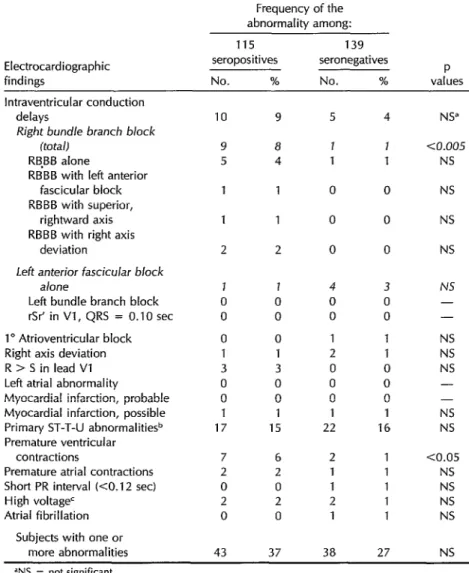
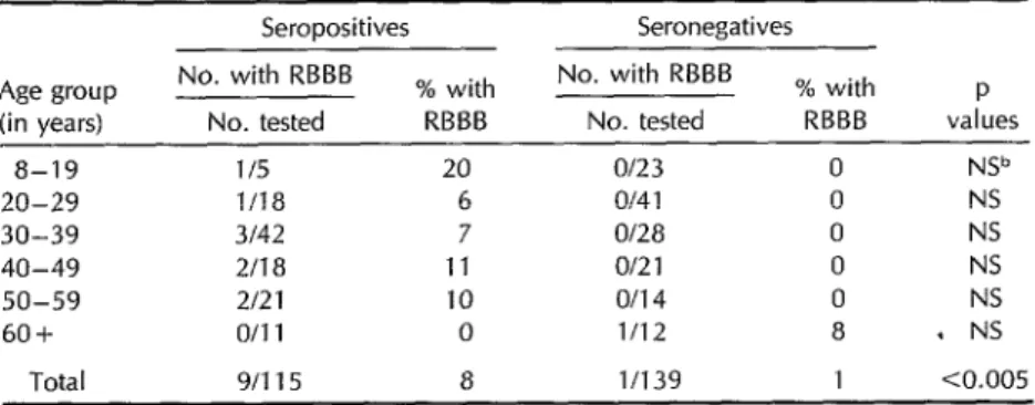
plete right bundle branch block (RBBB) in the longitudinal study. All 42 of the with or without other findings in sero- initially seropositive subjects remained positive versus seronegative subjects. seropositive over the eight-year period, Overall, RBBB was found in nine (7.8%) and all 15 of the initially seronegative of the seropositives but only one (0.72%) subjects likewise remained seronegative of the seronegatives. (Table 7).
Another significant difference (p < 0.05) was observed with respect to premature ventricular contractions, which were found in seven (6.1%) of the seroposi- tives as compared to two (1.4%) of the seronegatives.
Demographic and Laborato y Findings for Subjects with RBBB
Of the 10 subjects with RBBB, nine were seropositive. Of these seropositives, two were between the ages of 8 and 29 years, five were 30 to 49 years old, and two were 50 years of age or older; three of the nine were men and six were women. The one seronegative subject with RBBB was a 66-year-old man with a negative history for Chagas’ disease and no ab- normalities on physical examination.
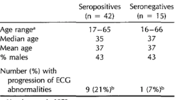
Of the 57 ECG pairs, 18 showed no changes, 29 showed doubtful changes, and 10 revealed definite changes. As Table 7 indicates, most of these changes oc- curred among the seropositives. Specifi- cally, nine (21.4%) of the 42 seropositive pairs showed definite changes (equiva- lent to 2.7% per year), as compared to only one (6.6%) of the 15 seronegatives. However, this distribution was not sta- tistically significant (p > 0.05).
Longitudinal
Study of 57 Subjects:
ECG Changes, 1973-1981
A total of 57 people (all over the age of 15 years in 1973) who received an ECG in 1973 and again in 1981 were included
Of the definite changes observed in the nine ECG pairs from seropositive sub- jects, three were changes characteristic of Chagas’ disease, two involving ventric- ular premature beats and one involving an increase of the R:S ratio in lead Vl. The other six changes were not charac- teristic of Chagas’ disease, three involv- ing QRS axis shifts (two rightward and one leftward, the axis always remaining within normal limits); two involving mild T contour changes; and one involving changes suggestive of an interval inferior and posterior wall myocardial infarction. The one seronegative subject with a def- inite change showed a change of T con- tour from upright to inverted in AVL. No
Table 5. Frequency of electrocardiographic abnormalities among 115 seropositive and 139 seronegative persons.
Frequency of the abnormality among:
Electrocardiographic findings
115 139
seropositives seronegatives No. % No. %
P values lntraventricular conduction
delays
Right bundle branch block (tota/)
RB,BB alone
RBBB with left anterior fascicular block RBBB with superior,
rightward axis RBBB with right axis
deviation
Left anterior fascicular block a/one
Left bundle branch block rSr’ in Vl, QRS = 0.10 set 1” Atrioventricular block Right axis deviation R > S in lead Vl
Left atrial abnormality
Myocardial infarction, probable Myocardial infarction, possible Primary ST-T-U abnormalitie+ Premature ventricular
contractions
Premature atrial contractions Short PR interval (CO.12 set)
High voltage’ Atrial fibrillation
Subjects with one or
more abnormalities
10 9 5 1 1 2 J 0 0 0 1 3 0 0 1 17 7 2 0 2 0 43 9 8 4
1 0
1 0
2 0
4 NS”
J aJ.005
1 NS
0 NS
0 NS
0 NS
1 0 0 0 1 3 0 0 1 15 4 0 0 1 2 0 0 0 1 22
6 2 2 1 0 1 2 2 0 1
3 NS 0 - 0 - 1 NS 1 NS 0 NS 0 - 0 - 1 NS 16 NS
1 CO.05 1 NS 1 NS 1 NS 1 NS
37 38 27 NS ‘NS = not significant.
%T-T-U abnormalities occurring in the presence of normal QRS complexes. cPossibly due to left ventricular hypertrophy.
subject exhibited a new bundle branch numbers of bugs positive when tested
block. individually ranged from 2 to 23.
Isolation
of T. cmzi from
Seropositives
DISCUSSION
Of 40 seropositive subjects tested by xenodiagnosis in 1981, seven (18%) yielded positive results. Among the batches of bugs used to test these seven people, the
High Prevalence of Infection
but
Absence of Transmission
in Chila
The 1971 serosurvey in Chila found a T. cvtlzi antibody prevalence in adults of
Table 6. Frequency of complete right bundle branch block (RBBB) by age group and serologic status for 254 persons on whom ECCs were conducted in 1973 and/or 1981 .B
Seropositives Seronegatives Age group
(in years)
a-19 20-29 30-39 40-49 50-59 60+
No. with RBBB No. tested
% with RBBB
No. with RBBB No. tested
% with P RBBB values l/5
l/la 3142 2118 2121 o/11
20 Of23 6 Of4 1 7 of28 11 o/21 10 o/14 0 1112
0 NV 0 NS 0 NS 0 NS 0 NS
a . NS
Total g/115 a l/139 1 co.005 “If ECCs were conducted durmg both time periods, the subject’s age was that at the first test. bNS = not significant.
67%. This finding approximates the 76% prevalence reported in nearby Cerro de1 Aire (20). These are both high preva- lences for Mexico; however, they are not unlike high prevalences reported from South America.
In contrast, our serosurveys of Chila children revealed a near-absence of T. cruzi antibody, a finding similar to those we obtained with sera from young people in Cerro de1 Aire and other communities in the region (9, 10).
This paucity of antibody in young chil- dren indicates a near cessation of T. cruzi transmission to humans in the region from approximately 1955 to 1981. This corre- lates with onset of residual insecticide spraying for malaria control and with dis- appearance of triatomine vector(s) from Chila and other communities in the Pa- cific coastal study region of Oaxaca (9, 10). A review of malaria campaign rec- ords through 1981 has shown that DDT was used almost exclusively for spraying within the region after 1962 (31). Initially, the insecticide was applied every four months. Beginning in 1976, the schedule was changed to twice-yearly spraying in Chila and other communities, and in some areas spraying was discontinued.
Extensive searches by us for vectors in
the region produced only rare specimens of Rhodnius prolixus and Triatoma dimidiata (22, 13). Villagers told us that they had not seen the bugs for about 10 years. We presume the indigenous vectors before
onset of spraying were 7’. dimidiata and
R. prolixus (9) and T. mazzottii (32).
Clinical Manifestations
of Chagas’
Disease in Chila
Acute Chagas’ disease probably occurs in Chila but is not recognized for lack of any trained medical observers in the
community. However, in response to our inquiries about possible clinical findings, chagoma-like or Romana-like lesions were reported more frequently by seropositive subjects than by seronegatives.
In the chronic myocarditis of Chagas’ disease, cardiomegaly and heart failure may occur; and the conduction system may be affected, leading to a variety of arrhythmias and conduction disturban- ces. The histories we obtained from chronically infected subjects showed fa- tigue and orthopnea to be significantly more frequent among seropositives than seronegatives. However, the physical ex- aminations showed no significant differ- ences in findings of the sort that may
Table 7. Longitudinal study: Electrocardio- graphic changes between 1973 and 1981 for paired ECGs from 57 subiects.
Seropositives Seronegatives (n = 42) (n = 15) Age range” 17-65 16-66
Median age 35 37
Mean age 37 37
% males 43 43
Number (%) with progression of ECC
abnormalities 9 (21%)b 1 (7%)b ‘Age in years in 1973.
bp > 0.05.
occur in chronic Chagas’ disease. In ad- dition, sudden and unexpected death of young adults, a feature of the disease in some parts of South America, does not appear to occur in Chila. Although chronic infection may also result in intestinal mo- tility disturbances or in dilatation of the esophagus, colon, or other organs in cen- tral Brazil and some other regions of South America, our history and physical ex- amination findings do not suggest that the infection in Chila causes the digestive form of the disease.
Electrocardiographic
Findings
Cross-sectional Study
RBBB is a characteristic ECG feature of chronic Chagas’ disease (13). Pinto-Dias (33) has commented that an endemic re- gion for Chagas’ disease could be rec- ognized electrocardiographically by either one of two findings: (1) a prevalence of RBBB higher than 2% and (2) a marked disproportion in the frequency of right and left bundle branch block. In this study, the frequency of RBBB was 4% among all people tested and 8% among seroposi- tives; while the ratio of RBBB to LBBB was 9:l among seropositives and 1:4 among seronegatives.
In addition to being a feature of Cha- gas’ disease etiology, RBBB can occur as an expression of coronary artery disease (generally in people over age 60), in pa- tients with hypertension, on rare occa- sions as a manifestation of rheumatic heart disease, and as an isolated finding in the absence of other cardiac abnormalities. In Chila, the relatively young age of most people with RBBB and the near absence in the population of hypertension or murmurs indicative of rheumatic heart disease suggests that these other poten- tial causes of a high RBBB frequency were not operative.
Longitudinal Study
Our eight-year prospective observa- tions of paired ECGs from 57 subjects showed electrocardiographic changes ap- pearing among seropositives at a rate of 3% per year. These changes, although distinct, did not include new atrioven- tricular blocks; and some of the changes were not characteristic of Chagas’ dis- ease. For purposes of comparison, it is worth noting that we found no ECG changes in 37 people from Cerro de1 Aire who we followed prospectively for seven years (10).
Community-based longitudinal studies of Chagas’ disease conducted in other counmes have shown progression of ECG abnormalities in seropositive persons that have often included development of atrioventricular blocks (13). More specif- ically, in Venezuela, studies have found 3.1% yearly increases in ECG abnormal- ities over a seven-year followup period (2.5) and 0.5% yearly increases over a four- year followup period (24). In Brazil, stud- ies have found 3.2% yearly increases over a lo-year followup period (28), 5.8% yearly increases over a six-year followup period (28), and other yearly increases of 2.6% (II, 12, 26) and less than 5% (20). In Chile, studies have found 2.3% yearly increases
. 1)
_ * -. P ’ .--.A
Typical homes in the study area display walls of wood poles and mud with roofs
of palm thatch and tile.
over a four-year followup period (29), 2.5% yearly increases over a four-year fol- lowup period (29), and yearly increases of 10% (27).
We conclude from the cross-sectional and longitudinal ECG studies in Chila and from the T. cruzi isolations from sero- positive subjects that T. crtlzi infection in this region of Oaxaca can induce a sub- stantial degree of cardiac electrical ab- normality. Whether the myocardial dam- age responsible for these abnormalities results in significant morbidity or in early death is not yet established. Our knowl- edge is also incomplete with regard to the frequency and severity of both acute and congenital T. cruzi infections.
Given the pathogenic potential of the infection in Chila, the serendipitous but effective control of T. cruzi transmission as a result of the malaria control pro- gram, and the reduction in the scope of that program in some regions, it is es- sential that monitoring designed to de- tect repopulation of houses by the tria- tomid bugs be maintained and that serologic surveillance of residents con- tinue, particularly through serial surveys of children, in order to recognize re- sumption of transmission of the infection to humans.
Acknowledgments. This study is part of a collaborative research program for the study of Chagas’ disease carried out by the University of California, San Fran- cisco, California, United States, and (a) in 1971-1973 by the Universidad “Benito JuBrez” de Oaxaca, Oaxaca, Mexico, and (b) in 1981 by the Centro de Investiga- ciones Ecol6gicas de1 Sureste (CIES), San Cristobal de las Casas, Chiapas, Mexico. The research was supported in part by grants from the following four institu- tions: in 1971-73 from the National In- stitutes of Health, U.S. Public Health Service (grants AI-10051 and HD-06922), and in 1981 from Mexico’s Consejo Na-
cional de Ciencia y Tecnologfa, the U.S. National Science Foundation, and the World Health Organization’s Special Pro- gram on Research and Training in Trop- ical Diseases.
For their invaluable help in making this study possible, we wish to thank Dr. Fer- nando BeltrBn, former Director of CIES, and Dr. Nicholas Petrakis, former Chair- man, Department of Epidemiology and International Health, University of Cali- fornia. We are also grateful to Dorothy Allain of the U.S. Centers for Disease Control, Atlanta, Georgia, U.S.A., and to Rita Williams of the University of Cali- fornia, San Francisco, California, U.S.A., for their valuable assistance during dif- ferent phases of the study.
REFERENCES
1.
2.
3.
4.
5.
6.
7.
Tay J, Ontiveros D, Ortega M, Torres J. Estado actual de 10s conocimientos sobre infeccidn en vertebrados por la enferme- dad de Chagas en Mkxico. Bol Of
Sanif
Pmam. 1969;67:310-14.
Ortega MF, Beltran Herndndez F, Zavala JJ. Enfermedad de Chagas en Chiapas: estudios clinicos epidemiol6gicos. Salud Ptiblica Mex. 1976;18:837-43.
Tay J, Salazar-Schettino PM, Velasco M, de Haro I, Garcia Ybfiez Y, Gutibrrez M. Estudio epidemiol@ico de la enfermedad de Chagas en el estado de Jalisco, repiib- lica mexicana. Salud Pliblica Mex. 1979; 21:145-49.
Tay J, Salazar-S PM, Bucio MI, Z&ate R, Z&rate L. La enfermedad de Chagas en la reptiblica mexicana. Salud Ptiblicu Mex. 1980;22:409-50.
Salazar-Schettino PM, de Haro Arteaga I, Uribarren Berrueta T. Chagas’ disease in Mexico. Parasitology Today. 1988;4:348-52. Andersson N, Morales A, Nava E, et al. Trypanosoma cruzi infection in the Mexican state of Guerrero: a seroepidemiological (ELISA) survey of 20 communities. J Trap
Med Hyg. 1990;93:341-46.
Tay J, Salazar Schettino PM, Ontiveros A, et al. Epidemiologic study of Chagas’ dis- ease in a town in Oaxaca, Mexico. Bull Pan Am Health Organ. 1986;20(4):358-65.
8. 9. 10. 11. 12. 13. 14. 15. 16.
Goldsmith RS, Kagan IG, Reyes-GonzBlez MA, Cederio Ferreira J. Seroepidemiol- ogic studies in Oaxaca, Mexico: search for parasitic antibody using the indirect hem- agglutination test. Bull Pan Am Health Or- gan. 1972;6(2):39-52. [Spanish] Bol Of Sanif Punam. 1971;69:500-18.
Goldsmith RS, Kagan IG, Z&rate R, Reyes- Gonz&lez MA, CedeAo Ferreira J. Epide- miologic studies of Chagas’ disease in Oaxaca, Mexico. Bull Pan Am Health Organ. 1978;12(3):236-50. [Spanish] Bol Of Sanif Punam. 1979;87(1):1-17.
Goldsmith RS, ZBrate RJ, Z&-ate LG, Ka- gan I, Jacobson LB. Clinical and epide- miologic studies of Chagas’ disease in ru- ral communities in Oaxaca State, Mexico, and a seven-year follow-up: I. Cerro de1 Aire. Bull Pan Am Health Organ. 1985; 19(2):120-38. [Spanish] Bol Of Sanif Panam. 1986;100(2):145-69.
Mota EA, Guimaraes AC, Santana 00, Sherlock I, Hoff R, Weller TH. A nine- year prospective study of Chagas’ disease in a defined rural populaticn in Northeast Brazil. Am J Tvop Med Hyg. 1990;42(5):429-
40.
Maguire JH, Mott KE, Lehman JS, et al. Relationship of electrocardiographic ab- normalities and seropositivity to Trypa- nosoma cruzi within a rural community in Northeast Brazil. Am Heart 7. 1983;105: 287-94.
Prata A, Macedo V. Morbidity of Chagas’ heart disease. Mem Insf Oswaldo Cruz. 1984;79(suppl):93-96.
Puigbo JJ, Nava Rhode JR, Garcia Barrios H, Gil YGpez C. A 4-year follow-up study of a rural community with endemic Cha- gas’ disease. Bull WHO. 1968;39:341-48. Pifano CF. La miocardiopatia chagasica crbnica en el medio rural venezolano. Gac Med Caracas. 1977;85(1,3):17-30.
Maguire JH, Mott KE, Hoff R, et al. A three-year follow-up study of infection with Trypanosoma cruzi and electrocardi- ographic abnormalities in a rural com- munity in northeast Brazil. Am ] Trap Med Hyg. 1982;31(1):42-47.
17. Moleiro F, Mendoza I. Miocardiopatia chr6nica chaggsica: un estudio epide- miol6gico utilizando metodos electrofi- siol@icos de exploracibn clinica. Acfa Cienf Venez. 1980:31:66-72. 18. 19. 20. 21. 22. 23. 24. 25. 26. 27. 28.
Laranja FS. Perspectiva longitudinal dos :onhecimentos clinicos sobre a doenga de Chagas (abstract). Arq Bras Cardiol. 1979:57. Apt W, Arribada A, Cabrera L, Sandoval 1. Natural history of chagasic cardiopathy in Chile. Follow-up of 71 cases after 4 years. 1 Trop Med Hyg. 1983;86:217-22. Prata AR. Natural history of chagasic car- diomyopathy. New approaches in American trypanosomiasis research: proceedings of a symposium. Washington, DC: Pan Amer- ican Health Organization; 1976; 191-94. (PAHO scientific publication 318). Dias JCP, Kloetzel K. The prognostic value of the electrocardiographic features of chronic Chagas’ disease. Rev Insf Med Trap Sfio Paula. 1968;10:158-60.
Macedo V. Forma indeterminada da doenfa de Chagas. J Bras Med. 1980;38: 34-40.
Caeiro T, Palmer0 HA, Bas J, Iosa D. Es- tudio de la sobrevida de una poblaci6n con enfermedad de Chagas crbnica. Med- icina (Buenos Aires). 1982;42:15-21. Moleiro F, Aselmi A, Pifano CF, Ruesta V. La din&mica epidemiol6gica de la en- fermedad de Chagas en el valle de 10s Naranjos, estado Carabobo, Venezuela: III, EvaluaciBn longitudinal de1 daiio mio- cdrdico en cases de enfermedad de Cha- gas en fase cr6nica de valle de 10s Nar- anjos, Estado Carabobo, Venezuela. Arch Venez Med Tvop Parasifol Med. 1973;5: 47-83.
Espinosa R, Carrasco HA, Belandrial F, et al. Life expectancy analysis in patients with Chagas’ disease: prognosis after one decade (1973-1983). lnf l Cardiol. 1985;s: 45-56.
Maguire JH, Hoff R, Sherlock I, et al. Car- diac morbidity and mortality due to Cha- gas’ disease: prospective electrocardi- ographic study of a Brazilian community. Circulation. 1987;75(6):1140-45.
Arribada A, Apt W, Ugarte JM. A four- year followup survey of chagasic cardi- opathy in Chile. Bull Pan Am Healfh Organ. 1986;20(3):245-266.
Rodrigues Coura J, Pereira JB. A follow- up evaluation of Chagas’ disease in two endemic areas in Brazil. Mem Insf Oswald0 Cruz. 1984;79(suppl):107-12.
29. Brenner Z. Pathogenesis and immuno- tus of malaria eradication programs. Ep- pathology of chronic Chagas’ disease. Mem idemiol Bull (PAHO). 1980;1(6):1-5. lnsf Oswald0 Cruz. 1987;82(suppl):205-13. 32. Rojas JC, Malo EA, Espinoza-Medinilla E, 30. Kagan IG, Goldsmith RS, Z&rate- Ondarza RN. Sylvatic focus of Chagas’ Castaneda R, Allain DS. Evaluation of disease in Oaxaca, Mexico. Ann Trop Med serologic tests for studies on Chagas’ Parasifol. 1989;83(2):115-20.
disease. Bull Pan Am Health Organ.
1978;12(4):341-48. [Spanish] BoZ Of Sanif 33. Pinto-Dias JC, Kloetzel K. The prognostic Paxam. 1979;87(4):309-18. value of the electrocardiographic features of chronic Chagas’ disease. Rev Insf Med 31. Pan American Health Organization. Sta- Trop Slio Paula. 1968;10(3):158-62.
444
Cholera in the Americas
As of 27 February 1992, a cumulative total of 439,839 cases of cholera had been recorded in the Americas since the epidemic began in early 1991. Nearly 49,000 of these cases have occurred in 1992, resulting in 24,000 hospitalizations and 296 deaths. To date, 18 countries have re- ported cases, four of them (Argentina, Belize, Costa Rica, and French Guiana) for the first time since the beginning of 1992.
Source: Pan American Health Organization, Health Situation and Trend Assessment
Program.