E
PIDEMIOLOGIC
STUDY OF CHAGAS’ DISEASE IN A TOWN
IN OAXACA, MEXICO1
Jorge Ey, 2 Paz Ma. SaZazar Schettino, 3 Agonso Ontiveros, 3
Jesh Jimhzez, 3 Irene de Haro Arteaga,,’ Yohda Garcga YZfiez, 3
and Manued Guti~rrex Quirox3
I
NTRODUCTION Since 1940, when Mazzotti first reported human cases of Chagas’ disease in Mexico (observed at the town of Teojomulco in Oaxaca State--I), over150 parasitologically confirmed acute and chronic cases have been reported, most of them in Mexico’s Pacific zone (2).
During this period there have been seven epidemiologic investigations of Chagas’ disease in Mexico: the first by Biagi and colleagues in 1958 at Tu- tuapan, Mexico State (3) ; the second by Biagi and Tay in 1964 at Tetitlan, Guer- rero State; the third by Tay and col- leagues in 1966 in Michoacan State (4); the fourth by Zavala and colleagues (5); the fifth by Ortega and colleagues in 1976 in Chiapas State (6); the sixth by Tay, Salazar, and colleagues in Jalisco State (7); and the seventh the investiga- tion in Oaxaca State that is being re- ported here.
’ This article will also be published in Spanish in the 1987 Bole& de la Ofzcina San&a& Panamericana. ’ Chief, Parasitology Laboratory, Department of Human
Ecology, Faculty of Medicine, National Autonomous University of Mexico (Universidad National Autdnoma de Mexico-UNAM), Mexico City, Mexico.
i Researcher, Parasitology Laboratory, Department of Human Ecology, Faculty df Medicine, UNAM.
Prior to this study, 11 clinical cases of Chagas’ disease had been re- ported in the state of Oaxaca. These 11
cases included two of only three chronic cases parasitologically confirmed in Mex- ico, the first being detected in 1965 by Biagi and Arce in Tututepec (8), and the other being discovered in 1975 by Sa- lazar and colleagues at San Juan Colo- rado (9).
In addition, chagasic infec- tions of Oaxaca residents have been de- tected serologically. Biagi and Arce de- tected two cases this way in 1965 (8); Goldsmith and colleagues reported 47 cases in 1971 (10); and Marcuschamer and Reyes studied one case in 1978 (11).
M
ATERIALSAND METHODS
The study reported here was done at the town of Miahuatlan de Porfi- rio Diaz, municipal seat of Miahuatlan in Oaxaca State. The municipality is situ- ated in the valleys of Oaxaca State, being separated from the Pacific Coast to the south only by the neighboring munici- pality of Pochutla. The town of Miahuat- lan, located 123 km from the state capi- tal (the city of Oaxaca), is directly south of that city and about halfway between Oaxaca and the Pacific; it has a warm cli- mate with a cool winter and a mean an- nual temperature of 18°C. The vegeta- tion in most of the area is of the desert type, with predominantly xerophilous and herbaceous low-growing plants, al- though tropical forest is present in the southern part.
The study dealt with 85 pa- tients from the population served by the rural hospital of the town of Miahuatlan These patients, all of whom were seen at the hospital, were selected because they had epidemiologic or clinical histories suggesting Chagas’ disease. Fourteen of these patients had clinical pictures sug- gestive of the acute phase of the disease (Romana’s sign, a febrile syndrome un- der observation, myocarditis, or other symptoms). From each of these patients we obtained a complete clinical history, blood specimens (for smears, thick films, growth in NNN culture medium, and se- rodiagnosis), and an electrocardiogram; each subject was also given chest telera- diography or a heart X-ray series, as ap- propriate.
The other 71 patients, exam- ined for possible chronic infections, in- cluded people who were known to have been bitten by triatomine vectors or who presented suggestive clinical pictures (in- volving myocardiopathy, heart rhythm alterations, or megaviscera). Like the other 14 patients, these subjects pro- vided complete clinical histories and blood specimens for serology, and were given an electrocardiogram and chest teleradiography or a heart X-ray series. In addition, in cases where Chagas’ disease was suspected in hospitalized patients, biopsy or necropsy specimens were ob- tained for histopathologic examination.
It should be mentioned that routine laboratory examinations could only be performed on the hospitalized patients and some of the patients with confirmed cases. Patients with acute cases confirmed by finding T. cmzi para- sites were treated with Nifurtimox at 8 mg per kg of body weight per day and 2 were given follow-up electrocardiograms. z 2
All of the specimen material was dispatched to the laboratories of the
2
Department of Human Ecology at the 3
UNAM Medical School in Mexico City, where it was processed as follows: The g smears were stained with Giemsa and ex- 8 amined. The cultures were examined be- ginning at 15 days and thereafter every $ 15 days for two months. The sera were
2
tested by complement fixation (CF) to 2
100% hemolysis, in accord with modifi-
cations introduced by the United States g Centers for Disease Control (CDC) (13). 8 The organ specimens were embedded in
g
paraffin, and at least 10 serial sections $
were made of each organ.
2 w Also, outside of this study ’ population, a seroepidemiologic survey was performed on 52 children of both
2
sexes selected at random from the pri- z mary school of San Felipe Yegachi, a lo- cality near Miahuatlafi with a total popu-
rural living conditions. The results of this survey were then compared with results obtained from a collection of serum sam- ples taken from 52 children of the same age group in Mexico City.
Finally, 14 adult male sera ob- tained from blood given for transfusion in the rural. hospital of Miahuatlan were examined, and the results were com- pared with those yielded by an equal number of sera obtained from blood given for use in Mexico City.
Triatomine bugs, used for xenodiagnosis of four acute cases from which T. crmzi had been isolated, were examined on day 15 after being fed, and thereafter every 10 days. (This examina- tion consisted of collecting fecal speci- mens from the bugs and examining them microscopically for i’Y cmzi para- sites.) After three months the bugs were killed and their intestines were macer- ated and subjected to microscopic exami- nation. If none of these examinations de- tected parasites, the bugs in question were considered negative for 1: cmzi.
As part of a complementary study, intradomiciliary insect vectors of Chagas’ disease were collected at 10 Mia- huatlgn residences, including those of a number of subjects positive for ZY cmzi.
RE
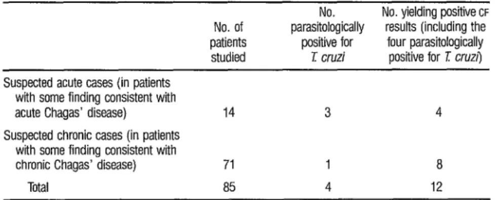
SULTS AND DISCUSSIONAs shown in Table 1, three (2 1% ) of the 14 Miahuatlgn patients presenting signs consistent with acute Chagas’ disease were found positive for I: mm’ through visual observation of the parasite in fresh blood and in smears; the parasite was also detected through inocu- lation into rats and into NNN culture me- dia. However, only one of the 7 1 patients with indications of chronic Chagas’ dis- ease ( 1.4 % ) yielded T. crmxi organisms. (This was to be expected, since it is recog- nized that the parasite is much harder to detect after the acute phase of the disease has passed.) In this case, the parasite was detected as groups of i’: cmzi amastigotes in histologic sections stained with hema- toxylin-eosin.
Table 1 also shows that one more acute case and seven more chronic cases-in addition to the four cases para- sitologically confirmed-yielded positive
TABLE 1. Twelve cases of ?I cruzi infection detected among the 85 Miahuatlan patients studied, ether by observation of the parasite or by procurement of a posftiie response to the complement fixation (CF)
test.
Suspected acute cases (in patients with some finding consistent with acute Chagas’ disease)
Suspected chronic cases (in patients with some finding consistent with chronic Chagas’ disease)
Total
No. of patients studied
14 71 85
No. No. yielding positive CF
parasitologically results (including the positive for four parasitologically
TABLE 2. Clinical, parasitologic, and serologic indications of chagasic in- fection among the 12 positive study subjects.
No. of study Clinical, parasitologic, and serologic findings
Patients positive by CR but without parasitologic confir- mation of infection
subjects No clinical evidence of Chagas’ disease
Clinical evidence of Chagas’ disease
Patients positive by CF with parasitologic confirmation of infection
1 7 Electrocardiographic changes observed, together
with an anatomic and clinical picture consis- tent with Chagas’ disease
Enteromegaly (megasigmoid) observed, together with an anatomic and clinical picture consis- tent with Chagas’ disease
Total cases
3 1 12 CF test results. Table 2 presents summary
data on the 12 cases involved.
As indicated in Table 3, CF
tests for lY cmzi antibodies in 5 2 children 6 to 14 years old from the Miahuatlan area were positive in 46 (88%) of the cases. In contrast, all 52 sera from Mexico City children in the same age group were negative. The high percentage of posi- tive reactions to the 1: crzzi antigen among the Miahuatlan children suggests high risk and a high frequency of expo- sure to the parasite in the area.
Table 4 shows the CF results obtained with blood samples from do- nors in Miahuatlan and Mexico City. Again, a high proportion (78%) of the MiahuatlSn sera tested positive, while all of the Mexico City sera were negative. These results give further grounds for considering the Miahuatlan region of Oaxaca as an area quite possibly endemic for Chagas’ disease.
Of the four cases rated acute, three were confirmed by finding the par- asite. The three patients involved were two boys 9 and 11 years of age (Photos 1 and 2) and a girl 14 years old, all with
TABLE 3. CF test results obtained with sera tom 52 San
Felipe Yegachi schoolchildren 6-14 years old and 52 Mexico City children in the same age group.
Sera positive No. of by CF
sera No. % Miahuatlan children 52 46 88% Mexico City children 52 0 0%
TABLE 4. CF test results obtained with sera from 14 adult blood donors in Miahuatlin and 14 others in Mexico CQ. Sera positive No. of by CF
PHOTOS 1 AND 2. Two of the Miahuatf4n study subpcts with acute Chagas’ disease, both showing Ro- maiia’s sign.
Romalia’s sign. These subjects had posi- tive CF titers of 1:8, 1:32, and 1:64, re- spectively; electrocardiographic changes in- dicating an incomplete block of the right branch of the bundle of His; and cultures positive for T. crzlzi in NNN media.
TABLE 5. Triatomine bugs collected in and around Miahuatlan residences.
No. collected Alive Dead
No. of live bugs positive
for r cruzi
All eight chronic cases yielded positive CF test results with titers ranging
from 1:32 to 1:128; of these eight, six had clinicial pictures and/or ECGs con- ” sistent with chagasic cardiopathy; four were asymptomatic; and two had left and right cardiac insufficiency with pre- dominance of the latter (Photos 3 and 4).
Adults:
Males 23 11 21 Females 18 9 17 Nymphs:
Males 8 4 6 Females 8 6 9 Total 57 30 53
One of the chronic patients had the first case of Chagas’ disease in- volving a megasigmoid (Photo 5) that had ever been reported in Mexico. This patient, a male campesino 51 years old with a history of triatomine bites, came to the hospital with a picture of mild in- testinal obstruction. Upon surgery, a large volvulus of the sigmoid was found, resected, and examined histopathologi- tally. Aggregations of 1: G~ZLZZ’ amasti- gotes were found in histologic sections. The patient’s serum tested positive for ZY crzlzt’ by CF at a titer of 1: 64.
species (never before reported in Oaxaca State) so far south and so far away from the places in which it had been reported previously, appears to greatly extend the area of its known distribution.
S
UMMARY2
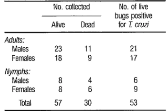
3As a supplement to this study, 87 triatomine bugs were collected in Miahuatlan and tested for T. crmzz’ (Table 5). Thirty of these insects were dead and 57 were alive. Of the live insects, 53
(92 %) were positive for T c?z.zti none of the dead insects tested positive. All the dead specimens and most (50) of the 57 live specimens were of the Triatoma bar- beri species and were captured by hand in human dwellings, which attests to their domesticity. The other seven living specimens, belonging to the species Tria- toma genstaeckeri, were collected around
homes, and five of these tested positive for i? CWZ~. The finding of this latter
A survey was made of resi- dents and hospital patients in Miahuat- Ian, Oaxaca, Mexico, for cases of Chagas’ disease. This survey focused primarily upon 85 patients attending the local hos- pital who had clinical pictures suggesting the disease, 14 with possible acute cases and 71 with possible chronic cases. In ad- dition, sera from 14 Miahuatlan blood donors were examined; seroepidemiolo- gic testing was performed with 52 sera from schoolchildren at another commu- nity nearby; and triatomine bugs were collected from Miahuatlan residences.
Each of the 85 patients pro- vided a complete clinical history and blood specimen, and each was given an ECG and a series of heart X-rays or chest teleradiography. In some cases biopsy or necropsy specimens were obtained. A va- riety of parasitologic and serologic tests performed with these specimens indi-
PHOTOS 3 AND 4. Two of the Miahuatfhn study subjjs with chronic Chagas’ disease showing cardiomegaty (grade III at left, grade IV at right).
PHOTO 5. Megacolon surgically removed from a MiahuatUn study subject with chronic Chagas’ disease.
subjects with possible acute cases and eight of the 71 subjects with possible chronic cases. One of these latter subjects had the first case of Chagas’ disease in- volving a megasigmoid that had ever been reported in Mexico.
A high proportion (78%) of the sera from Miahuatlsn blood donors tested positive for Typanosoma mm’ an- tibodies by complement fEation, as did 88% of the sera from the 52 schoolchil- dren. A total of 57 triatomid vector in- sects were captured alive and tested for 1: cTZCZZ’. Most (92%) were positive for the parasite; and while most (50 of the 57) were specimens of Triatoma barberi, the capture of seven Trt;ztoomagerstaederiin- sects appears to greatly extend the known range of the latter. Overall, the survey results (especially those involving the schoolchildren) demonstrate a high risk of exposure to T crzlzi in this area and give grounds for considering the Mia- huatlan region of Oaxaca endemic for Chagas’ disease.
Rx
FERENCES1 Mazzotti, L. DOS cases de enfermedad de Cha- gas en el estado de Qaxaca: Nota preliminar. Gac Mh’Mex 70(4):417-420, 1940.
2 Tay, J., P. M. Salazar-Schettino, M. Bucio, R. Zarate, and L. Zarate. La enfermedad de Cha- gas en la Republica Mexicana. S&d Pz%ica Mexicana 22(4):409-450, 1980.
Biagi, F., E Gutm%r&arcia, F. Navarrete, J. %y, J. Portilla, and S. Olivares. Enfermedad de Chagas en Tutuap&n, estado de Mexico. Prensa MZdica Mexicana 22(11-12):463-465, 1958.
Tay, J., E Navarrete C., E. R. Corominas. and E Biagi. Enfermedad de Chagas en el munici- pio de Thxpan, estado de Michoacan, Mexico. Rev&a de la Facultaa’ de Medic&a (Mexico)
8:263-270, 1966.
Zavala V., J.. D. Arjona C., and R. Quintal A. Enfermedad de Chagas: Informe de un case clinico. Rev Invest Chn 251367-371, 1973. 6 Ortega, M., F. Belt&-Hernandez, and
J.J. Zavala. Enfermedad de Chagas en Chiapas: Estudios clinicos epidemioldgicos. SahdPzibhca MexiGana 28(5):837-843, 1976.
7
8
9
10
11
12
13
Tay, J., P M. Salatar-Schettino, M. Velasco C., I. de Haro A., Y. Garcia-Yaiiez, and M. Gu- tierrez. Estudio epidemiol6gico de la enferme- dad de Chagas en el estado de Jalisco, Re- ptiblica Mexicana. Sal’zld Pzl’bha Mexzkana 21:145-149, 1979.
Biagi, E, and G. Arce. Los dos primeros cases de miocarditis chaebica comorobados en MC- xic~.Arch Inst C&iol Me; 35(5):611-623, Salatar-Schettino, P M., J. Castrejdn, H. Rodriguez, and J. Tay. Tercer case comprobado de miocarditis chagQica cr6nica en Mexico. Prensa M&&a Mexicana 64( 5-6): 115- 120, 1979.
Goldsmith, R. S., I. G. Kagan, M. A. Reyes- Gonzalez, and J. Cedefio-Ferreira. Estudios se- roepidemiologicos reahzados en Oaxaca, Mi- xico: I. Encuesta de anticuemos parasitarios mediante la prueba de hemaglutmaci6n in- directa. Bd Of Sanit Panam 71:500-5 18. _I
1971.
Marcuschamer, M. J., and L. P. Reyes. En- fermedad de Chagas en Mexico: Informe de 5 cases comprobados. Arch Inst Car&o/ Mex
48:952, 1978.
lay, J., 0. Goycoolea, and F. Biagi. Observa- ciones sobre enfermedad de Chagas en la Mix- teca Baja: Nuevo case human0 en la Repdblica Mexicana. BolOfSanitPanam 51(4):322-327, 1961.