CLINICAL
AND EPIDEMIOLOGIC
STUDIES OF CHAGAS’ DISEASE
IN RURAL COMMUNITIES
IN OAXACA STATE, MEXICO,
AND A
SEVEN-YEAR
FOLLOW-UP:
I. CERRO DEL AIRE’. 2
R. S. Goldsmith,3 R. J. Zzirate,4 L. G. ZBrate,4 I. Kagan,’ and L. B. Jacobson6
In 1971, serologic studies in the Mexican state of Oaxaca revealed unusually high chagasic infection in certain communities. This article reports the results of 1973 and 1980 follow-up work performed in one of those communities, Cerro de1 Aire, which shows that the infection caused significant electrocardiographic abnormalities in seropositive persons (as compared to seronegative persons), without there having
been evident progression of those abnormalities as of the end of the follow-up period in 1980.
Introduction
The frequency and severity of Chagas’ disease in the Americas varies by geographic region (1). In Mexico before 1970 the infection was not thought to occur commonly or to cause signifi- cant cardiac pathology. Prevalence rates reported from several communities ranged from 7% to 13% (2-5). However, seroepidemiologic sur- veys conducted in 1971 in 60 rural communities on the Pacific side of Mexico’s Oaxaca State showed unusually high rates of Ttypanosoma cruzi infection (6-9). In particular, antibody prev- alence rates for adults living in the three com- munities of Cerro de1 Aire, Chila, andTataltepec
were 76%, 58%, and 51%, respectively. These rates were similar to some of the highest rates reported in South America.
Although many community-based cross-sec- tional studies of Chagas’ disease have been re- ported (IO-21), relatively few community-based longitudinal studies (22-28) have been de- scribed. Such longitudinal studies are needed to provide information about the natural history of the disease-which is not fully understood (22, 29).
This and companion reports present recent findings of both cross-sectional and longitudinal studies in several Oaxaca communities. These studies sought (1) to determine the pathologic
‘This article will also be published in Spanish in the Boleti’n de la Ojkina Sanitaria Panamericana.
*This study is part of a collaborative research program for the study of Chagas’ disease undertaken by the University of California, San Francisco, California, U.S.A.; the “Beni- to Juarez” University of Oaxaca (Universidad “Ben&o Judrez” de Oaxaca), Oaxaca, Mexico, in 1971-1973; and the Ecological Research Center of the Southeast (Centro de Invesfigaciones Ecolbgicas de1 Sureste-ClES), San Cris- tdbal de las Casas, Chiapas, Mexico, in 1980. The research was supported in part by the three institutions and by the following grants: Grants numbered Al-10051 and HD- 06922 from the National Institutes of Health, U.S. Public Health Service, in 1971-1973; and grants in 1980 from the National Council of Science and Technology of Mexico
(Consejo National de Ciencia y Tecnologia), from the Na- tional Science Foundation of the United States, and from the Special Program on Research and Training in Tropical Dis- eases, World Health Organization, Geneva, Switzerland.
3Professor of Tropical Medicine, Department of Epide- miology and International Health, University of California, San Francisco, California 94 143, United States.
4Researcher, Ecological Research Center of the Southeast (Centro de Investigaciones Ecoldgicas de1 Sureste), San Crist6bal de las Casas, Chiapas, Mexico.
‘Director, Parasitology Division, Centers for Disease Control, Atlanta, Georgia, United States.
6Attending Cardiologist, Department of Medicine, Pacific Medical Center, San Francisco; and Associate Clinical Pro- fessor, Department of Medicine, University of Cali- fornia, San Francisco.
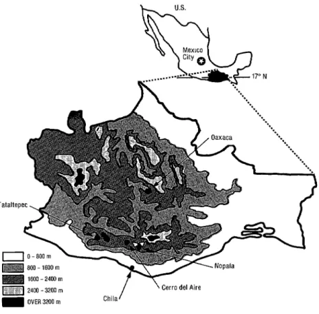
Figure 1. A map of Mexico’s Oaxaca State showing the location of Cerro de1 Aire.
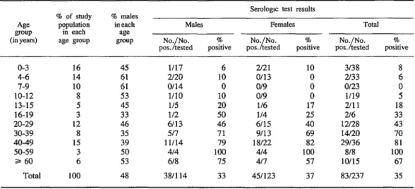
Table 1. Age and sex distribution of 248 subjects tested in Cerro de1 Aire in 1971 and age-specific indirect hemagglutination (MA) test results. A titer >128 was considered positive. This study population
included 89% of Cerro del Aire’s total 1971 population of 280 residents.
Age gmv (in years)
% of study % males population in each
in each a?3 age W”P group
IHA test results
Males Females Total
No. No.
/ % No No.
pos. tested positive pas. tested ;/ positive % p,“,n%,“,kd po%ve
o-3 2
4-6 10
7-9 9
10-12 10 13-15 8 16-19 10 20-29 16 30-39 16 40-49 10 50-59 5 2 60 4 Total 100
83 68 68 54 53 38 41 40 50 46 20 49
015 0
1117 6 o/15 0 2113 1.5 400 40 8110 80 7/l 6 44 14116 88 12/12 100 516 83 212 100 551122 45
O/l 0 O/6 0
018 0 1125 4
o/7 0 O/22 0
2/l 1 18 4124 17
319 33 7119 37
122 PAHO BULLETIN l Vol. 19, no. 2, 1985
impact of T. cruzi infection in humans, (2) to investigate the natural history of the disease, and (3) to confirm that serologically positive persons were parasitologically positive. This report sum- marizes the clinical, electrocardiographic, sero- logic, and parasitologic findings of cross-sec- tional and longitudinal studies conducted in the community of Cerro de1 Aire. The results of similar studies in three other communities--Chila, Tataltepec, and Nopala-will be reported later. Additional aims of the Cerro de1 Aire work were (1) to compare the sensitivity of xenodiag- nosis and two hemoculture methods-one using LIT medium (30)7 and the other employing MK2 tissue culture cells-for isolation of T. cruzi from serologically positive persons, and (2) to monitor resumption of Chagas’ disease transmis- sion that could be occurring as a result of a reduc- tion in the frequency of residual insecticide spraying by the malaria control program in and near Cerro de1 Aire.
In the 1971 survey (6, 7), only 1.4% of 1,289 children under 10 years of age tested in the 60 communities were seropositive. This low level of infection in young children, reflecting near- cessation of transmission of infection to humans in the region, was associated with concurrent disappearance of the vector and was attributed to the malaria control program that began in 1962. From 1976 onward, however, spraying was discontinued in some communities, and the frequency of spraying was reduced in others. Thus, one objective of the Cerro de1 Aire study was to monitor possible seroconversion in chil- dren and adults for an indication that the vectors were returning and transmission had resumed.
Methods
The Study Area
Oaxaca, one of Mexico’s southernmost states, is at about latitude 17” North and longitude 96O West (Figure 1). The community of Cerro de1
‘LIT medium= liquid liver-infusion-tryptose medium.
Aire (also known as La Canada de Atotonilco) is located on the Pacific side of the state at an alti- tude of about 800 meters. The annual rainfall is about 525 mm, with most of the precipitation occurring between May and October. The forest cover is deciduous.
Until 1980, Cerro de1 Aire was isolated and was about 10 kilometers from the nearest unim- proved road. Except for a few men, most of the inhabitants had not lived or traveled outside the local area. During 1971-1973, some 280 persons lived in the town; our 1971 sample of 248 per- sons (Table 1) represented 89% of the population (6, 7). By 1980 the population had grown to 1,200, with most of the new residents having come from the nearby community of Teotepec located about eight kilometers to the north and 200 m higher. Houses in Cerro de1 Aire usually have dirt floors, walls made of wood slats and adobe, and ceilings made of palm fronds or straw.
Field Trips to Cerro de1 Aire
In 1971, during the first field trip to Cerro de1 Aire, 248 persons were included in the serologic survey. The results of this survey, summarized in Table 1, have been reported previously (6-9).
In May 1973, during a second trip, the cardiac status of 130 persons seen in 197 1 was evaluated by clinical and electrocardiographic examina- tions, and new blood specimens were tested by indirect hemagglutination (IHA) serology.
Goldsmith et al. l CHAGAS’ DISEASE IN OAXACA 123
Table 2. Age and sex distribution of 237 subjects tested in Cerro de1 Aire in 1980 and age-specific serologic teat results. All 237 subjects were tested by indirect hemagglutination (MA) and direct agglutination (DA), and X6 subjects were tested by complement fmation (CF). A subject responding positively to any one test (with an IHA titer ~128, a DA titer ~256, or a CF titer 28) was considered seropositive. This study population included 20%
of Cerro de1 Aire’s total 1980 population of 1,200 residents. % males
in each w group
Serologic test results
M&S Females Total No No.
;/ % p~~~~~d poske p!%%d ”
pas. tested positive posmve
o-3 16 4-6 14 7-9 10 10-12 8 13-15 5 16-19 3 20-29 12 30-39 8 40-49 15 50-59 3 a 60 6 Total 100
45 61 61 53 45 33 46 35 39 50 53 48
l/l7 6 z21 10 3138 8
u20 10 o/13 0 2i33 6
0114 0 o/9 0 O/23 0
l/10 10 o/9 0 l/19 5
l/5 20 116 17 2/l 1 18
l/2 50 l/4 25 216 33
6113 46 6115 40 12128 43 511 71 9/13 69 14120 70 1 l/14 79 18122 82 29136 81 414 100 414 100 8/X 100
618 75 411 57 1005 67
38/l 14 33 451123 37 831237 35
tested in 1973, while 46 were tested for the first T. cruzi Isolation by Xenodiagnosis
time. and Hemoculture
Overall, during all of the cross-sectional studies, ECGs were obtained from 176 subjects, medical histories from 130 (52 in 1973 and 78 in 1980), and physical examinations from 101 (40 in 1973 and 61 in 1980).
Xenodiagnosis . Forty fourth-instar nymphs of the triatomid vector Rhodniusprolixus were used per patient. The bugs had not been fed for four weeks. Patient exposure time was 30 minutes. After four to seven weeks, the bugs fed on the patient were individually examined for T. cruzi infection by thoroughly and entirely triturating each bug in 1 ml of normal saline. The parasites that were isolated were identified as T. cruzi by their morphology and their infectivity for labora- tory mice.
Blood Collection, Storage, and Testing Blood specimens were collected in heparinized venipuncture tubes in 1971 and in heparinized capillary tubes thereafter. Specimens were cen- trifuged at about 700 RPM for 10 minutes and processed aseptically; the resulting plasma was stored on “wet” ice (H,O) in the field and at -2O’C in the laboratory. The T. cruzi antigen used was a saline extract of lyophilized epimas- tigotes, the latter having been delipidized with benzene before extraction (4, 5). The serologic methods employed in this study for the IHA, CF, and DA tests have been described elsewhere (4, 5, 31). Positive titers in these tests were 2 128 for IHA, 2256 for DA, and 38 for CF.
Hemocultures. The first hemoculture method employed closed test-tube tissue cultures of MKZ cells. The source of the MI& cell line, the com- position of the maintenance medium, and the mode of inoculation have been described pre- viously (6, 7). Two tissue-culture tubes were each inoculated with 2 ml of plasma.
124 PAHO BULLETIN l vol. 19, no. 2, 1985
cells recovered after centrifuging 10 ml of hepa- rinized blood at 2,000 rpm for 15 minutes; before use, the packed cells were washed once with LIT medium and recentrifuged.
Both sets of cultures were kept at ambient field temperatures for one week. Thereafter, in the laboratory, the LIT medium cultures were kept at 24’C and the MK2 cell cultures were kept at 37Y. The LIT medium cultures were examined over a fifteen-week period for the presence of flagellates by. fresh-film microscopy. The MK* tissue-cultures were examined at weekly inter- vals for approximately one month.
Electrocardiograms
ECGs were recorded with either Cambridge or Burdick portable ECG machines powered by a portable generator, The ECGs contained the six standard limb leads and 3-6 standard precor- dial leads. A cardiologist (LBJ) recorded the rhythm, rate, PQRSTU contours, PR interval, and the QRS axis, duration, and configuration of each ECG in accordance with standard criteria but without knowledge of the patient’s serologic status.
Results
Serologic Tests, 1971
In 1971, 248 blood specimens were collected and tested by IHA for antibody to T. cruzi (6, 7). The results of these tests, by age and sex of the subjects, are shown in Table 1. Only one of the 53 children tested who were under the age of 10 years was seropositive. For each of the age groups between 10 and 49 years the percent- age of seropositives increased progressively- from 17% to 100%. A total of 54% of the females and 45% of the males were seropositive, but this difference between the sexes was not statistical- ly significant. Overall, 50% of the 248 persons tested were seropositive, but a much higher pro-
portion (76%) of the 126 subjects 20 years of age or older were seropositive. These overall 1971 prevalences are very likely to have been within two percentage points of the town’s true prevalences because the sample tested included 89% of the population of Cerro de1 Aire.
Serologic Tests, 1980
In 1980, blood specimens were collected from 237 residents of Cerro de1 Aire (Table 2) and tested by IHA and DA; 86 specimens were also tested by CF. As indicated in the table, a specimen found positive by one or more of the tests was considered seropositive.
Of 182 persons tested for the first time in 1980, 42 (23%) were found seropositive by one or more of the three tests. Of the 59 persons 20 years of age or older, 34 (58%) were found to be seropositive. No significant differences between results for males and females were observed when the data were analyzed by sex.
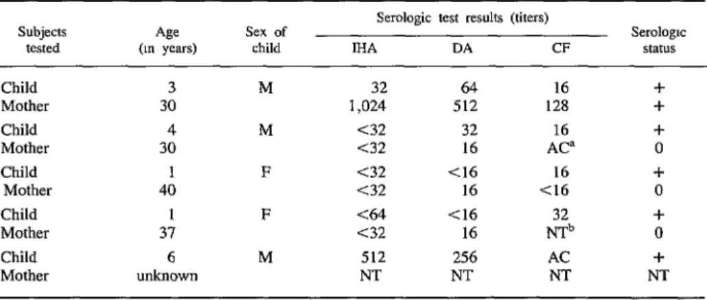
Of 38 children under three years of age, three (8%) were found to be seropositive; and of 33 children four to six years old, two (6%) were found to be seropositive; all of the children tested were born in Cerro de1 Aire. The test results for the five serologically positive children under age seven and the serologic status of their mothers are presented in Table 3. One of the mothers was seropositive and three were seronegative; no blood sample was available from the fifth mother.
Goldsmith et al. a CHAGAS’ DISEASE IN OA~ACA 125
Table 3. Serologic test results for the five 1980 study subjects under seven years old who were found to be seropositive, and corresponding serologic test results for their mothers. Subjects
tested (In years) Age Sex of child
Serologic test results (titers)
Serologic
IHA DA CF states
Child Mother Child Mother Child
Mother Child Mother Child Mother
3 M 32 64 16 +
30 1,024 512 128 +
4 M <32 32 16 +
30 <32 16 AC= 0
1 F <32 <I6 16 +
40 <32 16 <16 0
1 F <64 <16 +
37 ~32 16 $b 0
6 M 512 256 AC +
unkuown NT NT NT NT
“AC = anticomplementary. bNT = not tested.
Comparative Sensitivity of the IHA, DA, and CF Tests
Eighty-three of the 237 blood specimens col- lected in 1980 were positive by one or more of the three tests; 13 (5%) were positive only by the IHA test and 10 (4%) only by the CF test. None were positive by the DA test alone.
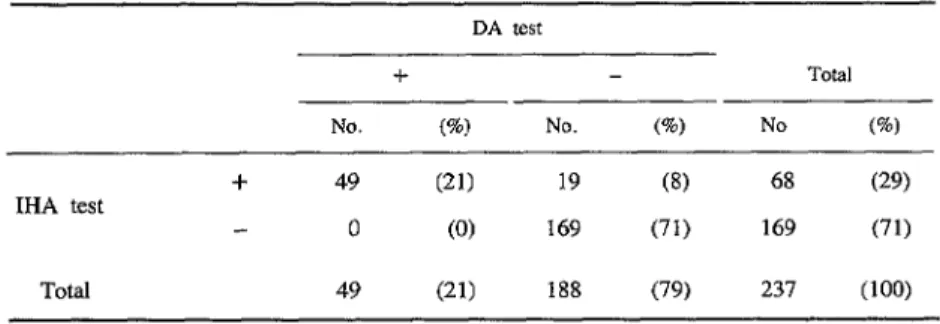
Of the 237 specimens examined by II-IA and DA tests (Table 4), 29% were positive by the MA test and 2 1% were positive by the DA test. Overall, 218 of the specimens were either posi- tive by both tests or negative by both tests, re- sulting in 92% agreement between the two methods.
Of the 86 sera examined by IHA and CF tests (Table 5), 43% were positive by the MA test and 45% were positive by the CF test. Overall, 62 of the specimens were either positive by both tests or negative by both tests, resulting in 72% agreement between the two methods.
Of the 86 sera examined by CF and DA tests (Table 6), 3 1% were positive by the DA test and 44% were positive by the CF test. Overall, 63 of the specimens were either positive by both tests or negative by both tests, resulting in 73% agreement between the two methods.
Cross-sectional Clinical and Electrocardiographic Studies
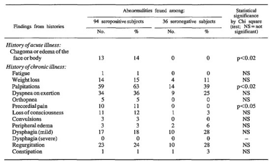
Comparison of clinicalfindings for seroposi- tive and seronegative subjects. Histories of symp- toms were provided by 130 subjects, 94 of whom were seropositive and 36 of whom were sero- negative (Table 7). Differences in the reported frequency of certain symptoms were statistically significant in the following instances: (1) chagoma or edema of the face or body was recalled by 13 (13.8%) of the seropositive subjects but by none of the seronegative subjects (pcO.02); (2) palpita- tions were reported by 59 (62.8%) of the seroposi- tive subjects but only by 14 (38.8%) of the sero- negative subjects (pcO.02); and (3) precordial pain was reported by 10 (10.6%) of the seroposi- tive subjects but by none of the seronegative sub- jects (lkO.05).
126 PAHO BULLETIN l vol. 19, no. 2, 198.5
Table 4. Comparison of indirect hemagglutination (MA) and direct agglutination (DA) test results obtained with the 237 Cerro del Aire sera collected in 1980. IHA titers 2128
and DA titers 2256 were considered positive. DA test
+ Total
No. (%I No. (%) No (%I IHA test + 49 (21) 19 (8) 68 (29)
0 (0) 169 (71) 169 (71) Total 49 (21) 188 (79 237 (100)
Table 5. Comparison of indirect hemagglutination (IHA) and complement-fixation (CF) test results obtained with 86 Cerro de1 Aire sera collected in 1980. IHA titers 2128 and
CF titers 28 were considered positive. CF test
+ Total
No. em No. @) No. 6) IHA test + 26 (30) 11 (13) 37 (43)
13 (15) 36 (42) 49 (57)
Total 39 (45) 47 (55) 86 (100)
Table 6. Comparison of complement-fixation (CF) and direct agglutination (DA) test results obtained with 86 Cerro del Aire sera collected in 1980. DA titers a256 and CF titers ~8
were considered positive. CF test
f Total
No. (%) No. (%) No. 6) DA test -I- 21 (24 6 (7) 27 (31)
17 (20) 42 (49) 59 (6%
Goldsmith et al. . CHAGAS’ DISEASE IN OAXACA 127
Table 7. A comparison of findings derived from medical histories of seropositive and seronegative subjects.
Findings from histories
Abnormalities found among:
94 seropositive subjects 36 seronegative subjects
NO. % NO. %
Statistical significance by Chi square (test; NS = not significant) History of acute illness:
Chagoma or edema of the face or body
History of chronic illness: Fatigue
Weight loss Palpitations Dyspnea on exertion Orthopnea Precordial pain Loss of consciousness Convulsions Peripheral edema Dysphagia (mild) Dysphagia (severe) Regurgitation Constipation
13
1 1 0
14 15 4
59 63 14
34 36 9
5 5 0
10 11 0
11 12 1
3 3 0
3 3 2
17 18 10
0 0 0
23 24 10
1 1 1
14 0 0
0 11 39 25 0 0 3 0 6 28 0 28 3
pco.02 NS NS pco.02
NS NS pco.05
NS NS NS NS NS NS
Table 8. A comparison of clinical findings derived from physical examination of seropositive and seronegative subjects.
Clinical findings
Abnormalities found among:
74 seropositive subjects 27 seronegative subjects
NO. % NO. %
Statistical significance (by Chi square (test; NS = not
significant)
Arrhythmia 1 1 1 4 NS
Cardiac murmurs 29 39 11 41 NS
Gallops 9 12 3 11 NS
Cardiomegaly 7 9 0 0 NS
Cardiac insufficiency 1 1 0 0 NS
Large bowel fecal mass 0 0 0 0 -
Comparison of electrocardiographicfindings for seropositive and seronegative subjects.
ECGs were obtained from 176 subjects, of whom 93 were tested only in 1973, 37 were tested in both 1973 and 1980, and 46 were tested only in 1980 (Table 9). Of the 176,111 were seropos- itive by one or more of the three serologic tests and 65 were seronegative by all three. The age
distribution of these subjects, grouped according to their serologic status, is shown in Table 10.
128 PAHO BULLETIN l vol. 19, no. 2, 1985
Table 9. Electrocardiographic abnormalities found among 111 seropositive and 65 seronegative subjects. These findings were derived from ECGs recorded in 1973 (in 93 subjects),
in 1980 (in 46 subjects), and in both years (in 37 subjects).
Specific electrocardiographic abnormality
Abnormalities found among:
111 seropositive subjects 65 seronegative subjects
NO. % NO. %
Statistical significance
(by Chi square test; NS = not
significant) Intraventricularconduction delays (IVCDs) 18 16
Right bundle branch block (total) 13 I2
RBBB alone 8 7
RBBB with lefl anterior fascicular block 4 4 RBBB with superior, rightward axis 1 I Left anteriorfascicular blockalone 3 3 Left bundle branch block I 1 r.Sr,‘inV,, eRS=O.lOsec 1 1 lo atrioventricularblock 2 2
Right axis deviation 1 1
R>S in lead V, 5 5
Left atria1 abnormality 0 0 Myocardial infarction, probable 0 0 Myocardial infarction, possible 3 3 Primary ST-T-U abnormalities” 20 18 Prematureventricularcontractions 7 6 Premature atria1 contractions 7 6 Short PR interval (<O.l2sec.) 3 3 Left ventricular hypertrophy 1 1 Subjects with one or more abnormalities 51 46
3 5 pco.05
I 2 p-Co.02
I 2 NS
0 0 NS
0 0 NS
2 3 NS
0 0 NS
0 0 NS
0 0 NS
1 2 NS
1 2 NS
1 2 NS
1 2 NS
0 0 NS
6 9 NS
0 0 pco.05
3 5 NS
1 2 NS
0 0 NS
14 22 p<o.o05 ‘ST-T-U abnormalities occurring in the presence of normal QRS complexes.
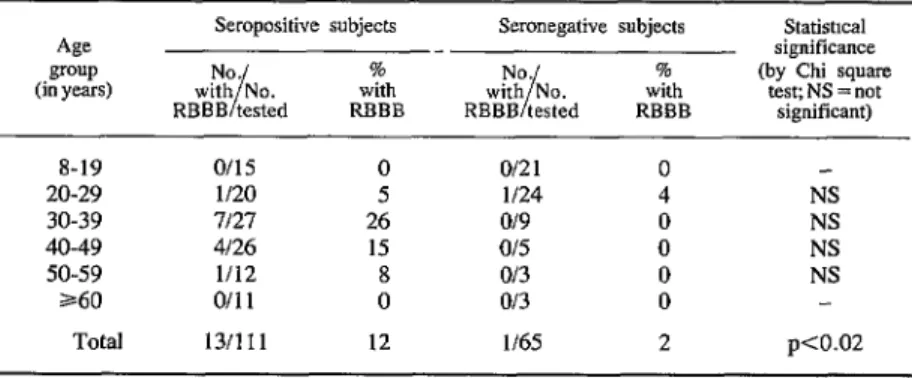
Table 10. Right bundle branch block by age group and serologic status for 176 persons on whom ECGs were conducted in 1973 and/or 1980. If ECGs were conducted
in both years, the subject’s age group was determined according to his or her age at the time of the 1973 test.
Age group (in years)
Seropositive subjects
NO. %
with No.
I with RBBB tested RBBB
Seronegative subjects
NO. %
with No.
I with RBBB tested RBBB
Statistical significance (by Chi square
test; NS = not significant)
8-19 o/15 0
20-29 1120 5 30-39 7127 26 40-49 4126 15 50-59 l/12 8
360 o/11 0
Total 13/111 12
o/21 0 l/24 4
o/9 0
o/5 0
o/3 0
o/3 0
l/65 2
Goldsmith et al.
lCHAGAS DISEASE IN OAXACA
129
130 PAHO BULLETIN l vol. 19, no. 2, I985
(16%) of the seropositives as compared to three (5%) of the seronegatives (pcO.05). Right bun- dle branch block (RBBB) alone or with other findings occurred in 13 (12%) of the seroposi- tives as compared to one (2%) of the seronega- tives (p<O.O2). In the latter seronegative sub- ject, RBBB occurred without other IVCDs. Among those with IVCDs, RBBB accounted for
13 of 18 IVCD abnormalities in the seropositives and for one of three IVCD abnormalities in the seronegatives. Premature ventricular contractions (PVCs) occurred in seven (6%) of the seroposi- tives as compared to none of the seronegatives (p<O.O5). All of the subjects with PVCs were found to have at least one other ECG abnormal- ity.
Other ECG abnormalities were found to occur at different frequencies in the seropositive and seronegative subjects, but these differences were not statistically significant. RBBB alone occurred in eight (7%) of the seropositive and one (2%) of the seronegative subjects. RBBB with left an- terior fascicular block (LAFB) occurred in four (4%) of the seropositives and none of the sero- negatives. R>S in lead V, occurred in five (5%) of the seropositives and in one (2%) of the sero- negatives. Primary ST-T-U abnormalities occur- red in 20 (18%) of the seropositives and in six (9%) of the seronegatives.
Findings for subjects with RBBB. Of the 13 seropositive subjects with RBBB, one was be- tween 20 and 29 years of age, seven were be- tween 30 and 39, four were between 40 and 49, and one was between 50 and 59 (see Table IO). Seven of the subjects were men and six women. The 13 were all positive by IHA, with IHA titers ranging from 128 to 4,096. Of the 11 tested by DA, all were positive with DA titers ranging from 256 to 4,096. Finally, of eight tested by CF, five were positive with titers ranging from 16 to 128. Parasitologic isolation from eight of these subjects was attempted by three methods, but was successful in only one case (by xenodiagnosis); these eight subjects were part of the group of 33 from whom parasite isolation was attempted.
The one seronegative person with RBBB was
a man 27 years of age who had a negative history for Chagas’ disease; physical examination of this subject revealed no abnormalities.
Longitudinal Study: Progression of Electrocardiographic Abnormalities
Comparison of the ECGs obtained from the same 37 subjects in 1973 and 1980 revealed no significant differences between the earlier and later ECGs. Rather, there were merely minor nonspecific changes in T wave contours and in the appearance or disappearance of atria1 and ventricular premature beats.
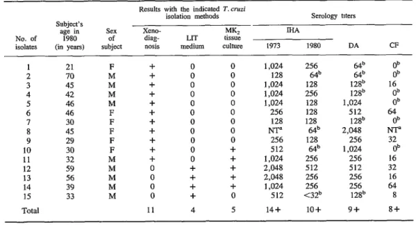
Isolation of T. cruzi
Isolation of T. cruzi was attempted by three methods. This was done in order to confirm that serologically positive subjects were parasitolog- ically positive and to compare the sensitivity of the isolation methods used. Thirty-three persons who were seropositive in 1971 and 1973 were seen again in 1980 and tested for T. cruzi infec- tion by xenodiagnosis, inoculation of LIT medium with plasma, and inoculation of MI& tissue cultures with serum. T. cruzi was isolated from 15 (45%) of the 33 persons tested (Table 11). Nine of the 15 isolates were obtained by xenodiagnosis alone, two were obtained by xenodiagnosis and the tissue-culture method, three were obtained by both culture systems, and one was obtained by LIT medium alone. Overall, 11 (73%) of the 15 isolates were ob- tained by xenodiagnosis, five (33%) by the MK2 tissue-culture system, and four (27%) by the LIT medium.
Goldsmith et al. l CHAGAS' DISEASE IN OAXACA 131
Table 11. Methods used to isolate T. cruzi from 15 of 33 seropositive subjects, showing the age and sex of each subject yielding an isolate, the results obtained with each isolation method,
and the subject’s serologic status.
No. of isolates
Subject’s age in
1980 (in years)
SC% of subject
Results with the indicated T.cruzi isolation methods
xeno- Mb
diag- LIT tissue nosis medium culture
Serology nters MA
1973 1980 DA CF 1 21
2 70 3 45 4 42 5 46 6 46 7 30 8 45 9 29 10 30 11 32 12 59 13 56 14 39 15 33 Total F M M M M F F F F F M M M M M
+ 0 0 1,024 256
+ 0 0 128 64b
+ 0 0 1,024 128
+ 0 0 1,024 256
+ 0 0 1,024 128
-I- 0 0 256 128
+ 0 0 128 128
-I- 0 0 m 64b
+ 0 0 256 128
+ 0 + 512 64’
+ 0 + 1,024 256
0 + + 2,048 512
0 + + 2,048 256
0 + + 1,024 256
0 + 0 512 <32b
11 4 5 14+ 10-k
128b 128b 1,024 512 12P 2,048 256 1,024 256 512 256 256 128b Ob Ob 16 ;Fi 64 Ob NT” 32 Ob 16 32 16 64 8 9+ 8+ “NT = not tested.
bResults negative.
Discussion and Conclusions
Prevalence of T. cruzi
Between 1962 and 1971 the transmission of T. cruzi to humans nearly ceased in Cerro de1 Aire, as evidenced by the very few seropositive reactors found among children less than 10 years old in the 1971 survey (6, 7). However, before 1962 and the fortuitous onset of transmission control, chagasic infection was holoendemic (6, 7). Indeed, age-specific infection rates in the 1971 survey indicated that, in the absence of control, more than 50% of the population became
infected by their early teens, and also that with advancing age and continuing exposure to in- fected triatomines, most of the remaining unin- fected persons eventually became infected.
The near-absence of antibody in young chil- dren correlates with both the commencement of
residual insecticide spraying by a malaria control program and the disappearance of the triatomine vector or vectors from Cerro de1 Aire and other communities in the Pacific Coast study region of Oaxaca (6, 7). Review of malaria campaign records through 1980 showed that DDT had been used almost exclusively in spraying in the region since 1962 (34).
This is especially interesting because DDT is not generally considered an effective agent for control of triatomines (35). Initially, the insec- ticide was applied every four months, but begin- ning in 1976 the schedule was changed to twice yearly in Cerro de1 Aire and other communities, and in some areas spraying was discontinued entirely.
132 PAHO BULLETIN . vol. 19, no. 2, 1985
one specimen of Rhodnius prolixus in Cerro de1 Aire and one specimen of Triatoma dimidiata in the nearby town of Nopala. On showing vil- lagers bug specimens in 1973, they reported that the bugs had not been seen for some seven to 10 years. As previously discussed, we presume that the vectors present in the region before the onset of spraying were Triatoma phylossoma spp., T. dimidiata, and R. prolixus (6, 7). No systematic search for vectors was made in 1980.
In the 1980 serologic survey, 94 children under 10 years old who were born in Cerro de1 Aire were tested serologically for T. cruzi anti- bodies. Five of these children, ranging from one to six years of age, were seropositive. Four mothers of these five seropositive children were also tested; three were seronegative, indicating that the infections in their children were not congenitally transmitted but had been acquired postpartum as a result of vector transmission. Thus, during the period 197 1 - 1980 vector trarrs- mission of Chagas’ disease in Cerro de1 Aire appears to have continued at a low level.
Comparative Sensitivity of the Serologic Tests
The IHA and DA tests probably measure dif- ferent antibodies. The IHA detects IgG immuno- globulins more efficiently than IgM immuno- globulins, while the DA measures IgM more efficiently than IgG. For this reason, in a popu- lation with only chronic infections, such as that in Cerro del Aire, IHA tests should be and were more sensitive than DA tests (see Table 4). CF is the test of choice for detecting chronic Chagas’ disease; like IHA, CF efficiently measures IgG but not IgM. In this study, the CF test was also more sensitive than the DA test and was as sen- sitive as the IHA test (see Tables 5 and 6).
In determining the sensitivity and specificity of serologic tests, a cutoff level must be selected to differentiate between positive and negative titers. The cutoff levels used in this study were taken from routine diagnostic serology at the U.S. Centers for Disease Control (4, 5, 31). The fact that reinfection had not occurred in this
population for about 18 years (1962-1980) may have contributed to the antibody decay seen over this period for IHA titers (see Table 11). Decay of DA antibody presumably also occurred, as exemplified by the cases of six subjects listed in Table 11 who had negative DA titers (64 or
128) but after isolation tests were shown to be parasite-positive. The concordance between DA and IHA tests was 92%; the concordance between CF and IHA, and CF and DA tests were 72% and 73%, respectively. A higher concordance for the latter pairs might have been obtained had we arbitrarily lowered the cutoff level of the IHA and DA tests to compensate for IHA and DA antibody decay.
Clinical Manifestations of T. cruzi Infection in Cerro de1 Aire
The clinical manifestations of Chagas’ disease vary in different regions of its geographic distri- bution (1). In cases of chronic infection, the heart is the most commonly afflicted organ. Severe chronic myocarditis can progress to cardiomega- ly and heart failure. In another form of the infec- tion, the conduction system is affected; this can lead to a variety of arrhythmias that can result in sudden death before the onset of clinically appar- ent hemodynamic abnormalities. In this study of chronically infected subjects, the physi- cal examinations yielded findings with regard to cardiomegaly, congestive heart failure, arrhyth- mias, cardiac murmurs, and gallops that were not significantly different for seropositive and seronegative subjects. The subjects’ histories, however, indicated that palpitations and precor- dial pain were significantly more frequent among seropositive persons.
Goldsmith et al. l CHAGAS' DISEASE IN OAXACA 133
death of young adults, a feature of the disease in some areas of South America, does not appear to occur in Cerro de1 Aire. In some regions of South America, particularly central Brazil, infection may also result in motility disturbances of the esophagus and distal colon or in “mega” syn- dromes of these organs. However, the findings we obtained from histories and physical exami- nations in Cerro de1 Aire do not suggest that the infection causes pathologic changes in the intes- tinal tract.
Electrocardiographic Findings
Intraventricular conduction delays and arrhyth- mias are cardinal ECG features of chronic chagasic cardiomyopathy. Complete right bun- dle branch block (RBBB) is the most frequent finding; left anterior fascicular block is reported to coexist at about the same frequency. Other common ECG findings include atrioventricular (AV) block, premature ventricular contractions (PVCs), and ST-T-U abnormalities (II, 36-38).
In Cerro de1 Aire, ECG abnormalities statis- tically more frequent among seropositives than seronegatives were RBBB (with or without other abnormalities), IVCDs in total, and PVCs. For RBBB in particular, 12% of the seropositives but only 2% of the seronegatives had the finding; this difference is statistically significant (pcO.02).
Pinto-Dias (36) has commented that a region endemic for Chagas’ disease could be recog- nized electrocardiographically by one of two findings: (1) a prevalence of RBBB higher than 2% and (2) a marked disproportion between right and left bundle branch block. In his work, the ratio of RBBB to LBBB was 28:l. In our study, among seropositives, it was 13: 1.
In addition to being caused by Chagas’ dis- ease, RBBB can occur as an expression of atherosclerotic heart disease (generally in people over age 60), in people with hypertension, as a rare manifestation of rheumatic heart disease, and as an isolated finding in the absence of other cardiac abnormalities. In Cerro de1 Aire, the relatively young age of most people tested by
ECG and the near-absence in the population of hypertension or murmurs suggestive of rheumat- ic heart disease rules against these other potential causes as providing an explanation for the high frequency of RBBB.
Since 46% of the seropositive persons tested by ECG had one or more abnormalities, com- pared to 22% of the seronegatives (p<O.O05), it can be concluded that a relatively large propor- tion of the persons infected with T. cruzi in Cerro de1 Aire exhibited cardiac pathology. Nevertheless, limitations of the sampling methods do not permit extrapolation from these findings for the purpose of determining a preva- lence rate for chagasic cardiopathy.
Progression of the Disease Over a Seven-Year Period
134 PAHO BULLETIN l vol. 19, no. 2, 1985
seropositive subjects from the Limiri Valley (also in Chile) who were followed for four years. It is possible that the Cerro de1 Aire strain of T. cruzi is less pathogenic, and that as a result cardiac pathology proceeds at a much slower rate. A second hypothesis takes into considera- tion a difference between the Cerro de1 Aire study area and the areas of the other longitudinal studies. In Cerro de1 Aire, transmission of the infection was nearly nonexistent throughout the study period, whereas in the other studies trans- mission apparently continued. So, although T. cruzi infection persists in humans indefinitely, repeated reinfection (39-42) could possibly be an important factor in the mechanism responsi- ble for progression of cardiac disease. With re- gard to the time when cardiac damage occurs, our epidemiologic findings also support a view on pathogenesis that most of the damage occurs during the acute phase of the disease (43) and possibly also at times of new infections, but not as a result of persistent infection in the chronic stage.
We conclude from the cross-sectional and lon- gitudinal ECG studies and from the T. cruzi isolations from seropositive patients in Cerro de1 Aire that infection in this region of Oaxaca can induce a substantial degree of cardiac electrical abnormality. Whether the damage responsible for these abnormalities results in significant mor- bidity or in early death is not yet established. Our knowledge is also incomplete with regard to the frequency and severity of both acute and congenital T. cruzi infections.
Given the pathogenic potential of the infection in Cerro de1 Aire, the serendipitous but effective control of T. cruzi transmission through the malaria control program, and the relatively re- cent administrative decision to decrease insec- ticide spraying in the region, it is essential that sylvatic searching for the vector be continued, and also that monitoring continue for repopula- tion of houses by the bugs. In addition, serologic surveillance, particularly through serial surveys in children, should be continued so that the re- sumption of T. cruzi transmission to humans can be detected.
Isolation of T. cruzi
Although most investigators accept positivity in serodiagnostic tests as evidence of infection with T. cruzi (I), confirmation was sought by isolation of the organism from seropositive per- sons. Well-documented cross-reactions with T. cruzi antigen have been observed only with Leish- mania; however, leishmaniasis does not occur in our study region of Oaxaca. In 1974 (8, 9) we reported finding no cross-reactions with T. rangeli in Oaxaca sera; to date this latter parasite has not been recognized in Oaxaca residents or in Rhodnius prolixus specimens from Oaxaca.
Of the 33 serologically positive persons from whom T. cruzi isolation was attempted, 45% yielded T. cruzi isolates by one of the three methods employed. Xenodiagnosis, in which each bug was examined individually for T. cruzi infection, was the most sensitive method, being nearly twice as effective as the two hemoculture methods combined (see Table 10). An evalua- tion of the sensitivity obtained by examining the gut contents of each individual bug for T. cruzi, as compared to the sensitivity obtained by the usual method of pooling gut contents (25, 29), will be reported separately. Generally, for chronically infected patients, the isolation of T. cruzi by xenodiagnosis is seldom successful in over 60% of the cases, as compared to a success rate of about 30% for hemoculture. In the hands of some workers, however, these ratios have been reversed (I, 30,44,45). Thus, our findings correspond to the usual experience that xeno- diagnosis is the more sensitive procedure, while indicating that xenodiagnosis plus hemoculture is more sensitive than either method alone.
Goldsmith et al. l CHAGAS' DISEASE IN OAXACA 135
cells, this being the maximum volume of whole strains of T. cruzi from subjects not yielding blood we could conveniently obtain by venipunc- isolates by xenodiagnosis, and LIT medium ture under outpatient conditions .) Nevertheless, alone also succeeded in isolating the parasite the LIT medium method (as well as the MI& from one subject negative by both of the other cell culture method) permitted isolation of three isolation methods.
ACKNOWLEDGMENTS
For their invaluable help in making this study possible, we wish to thank Dr. Fernando Bel- t&, Director of the Centro de Investigaciones de1 Sureste (CIES), and Dr. Nicholas Petrakis, Chairman of the Department of Epidemiology and International Health at the University of California. We also remain indebted to the late Dr. Ralph Audy, former Director of the Depart- ment of Epidemiology and International Health and the George Williams Hooper Foundation at
the University of California, and to the late Dr. Gerard0 Varela, former Director of the Institute of Health and Tropical Diseases in Mexico City. In addition, we are grateful to Dr. Gerard0 Morales of CIES; to Dorothy Allain of the Cen- ters for Disease Control in Atlanta; and to Elena Bleumers and Rita Williams of the University of California, San Francisco, for their valuable as- sistance during different phases of the study.
SUMMARY
In 197 1, serologic surveys conducted in the Mexi- can state of Oaxaca revealed unusually high levels of antibody to the Chagas’ disease agent, Trypanosoma cruzi, in certain communities. The survey in one of these communities, Cerro de1 Aire, showed that of 248 persons tested who were over 20 years of age, 76% were seropositive. However, only 2% of those under 10 years of age were seropositive, and the absence of the vector indicated that transmission of the infection had not occurred in the community for about 10 years. Further studies were undertaken in 1973 and 1980 to (1) determine the pathologic impact of these infections in humans, (2) investigate the nat- ural history of the disease, (3) confirm that people yielding positive serologic responses were parasito- logically positive, and (4) compare T. cruzi isolation methods.
Of 237 residents (124 under age 16) in Cerro de1 Aire tested by indirect hemagglutination, direct agglutination, and complement fixation tests in 1980, 35% were seropositive. Clinical and electrocardio-
graphic (ECG) examinations showed significant dif- ferences between 111 seropositive and 6.5 seronega- tive persons. Seropositive persons who provided med- ical histories reported chagoma-like or Romafia-like lesions more frequently than did seronegative persons (pcO.02). Differences were also significant for pre- cordial pain (p<O.O5) and palpitations (pcO.02). ECG abnormalities of one or more types occurred in 46% of the seropositive persons but only in 22% of the seronegative persons (p<O.OOS). Right bundle branch block occurred in 12% of the seropositives and 2% of the seronegatives (p<O.O2), and premature ventricular contractions occurred in 6% of the sero- positives but in none of the seronegatives (pcO.05). Histories of sudden, unexpected deaths of young adults and of intestinal tract abnormalities were not found.
136 PAHO BULLETIN l Vol. 19, no. 2, 1985
progression could relate to the lack of reinfection with T. cruzi during this interval as a result of antimalarial DDT spraying (since 1966) that greatly reduced local populations of triatomid vectors and resulted in a near- cessation of transmission of the infection.
T. cruzi was isolated from 45% of 33 seropositive persons. Xenodiagnosis carried out by dissection of individual bugs was more than twice as sensitive as the combined use of LIT medium and MK, tissue culture for these isolations.
Further information is needed from Cerro de1 Aire as to whether the T. cruzi infections there cause sig- nificant morbidity, premature deaths, or congenital malformations. In view of the administrative decision to decrease insecticide spraying in the community in 1976, monitoring for repopulation of houses by the vector and serologic surveillance-particularly of children-should continue so that resumption of T. cruzi transmission to humans can be detected.
REFERENCES
(1) World Health Organization. Memoranda: Im- munology of Chagas’ disease. Bull WHO 50:459-472, 1974.
(2) Tay, J., D. Ontiveros, M. Ortega, and J. Tomes. Estado actual de 10s conocimientos sobre infection en vertebrados por la enfermedad de Chagas en Mexico. Bol OfSanit Panam 67:310-314, 1969.
(3) Tay, J., S.P.M. Salazar, M. I. Bucio, R. Za- rate, and L. Z&rate. La enfermedad de Chagas en la Republica Mexicana. Salud Pliblica Mex 22:409-450, 1980.
(4) Goldsmith, R. S., I. G. Kagan, M. A. Reyes- Gonzalez, and J. Cedetio Ferreira. Estudios seroepi- demiologicos realizados en Oaxaca, Mexico: I. En- cuesta de anticuerpos parasitarios mediante la prueba de hemagglutinacidn indirecta. Bol Of Sanit Panam 69:500-518, 1971.
(5) Goldsmith, R. S., I. G. Kagan, M. A. Reyes- Gonzalez, and J. CedetioFerreira. Seroepidemiologic studies in Oaxaca, Mexico: Search for parasitic anti- body using the indirect hemagglutination test. Bol Of Sanit Panam (English edition) 6(2):39-52, 1972.
(6) Goldsmith, R. S., I. G. Kagan, R. Ztiate, M. A. Reyes-Gonzalez, and J. Cedeiio Ferreira. Epi- demiologic studies of Chagas’ disease in Oaxaca, Mexico. Bull Pan Am Health Organ 12(3):236-250, 1978.
(7) Goldsmith, R. S., I. G. Kagan, R. Z&rate, M. A. Reyes-Gonzalez, and J. CedeAo Ferreira. Estudios epidemiologicos de la enfermedad de Chagas en Oaxaca, Mexico. BoZOfSunitPanam 87:1-19, 1979.
(8) Kagan, I. G., R. S. Goldsmith, R. Ztiate-Cas- tafieda, and D. S. Allain. Evaluation of serologic tests for studies on Chagas’ disease. Bull Pan Am Health Organ 12(4):341-348, 1978.
(9) Kagan, I. G., R. S. Goldsmith, R. Ztiate-Cas- tatieda, and D. S. Allain. Evaluaci6n de pruebas serologicas utilizadas para estudiar la enfermedad de Chagas. Bol Of Sanit Panam 87:309-318, 1979.
(IO) Dias, E., F. S. Laranja, and J. Pellegrino. Estudos sobre a importancia social da doen9a de Chagas: I. Inquerito clinico-epidemiol6gico feito nas vizinhancas de bambui, Oeste de Minas. Brazil- Mkdico 62:412-413, 25 December 1948.
(II) Puigbo, J. J., J. R. Nava Rhode, H. Garcia Barrios, J. A. Suarez, and C. Gil Yepez. Clinical and epidemiological study of chronic heart involve- ment in Chagas’ disease. Bull WHO 34:655-669,
1966.
(12) Maekelt, G. A. EvaluaciBn estadistica de 10s resultados de encuestas epidemiologicas realizadas en Venezuela respect0 a la etiologia chagasica de las miocardiopatias cronicas rurales. Arch Venez Med Trop Par&t Midicu 5:107-l 15, 1973.
(13) Moleiro, F., A. Anselmi, F. Pifano, and V. Ruesta. La dinamica epidemiologica de la enfermedad de Chagas en el Valle de 10s Naranjos, Estado Carabo- bo, Venezuela. Arch Venez Med Trop Parasitol Med 5:47-81, 1973.
(14) Zeledbn, R., G. Solano, L. Burstin, and J. C. Swartzwelder. Epidemiological pattern of Chagas’ disease in an endemic area of Costa Rica. Am J Trop Med Hyg 24:214-225, 1975.
(15) Anibada, C. A., W. Apt, J. M. Ugarte, and J. Sandoval. Cardiomiopatia chagasica en el valle de Elqui: Estudio epidemioldgico y electrocardiografico. Rev Med Chil 107:9-15, 1979.
Goldsmith et al. l CHAGAS' DISEASE IN OAXACA 137
poblados de1 estado Cojedes, Venezuela. Bol Dir Malariol Saneam Ambient l&3-15, 1978.
(17) Hoff, R., K. E. Mott, J. F. Silva, V. Menezes, J. N. Hoff, T. V. Barrett, and I. Sherlock. Prevalence of parasitemia and seroreactivity to Trypanosoma cruzi in a rural population of northeast Brazil. Am J Trop Med Hyg 28:461-466, 1979.
(18) Mendivil, G. T., E. Schenone, J. Princich, S. Finkielman, A. Bustamante, E. Duarte, L. Rold&n, and J. 0. Gorodner. Alteraciones electrocardiogra- ficas en jovenes con pruebas seroldgicas positivas para Chagas y residentes en area endemica. Medicina (Buenos Aires) 39:345-350, 1979.
(19) Apt, B. W., A. Anibada C., A. Arribada M., J. Sandoval, and J. M. Ugarte. Cardiopatia chagasica en el vaiie de1 Rio Limari. Estudio seroepidemiologi- co, clinico y electrocardiografico. Rev Med Chil
108:203-209, 1980.
(20) Macedo, V., A. Prata, G. Rodrigues da Silva, and E. Castilho. Prevalencia de altera@es electrocar- diograficas em chagitsicos (Informa@es preliminares sobre o Inquerito Electrocardiografico National). Arq Bras Cardiol 38:261-264, 1982.
(21) Maguire, J. H., K. E. Mott, J. S. Lehman, R. Hoff, T. M. Mufiiz, A. C. Guimaraes, I. Sherlock, and R. H. Morrow. Relationship of electrocardio- graphic abnormalities and seropositivity to Trypano- soma cruzi within a rural community in northeast Brazil. Am Heart J 105:287-294, 1983.
(22) Prata, A. R. Natural History of Chagasic Car- diomyopathy. In: Pan American Health Organization. New Approaches in American Trypanosomiasis Re- search. PAHO Scientific Publication 318. Washing- ton, D.C., 1976, pp. 191-194.
(23) Puigbo, J. J., J. R. Nava Rhode, H. Garcia Barrios, and C. Gil Yepez. Cuatro atios de estudio longitudinal de una comunidad rural con endemicidad chagasica. Bol Of Sanit Panam 66:112-120, 1969.
(24) Pifano C., F. La miocardiopatia chagasica cr6nica en el medio rural venezolano. Gac Med Caracas 85:17-30, 1977.
(25) Maguire, J. H., K. E. Mott, R. Hoff, A. Guimar%es, J. T. Franca, J. A. Almeida de Souza, N. B. Ramos, and I. A. Sherlock. A three-year fol- low-up study of infection with Trypanosoma cruzi and electrocardiographic abnormalities in a rural com- munity in northeast Brazil. Am J Trop Med Hyg 3 1:42- 47, 1982.
(26) Moleiro, F., and I. Mendoza. Miocardiopatia cronica chagasica: Un estudio epidemioldgico utili- zando metodos electrofisiologicos de exploration clinica. Acta Cient Venez 31:66-72, 1980.
(27) Laranja, F. S. Perspectiva Longitudinal dos Conhecimentos Clinicos sobre a Doen9a de Chagas. TEMA Oficial do XXXV Congresso Brasileiro de Cardiologia Conferencia Magna, Funda9b Oswald0
CNZ, Rio de Janeiro, 9 July 1979, 57 (Abstract). (28) Apt, W., A. Arribada, L. Cabrera, and J. Sandoval. Natural history of chagasic cardiopathy in Chile: Follow-up of 71 cases after 4 years. J Trop Med Hyg 86:217-222, 1983.
(29) Marsden, P. D. Compendium of the Sym- posium. In: Pan American Health Organization. New Approaches in American Trypanosomiasis Research. PAHO Scientific Publication 318. Washington, D.C.,
1976, pp. 397-402.
(30) Chiari, E., J.C.P. Dias, M. Lana, and C. A. Chiari. Hemocultures for the parasitological diagnosis of human Chagas’ disease in the chronic phase. In: Anais Abstracts de Congresso Intemacional Sobre Doen9adeChagas,RiodeJaneiro, 1979,pp.N1-N5.
(31) Allain, D. S., and I. G. Kagan. An evaluation of the direct agglutination test for Chagas’ disease. J Parusitol 60:179-184, 1974.
(32) Rassi, A., V. Amato Neto, and R. L. de Oliveira. ObservaGbes sobre a hemoculture em meio LIT, para Trypanosoma cruzi, Segundo Mourgo e Mello (1975). RevZnstMedTropSao Paulo 23:57-60, 1981.
(33) Mourao, 0. G., and E. Chiari. Comprova@o parasitoldgica na fase cronica da doen9a de Chagas por hemoculturas seriadas em meio “LIT.” Revta Sot Bras Med Trop 9:215-219, 1975. (Abstract in Trop Dis Bull, p. 910, 1977.)
(34) Pan American Health Organization. Status of malaria eradication programs. Epidemiological Bulle- tin l:l-5, 1980,
(35) Pan American Health Organization. Report of a Study Group on Chagas’ Disease. PAHO Scien- tific Publication 195. Washington, D.C., 1970.
(36) Pinto-Dias, J. C., and K. Kloetzel. The prog- nostic value of the electrocardiographic features of chronic Chagas’disease. Rev Inst Med Trop Scio Paul0
10:158-162, 1968.
(37) Laranja, F. S., E. Dias, G. Nobrega, and A. Miranda. Chagas’ disease: A clinical, epidemiologic, and pathologic study. Circulation 14: 1035-1060, 1956.
(38) Rosenbaum, M. B., and A. J. Alvarez. The electrocardiogram in chronic Chagasic myocarditis. Am Heart J 50~492-527, 1955.
138 PAHOBULLETIN 0 vol. 19, no. 2, 1985
erence to Chugas’ Disease. Ciba Foundation Sym- posium 20. Excerpta Medica, Elsevier, Amsterdam, 1974, pp. 51-74.
(40) Muniz, J. Imunidade de Doerqa de Chagas. Anais do Congress0 International sobre a Doenga de Chugas. Rio de Janeiro, 1972, pp. 1003-1063.
(41) Neghme, A. R., and F. Schenone. Enfer- medad de Chagas en Chile: Veinte aiios de investiga- ci6n. In: Anais do Congress0 International sobre a Doeqa de Chagas (Vol. 3). Rio de Janeiro, 1962, p. 13.
(42) Gamham, P.C.C. The significance of inappa- rent infections in Chagas’ disease and other forms of trypanosomiasis. Mem Inst Oswald0 Cruz 75: 181- 188, 1980.
(43) Kijberle, F. Pathogenesis of Chagas’ Disease.
In: Tryponosomiasis and Leishmaniasis with Special Reference to Chagas’ Disease. Ciba Foundation Sym- posium 20. Excerpta Medica, Elsevier, Amsterdam,
1974, pp. 137-158.
(44) Minter-Goedbloed, E. Hemoculture Com- pared with Xenodiagnosis for the Detection of T. cruzi Infection in Man and in Animals. In: Pan Amer- ican Health Organization. New Approaches in Amer- ican Trypanosomiasis Research. PAHO Scientific Publication 318. Washington, D.C., 1976, pp. 245- 250.
(4.5) Minter-Goedbloed, E. The primary isolation by haemoculture of Trypanosoma (Schizotrypanum) cruzi from animals and from man. Trans R Sot Trop Med Hyg 72:22-30, 1978.
POLIOMYELITIS IN THE AMERICAS, 1983
The countries of the Americas reported 776 poliomyelitis cases to the World Health Organization in 1983, as compared to 840 in 1981 and 787 in 1982. Nineteen countries, mostly smaller ones, reported no cases in 1983. Others (including Brazil, the Dominican Republic, Ecuador, and Paraguay) reported a decline in the incidence of cases. On the other hand, some countries (includ- ing Colombia, El Salvador, Guatemala, Haiti, and Mexico) reported relatively high or increasing incidences. It is difficult to determine whether these latter reflect the actual epidemiologic situation or results obtained by improved surveillance and reporting efforts.
On the whole, the poliomyelitis situation in the Americas seems to be rela- tively stable, with a clear downward trend appearing in those countries with relatively high vaccination coverage of the population.