Responses in
Arabidopsis thaliana
Junhua Li1,2., Yingying Han1,4., Qingzhen Zhao3, Chunhua Li1, Qi Xie3, Kang Chong1, Yunyuan Xu1*
1Key Laboratory of Plant Molecular Physiology, Institute of Botany, Chinese Academy of Sciences, Beijing, China,2College of Life Sciences, Henan Normal University, Xinxiang, Henan, China,3Institute of Genetics and Developmental Biology, Chinese Academy of Sciences, Beijing, China,4Key Laboratory of Molecular Biology, College of Life Sciences, Heilongjiang University, Harbin, Heilongjiang, China
Abstract
Ubiquitination is an important post-translational protein modification that is known to play critical roles in diverse biological processes in eukaryotes. The RING E3 ligases function in ubiquitination pathways, and are involved in a large diversity of physiological processes in higher plants. The RING domain-containing E3 ligase AtRDUF1 was previously identified as a positive regulator of ABA-mediated dehydration stress response inArabidopsis. In this study, we report that AtRDUF1 is involved in plant responses to salt stress.AtRDUF1expression is upregulated by salt treatment. Overexpression ofAtRDUF1
inArabidopsis results in an insensitivity to salt and osmotic stresses during germination and seedling growth. A double knock-out mutant ofAtRDUF1and its close homologAtRDUF2(atrduf1atrduf2) was hypersensitive to salt treatment. The expression levels of the stress-response genesRD29B, RD22, and KIN1 are more sensitive to salt treatment inAtRDUF1
overexpression plants. In summary, our data show thatAtRDUF1positively regulates responses to salt stress inArabidopsis.
Citation:Li J, Han Y, Zhao Q, Li C, Xie Q, et al. (2013) The E3 Ligase AtRDUF1 Positively Regulates Salt Stress Responses inArabidopsis thaliana. PLoS ONE 8(8): e71078. doi:10.1371/journal.pone.0071078
Editor:Miguel A. Blazquez, Instituto de Biologı´a Molecular y Celular de Plantas, Spain
ReceivedMarch 14, 2013;AcceptedJune 26, 2013;PublishedAugust 12, 2013
Copyright:ß2013 Li et al. This is an open-access article distributed under the terms of the Creative Commons Attribution License, which permits unrestricted use, distribution, and reproduction in any medium, provided the original author and source are credited.
Funding:This work was supported by the Program from the Chinese Ministry of Agriculture (2011ZX0 8009-003-002), the Natural Science Foundation of China for the innovation team (31121065) and the Innovation Grant of CAS (KSCX2-EW-N-07-3). The funders had no role in study design, data collection and analysis, decision to publish, or preparation of the manuscript.
Competing Interests:The authors have declared that no competing interests exist.
* E-mail: xuyy@ibcas.ac.cn
.These authors contributed equally to this work.
Introduction
Ubiquitination is a mechanism of post-translational regulation. The ubiquitination cascade is catalyzed by ubiquitin-activating enzyme (E1), ubuiquitin-conjugating enzyme (E2) and ubiquitin protein ligase (E3). There are.1300 predicted E3 ligases in the
Arabidopsis genome, including .450 RING type E3s [1,2]. The vast majority of E3 uibiquitin ligases in the Arabidopsis genome facilitate the identification of specific substrates and their subsequent ubiquitination [1,3]. The E3 ubiquitin ligases are a huge and varied family of proteins and protein complexes which contain either a HECT domain or a U-box/RING domain. The HECT domain subfamily of E3s is relatively small inArabidopsis. The RING domain subfamily of E3s is large inArabidopsisand can be further divided into single subunit RING E3s, such as Constitutive Photomorphogenesis1 (COP1) [4], SEVEN IN ABSENTIA IN ARABIDOPSIS THALIANA 5 (SINAT5) [5], and Arm Repeat-Containing 1 (ARC1) [6], and multisubunit RING E3s including the SCF, CUL3-BTB, and APC complexes [7]. The RING E3s typically contain a cross-brace structure formed of eight Cys and His residues that coordinates two zinc ions [8,9,10]. E3 ligases are involved in various aspects of plant biological processes, including growth, development, and protec-tion from biotic and abiotic stresses [11,12].
To adapt to stressful conditions such as drought, cold, and salinity, plants have developed redundant and sophisticated response strategies which function throughout their life cycle [13,14,15]. Plants subjected to stress often accumulate abscisic acid
(ABA), an important phytohormone that can protect plants from damage induced by drought, salinity, and pathogenic attack [16,17]. The accumulation of compatible osmolytes such as proline under dehydration conditions allow cells to maintain osmotic balance with the extracellular space and help to protect the activities of the enzyme activity [18].
AtRDUF1 and AtRDUF2 are homologous proteins with a domain-of-unknown-function (DUF) 1117 motif in their C-terminal regions. Both proteins were identified as ABA-, salt-, and drought-inducible RING finger domain-containing E3 ligases [19]. A study using knock-out mutations revealed that AtRDUFs are positive regulators of ABA response and drought tolerance [19]. Here, through the use of overexpression and knock-out materials, we show that AtRDUF1 positively participates in the response of plants to salt stress.
Results
Characterization of AtRDUF1 protein
In order to identify stress-related genes, we analyzed several publicly available databases ofArabidopsismicroarray experiments. A gene family with three genes (At5g59550, At3g46620 and
At5g59550 and At3g46620 were up-regulated 5.5 and 2.9-fold, respectively, after salt treatment for 6–24 h. At5g59550 and
At3g46620were previously designated asAtRDUF2andAtRDUF1, respectively [19].
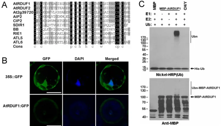
The AtRDUF1 protein contains a conserved C3H2C3-type RING domain, which shows similarity with many known proteins inArabidopsis(Figure 1A), including several proteins known to be involved in ABA and/or stress signaling pathways, such as ABI3-interacting protein 2 (AIP2) [21], SALT- AND DROUGHT-INDUCED RING FINGER 1 (SDIR1) [22] and ATLs [23,24]. One of these homologous proteins, RING finger protein for embryogenesis (RIE1), is required for normal development of seeds [25].
Subcellular studies using a 35S::AtRDUF1:GFP fusion protein inArabidopsisleaf protoplast cells showed that the fusion protein was mainly found in the cytosol and in the nuclei (Figure 1B). We examined whether AtRDUF1 is an E3 ligase usingin vitromethods. As shown in Figure 1C, in the presence of E1 and E2, autoubiquitination of MBP:AtRDUF1 can be detected in the presence of E1 and E2 by both nickel-horseradish peroxidase as well as by anti-MBP antibody assay, indicating that AtRDUF1 is an active E3 ligase. A mutant allele with substitution of metal ligand positions Cys-3, His-4, and His-5 of the RING motif with Tyr (CH/Y) was completely inactive (Figure 1C), indicating that an intact RING motif is essential for the E3 ligase activity of AtRDUF1.
Expression pattern ofAtRDUF1
To investigate the tissue-specific expression pattern of AtR-DUF1, a fusion gene comprising the nativeAtRDUF1promoter, a 1.3-kb fragment upstream of the start codon ofAtRDUF1 CDS, and theb-glucuronidase (GUS) gene [26] coding sequence as the reporter gene were constructed and transformed into wild-type
Arabidopsis. Histochemical staining showed that AtRDUF1 ex-pressed abundantly in seeds, but was also locally detectable in flowers, hypocotyls, leaves and roots (Figure 2). The staining was strong in immature seeds (Figure 2A), whereas in intact desiccated seeds, the GUS expression was only detectable at the funiculus attachment region (Figure 2B). In broken seeds, GUS staining was uniformly presented throughout the seed (Figure 2C), indicating that the limitation of GUS staining in intact seeds was due to blocking by the seed coat. To exclude possible false observations caused by the diffusion of the soluble intermediate of GUS substrates [27,28], seeds were dissected and stained separately. The GUS staining signal could be observed uniformly throughout the embryo, but only in the funiculus attachment region of the seed coat (Figure 2D). During germination, a reduction in GUS staining was detectable early on (Figure 2E–H), which is consistent with the decrease of AtRDUF1 expression detected by real-time qRT-PCR (Figure S2 in File S1). In 4-d-old seedlings, the GUS expression was mainly detected in the junction of the root and hypocotyl, in leaf tips, and around the meristem (Figure 2H). In 2-week old seedlings, the GUS staining was only detectable in leaf tips and root tips (Figure 2I and 2J). In reproductive tissue,
Figure 1. Analysis of the AtRDUF1 protein.(A) Alignment of the RING finger domains of the AtRDUF1 homologs inArabidopsis. Black and gray indicate 100% and$50% identities, respectively. (B) Subcellular localization of AtRDUF1:GFP fusion protein inArabidopsisleaf protoplast cells. Bars represent 20mm. The green and blue fluorescenece are GFP and 49,6-diamidino-2-phenylindole (DAPI) signals, respectively. (C) Verification of E3 ligase activity of AtRDUF1 by in vitro autoubiquitination assay. CH/Y represents the mutant form of the MBP:AtRDUF1 fusion protein, with substitution of metal ligand positions Cys-3, His-4, and His-5 of the RING motif with Tyr. The numbers at left denote the molecular masses of marker proteins in kilodaltons. Nichel-HRP (Ub), the nickel-horseradish peroxidase used to detect His-tagged ubiquitin. Anti-MBP, the anti-MBP antibody to detect maltose fusion proteins.
AtRDUF1::GUSactivity was observed at the junction of carpels and pedicels, as well as in stigma, anthers, and pollen, and low levels were detected in the vascular tissues of sepals and petals (Figure 2K–M).
AtRDUF1 positively regulates plant salt and osmotic stress responses
To investigate the function ofAtRDUFs, mutants with T-DNA insertions within the exons of the AtRDUFs were obtained and verified (Figure S1A–B in File S1). Expression of the mutant genes was not detected by RT-PCR with primers spanning the T-DNA in the respective homozygous mutants (Figure S1C in File S1). The mutants for AtRDUF1 and AtRDUF2were named atrduf1-2
and atrduf2-1, respectively (hereinafter called ‘‘atrduf1’’ and ‘‘atrduf2’’). The double mutant atrduf1atrduf2 was generated by crossing.
We generated transgenic Arabidopsis plants with constitutive expression of AtRDUF1, driven by cauliflower mosaic virus
(CaMV) 35S promoter. Six transgenic lines of AtRDUF1 were obtained (Figure S1D and S1E in File S1). The 35S::AtRDUF1
plants showed a wild-type growth phenotype under normal conditions.
The expression of theAtRDUFshad been shown to be induced by salt treatment [19]. We therefore tested whether AtRDUF1
plays a role in plant responses to salt. Seeds of wild-type (WT),
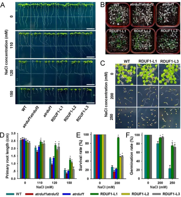
atrduf1atrduf2, atrduf1, and AtRDUF1 overexpression lines were germinated vertically in 1/2 MS medium. 3-day-old seedlings were transferred to 1/2 MS medium supplemented with 110, 120 or 150 mM NaCl. In the control condition, no significant difference in the length of primary roots was observed among any of the materials (Figure 3A and 3D). Under salt stress conditions, the primary roots of 35S::AtRDUF1 seedlings grew faster than did wild-type seedlings, and the atrduf1atrduf2double mutants showed inhibited growth (Figure 3A and 3D).
Germinatedatrduf1atrduf2,atrduf1and35S::AtRDUF1lines were transplanted to soil for two weeks, and watered with 200 mM Figure 2. Histochemical localization of GUS activity inAtRDUF1::GUStransgenic plants.(A) Developing seed at 12 days after pollination. (B) Desiccated mature seed. (C) Broken mature seed. (D) Dissected seed after imbibition. (E–H) Germinating seedlings at 1-day (E), 2-days (F), 3-days (G), and 4-days (H) after germination. (I) Leaf. (J) Root. (K) Flower. (L) Pollen. (M) Siliques. Bars represent 0.25 mm in A-D; 0.1 mm in J; 10mm in L; and 1 mm in E-I, K, and M.
NaCl solution for 15 days to induce salt stress. 35S::AtRDUF1
seedlings exhibited higher survival rates than did WT seedlings. Contrastingly, the survival rate of the salt treated atrduf1atrduf2
double mutant seedlings was lower than the WT control seedlings (Figure 3B and 3E).
After imbibition on 1/2 MS medium containing NaCl and transfer to a growth chamber for 10 days, the germination rates of
35S::AtRDUF1seeds were higher than that of WT seeds (Figure 3C and 3F). Therefore, the germination ability ofAtRDUF1 overex-pression seeds is insensitive to salt treatment. In our salt tolerance tests, no significant difference was observed between WT and the
atrduf1mutant.
Salinity causes ionic and osmotic stresses in plant cells. Germinated transgenic seedlings were transferred to MS medium supplemented with mannitol, a nonmetabolizable sugar, which is
known to be used as an osmotic agent in some studies [29,30,31]. The root growth of 35S::AtRDUF1 seedlings was less severely inhibited by mannitol than that of WT seedlings (Figure 4A and 4B). Therefore, the tolerance of 35S::AtRDUF1 seedlings to salt treatment is at least partly osmotic in nature.
In contrast with the accelerated water loss in detached leaves of the atrduf1 mutant [19], the water loss of rosette leaves in the overexpression lines was slower than that of WT leaves (Figure 4C). The overexpression plants also showed a sensitive response to ABA in terms of primary root length (Figure S3 in File S1). Our results support that AtRDUF1 positively participates in ABA-mediated dehydration stress responses.
We investigated the expression profiles of several stress-responsive genes in the AtRDUF1 overexpression lines grown under salt stress conditions. 10-day-old seedlings grown on 1/ Figure 3. Salt tolerance ofAtRDUFoverexpression plants and mutants.(A) WT,atrduf1atrduf2,atrduf1, RDUF1-L1 (Line 1 of35S::AtRDUF1
2 MS agar plates were sprayed with 200 mM NaCl solution, the seedlings were harvested after 1 h and 2 h, for extraction of total RNA. The result showed that in RDUF1-L1 seedlings, the transcription levels ofRD29B, RD22, and KIN1increased more than those of the WT after 2 h of salt treatment (Figure 5A), suggesting that AtRDUF1 may directly or indirectly interact with known abiotic stress response pathways. When theAtRDUF1::GUS
transgenic plants were subjected to salt treatment, the GUS staining was enhanced compared with control plants (Figure 5Ba– 5Bd), which is consistent with the real-time qRT-PCR results (Figure 5Be).
Discussion
In the course of our investigation of likely stress-related genes, we identifiedAtRDUFsfromin silicodata. Their putative proteins contain both RING finger and DUF1117 domains, and represent a novel E3 ligase family in plants. Real time qRT-PCR and promoter-GUS analyses confirmed that the transcription of
AtRDUF1 was indeed salt inducible (Figure 5B). We analyzed the phenotypes of wild-type as well as overexpression and loss-of-function mutants ofAtRDUFsfollowing salt treatment. Our results showed thatAtRDUF1positively regulates plant responses to salt treatment during both germination and post-germination growth (Figure 3). Furthermore,AtRDUF1also positively regulates plant tolerance to osmotic and dehydration stress (Figure 4).
The fluorescence of AtRDUF1:GFP fusion protein was detectable in our subcellular localization study (Figure 1B), and
35S::AtRDUF1transgenic plants showed obvious tolerance to salt, osmotic, and water loss stresses (Figure 3 and 4). In a similar report, a fusion protein approach was used to develop transgenic plants that overexpressed AtRDUF1. However, the authors were unsuccessful in detecting significant accumulation of the fusion protein, in spite of expression of significant amounts ofAtRDUF1
mRNA, so the plants were not extensively analyzed [19]. The discrepancy of detection may be attributable to distinctions in vector efficiency or differences in the sensitivity of the assay methods employed in these independent studies.
In addition to the results reported here,AtRDUF1andAtRDUF2
are also known to be up-regulated by chitin treatment [32]. Among the homologous proteins of the AtRDUFs subfamily, the E3 ligases KEG [12] and AIP2 [21] are negative regulators of ABA signaling, acting by targeting and degrading ABI5 and ABI3, respectively. The E3 ligase SDIR1 positively regulates ABA signaling, and thesdir1mutant is resistant to salt and drought stress [22]. Therefore, it is apparent that AtRDUFs and their homologous proteins are widely involved in plant adaptations to stress.
We were able to confirm that AtRDUF1 has E3 ligase activity by ubiquitination assays (Figure 1C). Ubiquitination has been shown to play an important role in the perception and transduction of various internal and external environmental signals [33,34]. To test whether AtRDUF1 affects the expression of known stress pathway genes, several marker genes in the stress-responsive pathways were analyzed, RD29B, RD22, and KIN1
showed a hypersensitive salt response in transgenic plants (Figure 5A).RD29Bis a cold-, high salt-, and dessication-inducible gene with two ABA-responsive elements (ABREs) present in its promoter region [35].RD22transcription is induced by salt and ABA treatment, but no ABRE was identified in its promoter Figure 4. Osmotic tolerance and detached leaf water-loss rates
ofAtRDUF1overexpression plants.(A) WT,atrduf1atrduf2,atrduf1, RDUF1-L1, RDUF1-L2 and RDUF1-L3 with or without osmotic stress treatment. 3-day-old seedlings were transferred to 1/2 MS medium containing 0, 200, or 350 mM mannitol, and vertically cultured for 6 d. Bars represent 1 cm. (B) Statistical comparison of root lengths of seedlings under the conditions described in (A). (C) Water loss rates of detached leaves. Detached rosette leaves from wild-type andAtRDUF1
overexpression seedlings were incubated for 6 h at room temperature.
region [22,36]. KIN1, which contains the C repeat/dehydration-responsive element (CRT/DRE) motif in its promoter, can be induced by cold, ABA and dehydration treatment [37]. According to our data and published results [19], AtRDUF1 may be involved in the up-regulation of stress responses inArabidopsisseedlings, in this respect, AtRDUF1, may be similar to SDIR1 and AtSAP5, both of which have been shown to be E3 ligases and are known in promoting stress gene expression and stress tolerance [22,38,39].
Glycerol, a compatible osmolyte, is used in defending against dehydration stresses in yeast, marine algae, insects, and amphib-ians [40,41,42,43]. Accumulation of intracellular glycerol was observed during the salt adaptation processes of many microor-ganism such as Aspergillus nidulans[44] and Aureobasidium pullulans
[45]. InArabidopsis, the results from studies in mutants with defects in storage lipid accumulation prior to seed maturation or lipid catabolism following germination [29,30,31] suggest that glycerol or glycerol-derived lipids could serve as compatible osmolytes in dehydration stress conditions in plants. As AtRDUF1 expressed primarily in embryos of matured seeds (Figure 2), we tested the correlation ofAtRDUF1expression with storage lipids (Text S1 in File S1). In young seedlings, the staining of GUS driven by the
AtRDUF1promoter partially coincided with staining of Sudan red 7B, a fat-soluble dye that stains lipids red, and the Sudan red 7B staining was darker in the35S::RDUF1 seedlings (Figure S4A in File S1). The levels of triacylglycerol (TAG), the primary seed oil in Arabidopsis were higher in seeds and young seedlings of
35S::AtRDUF1lines than in WT seedlings (Figure S4B in File S1).
The vegetative tissues of35S::RDUF1seedlings contained a higher content of triglycerides (the ester of glycerol) than did WT control tissues (Figure S4C in File S1). Finally, the oleosin maker gene
OleS3 and triglycerides content change in response to salt stress (Figure S4D and S4E in File S1). We speculate that a mechanism by which AtRDUF1 increases salt tolerance is through the positively regulation of either the delayed catabolism or the increased accumulation of the storage lipids.
In conclusion, our data show that AtRDUF1 is a functional E3 ligase and a positive regulator of the Arabidopsisresponse to salt stress. This study contributes to our understanding of the molecular factors involved in the responses of plants to abiotic stresses.
Materials and Methods
Plant materials
AllArabidopsisplants used in this study were of theColumbia (Col-0) ecotype. T-DNA insertion lines SALK_131634 (forAtRDUF1) and N471914 (forAtRDUF2) were obtained from ABRC [46] and NASC [47], respectively. Seedlings were grown under long-day conditions (16 h light/8 h dark) at 22uC, 40 to 60% RH and 63 mE?s21?m22light intensity.
Vector construction andArabidopsistransformation
The cDNA of AtRDUF1 was amplified and cloned into the pSN1301 expression vector [48] driven by the CaMV 35S Figure 5. Induction studies of salt-responsive genes andAtRDUF1.(A) Induction profiles of salt-responsive genes in wild-type andAtRDUF1
overexpression plants under salt stress. Transcript levels ofRD29B,RD22, andKIN1were determined by real-time qRT-PCR analysis of seedlings treated with 200 mM NaCl. Data represent means6SD. Mean values were normalized to the transcript levels of an internal control TUBULIN. Asterisks indicate significance (*, P,0.05 versus WT control). (B) Induction of AtRDUF1 expression by salt treatment. Compared with the mock treated plants (a and c), GUS expression was enhanced inAtRDUF1::GUSplants treated with 300 mM NaCl for 5 h (b and d). (c) and (d) are close-up views of partial regions of (a) and (b), respectively. Transcript expression values ofAtRDUF1were also determined by real-time qRT-PCR in 2-week-old plants treated with 300 mM NaCl (e). Bars represent 1 mm. Data represent means6SD. Mean values were normalized to the transcript levels of an internal control TUBULIN. Asterisks indicate significance (*, P,0.05 versus 0 h control).
promoter. The primers used for AtRDUF1 overexpression were R1BamHIF and R1KpnIR (all primer sequences used in this study are listed in Table S1 in File S1). The 1.3 kb promoter sequence of AtRDUF1 was amplified with primers R1PKpnIF and R1PBamHIR, and cloned into the pGUS1301 vector [49]. The constructed plasmid was introduced into Agrobacterium tumefaciens
strain C58.Arabidopsiswas transformed using the floral dip method [50]. Transgenic plants were first screened on medium containing 40 mg/l hygromycin and subsequently transferred to soil. To produce aAtRDUF1:GFPfusion gene driven by 35S promoter, the
AtRDUF1 CDS sequence with the stop codon deleted was amplified with primers R1GXhoIF and R1GKpnIR, and cloned in frame into pBI121GFP. To generate the MBP:AtRDUF1 fusion, the AtRDUF1 CDS was cloned in frame into pMAL-c2 (NEB, Berverly, MA, USA) with primers R1MBamHIF and R1MSalIR. A mutant allele with substitution of metal ligand positions Cys-3, His-4, and His-5 of the RING motif with Tyr (CH/Y) was introduced with primers R1mF and R1mR. DpnI mediated site-directed mutagenesis in the plasmid was performed as described previously [51].
Subcellular localization
Plasmid 35S::AtRDUF1:GFP and 35S::GFP were purified by the use of Tiangen kits according to the manufacturer’s protocols.
Arabidopsis protoplasts transformations were performed as de-scribed previously [52]. For detection of nuclei, samples were stained with DAPI at a final concentration of 1mg/mL.
E3 ubiquitin ligase activity assay
The MBP:AtRDUF1 fusion protein was expressed inEscherichia colistrain BL21 (DE3), and subsequently purified using amylose resin (NEB). The in vitro E3 ubiquitin ligase activity assay was performed as described previously [5]. Following the assay reactions, proteins were separated by SDS-PAGE, blotted, and probed by either HisDetector Nickel-HRP (KPL company, USA) for the detection of His-tagged ubiquitin or antibody to MBP (antiserum; NEB) for detection of the MBP-tagged AtRDUF1 protein. Results were visualized using chemiluminescence as per the instructions of the manufacturer (ECL; Amersham Pharmacia, Amersham, UK).
Stress and ABA treatment
After surface sterilization, seeds were imbibed at 4uC for 3 d in the dark. The seeds were sown on half-strength Murashige and Skoog medium [53] supplemented with 1% sucrose and 0.7% agar. 3-day-old seedlings were transferred to 1/2 MS medium containing NaCl, mannitol, or ABA. Primary root length was measured after 6 d. 4-day-old seedlings were transplanted to soil and cultured for two weeks, and treated with 200 mM NaCl for 15 days to induce salt stress. For the germination test, all seeds were harvested simultaneously and stored for 5 weeks after harvest. Germination was determined as penetration of the radicle through the seed coat 10 days after imbibition. Each assay was repeated three times. The data in the graphs (Figure 3D-F, 4B– C, S3B, S4C and S4E in File S1) were subjected to analysis of variance (ANOVA) and means were compared byt-test at the 5% level. All analyses were performed with version 13.0 of SPSS software.
The water loss assay was performed as described previously [54]. Eight rosette leaves per plant were detached from 3-week-old plants. The leaves were exposed to air and the fresh weights were measured at the indicated time points shown in Figure 4C. Water-loss rate is calculated as the ratio between water Water-loss and plant initial fresh weight, expressed in %.
GUS histochemical assays
Tissues of transgenic seedlings harboring AtRDUF1::GUS at various growth stages were used for the GUS activity assays. Fixation of the tissues by acetone and incubation in staining solution was performed as described by Sieburth and Meyerowitz [55]. For staining of pollen, the pollen was isolated from stamens by vortexing in acetone and enriched using centrifugation at rcf 3000.
Expression analysis
Total RNA was extracted from tissues with Trizol reagent (Invitrogen, Carlsbad, CA, USA), and treated with RNase-free DNase (Takara, Dalian, China) according to the manufacturer’s instructions. 2mg of total RNA was used for cDNA synthesis with avian myeloblastosis virus (AMV) reverse transcriptase (Promega, Madison, WI, USA) according to the manufacturer’s protocol. Real-Time qRT-PCR was performed as described previously [56]. Three independent experiments were performed. The relative quantification method (Delta-Delta CT) was used to evaluate variation in expression. Significance of differences was determined by x2-test. The primer sets for PCR were: R1realtimeF and R1realtimeR for AtRDUF1; RD29BF and RD29BR forRD29B; RD22F and RD22R forRD22; KIN1F and KIN1R for KIN1; TubulinF and TubulinR forTUBULIN; OleS3F and OleS3R for
OleS3; ACTINF and ACTINR forACTIN. Supporting Information
File S1 Text S1, lipid detection. Figure S1, verification of T-DNA insertion mutants ofAtRDUFsandAtRDUF1 overexpres-sion lines. Figure S2, relative quantification of AtRDUF1
transcription during germination assayed by real-time qRT-PCR.Figure S3, response ofAtRDUF1overexpression plants to ABA.Figure S4, effects of AtRDUF1 and salt treatment on plant lipids.Table S1, sequences of the oligonucleotides used in this study.
(DOC)
Acknowledgments
We thank Jia Zhao for plant care and Huanhuan Liu for help with microscope. The authors would like to thank two anonymous reviews for their valuable comments and suggestions for improving the quality of this paper.
Author Contributions
Conceived and designed the experiments: QX KC YX. Performed the experiments: JL YH QZ CL. Analyzed the data: JL YH QZ CL QX KC YX. Contributed reagents/materials/analysis tools: QZ QX CL. Wrote the paper: JL QZ KC YX.
References
1. Smalle J, Vierstra RD (2004) The ubiquitin 26S proteasome proteolytic pathway. Annu Rev Plant Biol 55: 555–590.
3. Dreher K, Callis J (2007) Ubiquitin, hormones and biotic stress in plants. Ann Bot 99: 787–822.
4. Lau OS, Deng XW (2012) The photomorphogenic repressors COP1 and DET1: 20 years later. Trends Plant Sci 17: 584–593.
5. Xie Q, Guo HS, Dallman G, Fang S, Weissman AM, et al. (2002) SINAT5 promotes ubiquitin-related degradation of NAC1 to attenuate auxin signals. Nature 419: 167–170.
6. Stone SL, Anderson EM, Mullen RT, Goring DR (2003) ARC1 is an E3 ubiquitin ligase and promotes the ubiquitination of proteins during the rejection of self-incompatibleBrassicapollen. Plant Cell 15: 885–898.
7. Moon J, Parry G, Estelle M (2004) The ubiquitin-proteasome pathway and plant development. Plant Cell 16: 3181–3195.
8. Barlow PN, Luisi B, Milner A, Elliott M, Everett R (1994) Structure of the C3HC4 domain by 1H-nuclear magnetic resonance spectroscopy. : A new structural class of zinc-finger. J Mol Biol 237: 201–211.
9. Borden KLB, Freemont PS (1996) The RING finger domain: A recent example of a sequence-structure family. Curr Opin Struct Biol 6: 395–401.
10. Borden KLB (2000) RING domains: Master builders of molecular scaffolds? J Mol Biol 295: 1103–1112.
11. Smalle J, Kurepa J, Yang PZ, Emborg TJ, Babiychuk E, et al. (2003) The pleiotropic role of the 26S proteasome subunit RPN10 in Arabidopsis growth and development supports a substrate-specific function in abscisic acid signaling. Plant Cell 15: 965–980.
12. Stone SL, Williams LA, Farmer LM, Vierstra RD, Callis J (2006) KEEP ON GOING, a RING E3 ligase essential forArabidopsisgrowth and development, is involved in abscisic acid signaling. Plant Cell 18: 3415–3428.
13. Shinozaki K, Yamaguchi-Shinozaki K (2007) Gene networks involved in drought stress response and tolerance. J Exp Bot 58: 221–227.
14. Ward JM, Hirschi KD, Sze H (2003) Plants pass the salt. Trends Plant Sci 8: 200–201.
15. Zhu JH, Dong CH, Zhu JK (2007) Interplay between cold-responsive gene regulation, metabolism and RNA processing during plant cold acclimation. Curr Opin Plant Biol 10: 290–295.
16. Finkelstein RR, Gampala SSL, Rock CD (2002) Abscisic acid signaling in seeds and seedlings. Plant Cell 14: S15–S45.
17. Lopez-Molina L, Mongrand S, Chua NH (2001) A postgermination develop-mental arrest checkpoint is mediated by abscisic acid and requires the AB15 transcription factor in Arabidopsis. Proc Natl Acad Sci U S A 98: 4782–4787. 18. Hasegawa PM, Bressan RA, Zhu JK, Bohnert HJ (2000) Plant cellular and molecular responses to high salinity. Annu Rev Plant Physiol Plant Mol Biol 51: 463–499.
19. Kim SJ, Ryu MY, Kim WT (2012) Suppression ofArabidopsisRING-DUF1117 E3 ubiquitin ligases, AtRDUF1 and AtRDUF2, reduces tolerance to ABA-mediated drought stress. Biochem Biophys Res Commun 420: 141–147. 20. Zimmermann P, Hirsch-Hoffmann M, Hennig L, Gruissem W (2004)
GENEVESTIGATOR. Arabidopsis microarray database and analysis toolbox. Plant Physiol 136: 2621–2632.
21. Zhang XR, Garreton V, Chua NH (2005) The AIP2 E3 ligase acts as a novel negative regulator of ABA signaling by promoting ABI3 degradation. Genes Dev 19: 1532–1543.
22. Zhang YY, Yang CW, Li Y, Zheng NY, Chen H, et al. (2007) SDIR1 is a RING finger E3 ligase that positively regulates stress-responsive abscisic acid signaling inArabidopsis. Plant Cell 19: 1912–1929.
23. Salinas-Mondragon RE, Garciduenas-Pina C, Guzman P (2000) Early elicitor induction in members of a novel multigene family coding for highly related RING-H2 proteins inArabidopsis thaliana. Plant Mol Biol 40: 579–590. 24. Serrano M, Parra S, Alcaraz LD, Guzman P (2006) TheATLgene family from
Arabidopsis thaliana and Oryza sativa comprises a large number of putative ubiquitin ligases of the RING-H2 type. J Mol Evol 62: 434–445.
25. Xu RQ, Li QSQ (2003) A RING-H2 zinc-finger protein geneRIE1is essential for seed development inArabidopsis. Plant Mol Biol 53: 37–50.
26. Yanagawa Y, Sullivan JA, Komatsu S, Gusmaroli G, Suzuki G, et al. (2005) Arabidopsis COP10 forms a complex with DDB1 and DET1 in vivo and enhances the activity of ubiquitin conjugating enzymes. Plant Cell Physiol 46: S242–S242.
27. De Block M, Van Lijsebettens M (1998)b-Glucuronidase enzyme histochemistry on semithin sections of plastic-embeddedArabidopsisexplants. Methods Mol Biol 82: 397–407.
28. Jefferson RA, Kavanagh TA, Bevan MW (1987) GUS fusions:b-glucuronidase as a sensitive and versatile gene fusion marker in higher plants. EMBO J 6: 3901. 29. Beisson F, Li YH, Bonaventure G, Pollard M, Ohlrogge JB (2007) The acyltransferase GPAT5 is required for the synthesis of suberin in seed coat and root ofArabidopsis. Plant Cell 19: 351–368.
30. Eastmond PJ (2004) Glycerol-insensitiveArabidopsismutants:gli1seedlings lack glycerol kinase, accumulate glycerol and are more resistant to abiotic stress. Plant J 37: 617–625.
31. Lu CF, Hills MJ (2002) Arabidopsis mutants deficient in diacylglycerol acyltransferase display increased sensitivity to abscisic acid, sugars, and osmotic stress during germination and seedling development. Plant Physiol 129: 1352– 1358.
32. Libault M, Wan JR, Czechowski T, Udvardi M, Stacey G (2007) Identification of 118Arabidopsistranscription factor and 30 ubiquitin-ligase genes responding to chitin, a plant-defense elicitor. Mol Plant-Microbe Interact 20: 900–911. 33. Hare PD, Seo HS, Yang JY, Chua NH (2003) Modulation of sensitivity and
selectivity in plant signaling by proteasomal destabilization. Curr Opin Plant Biol 6: 453–462.
34. Hellmann H, Estelle M (2002) Plant development: regulation by protein degradation. Science 297: 793–797.
35. Nakashima K, Fujita Y, Katsura K, Maruyama K, Narusaka Y, et al. (2006) Transcriptional regulation of ABI3- and ABA-responsive genes including RD29B and RD29A in seeds, germinating embryos, and seedlings of Arabidopsis. Plant Mol Biol 60: 51–68.
36. Yamaguchi-Shinozaki K, Shinozaki K (1993) The plant hormone abscisic acid mediates the drought-induced expression but not the seed-specific expression of rd22, a gene responsive to dehydration stress in Arabidopsis thaliana. Mol Gen Genet 238: 17–25.
37. Knight H, Veale EL, Warren GJ, Knight MR (1999) Thesfr6mutation in Arabidopsis suppresses low-temperature induction of genes dependent on the CRT/DRE sequence motif. Plant Cell 11: 875–886.
38. Hozain M, Abdelmageed H, Lee J, Kang M, Fokar M, et al. Expression of
AtSAP5in cotton up-regulates putative stress-responsive genes and improves the tolerance to rapidly developing water deficit and moderate heat stress. J Plant Physiol 169: 1261–1270.
39. Kang M, Fokar M, Abdelmageed H, Allen RD Arabidopsis SAP5 functions as a positive regulator of stress responses and exhibits E3 ubiquitin ligase activity. Plant Mol Biol 75: 451–466.
40. Lee R (1991) Principles of insect low temperature tolerance. In: Lee R, Denlinger D, editors. Insect at low temperature. New York: Chapman and Hall. 17–46.
41. Hohmann S (2002) Osmotic stress signaling and osmoadaptation in yeasts. Microbiol Mol Biol Rev 66: 300–372.
42. Pahlman AK, Granath K, Ansell R, Hohmann S, Adler L (2001) The yeast glycerol 3-phosphatases gpp1p and gpp2p are required for glycerol biosynthesis and differentially involved in the cellular responses to osmotic, anaerobic, and oxidative stress. J Biol Chem 276: 3555–3563.
43. Storey KB (1997) Organic solutes in freezing tolerance. Comp Biochem Physiol A: Physiol 117: 319–326.
44. Redkar RJ, Locy RD, Singh NK (1995) Biosynthetic pathways of glycerol accumulation under salt stress inAspergillus nidulans. Exp Mycol 19: 241–246. 45. Managbanag JR, Torzilli AP (2002) An analysis of trehalose, glycerol, and
mannitol accumulation during heat and salt stress in a salt marsh isolate of Aureobasidium pullulans. Mycologia 94: 384–391.
46. Alonso JM, Stepanova AN, Leisse TJ, Kim CJ, Chen H, et al. (2003) Genome-wide insertional mutagenesis ofArabidopsis thaliana. Science 301: 653–657. 47. Scholl RL, May ST, Ware DH (2000) Seed and molecular resources for
Arabidopsis. Plant Physiol 124: 1477–1480.
48. Zhou J, Li F, Wang J, Ma Y, Chong K, et al. (2009) Basic helix-loop-helix transcription factor from wild rice (OrbHLH2) improves tolerance to salt-and osmotic stress inArabidopsis. J Plant Physiol 166: 1296–1306.
49. Ge L, Chen H, Jiang JF, Zhao Y, Xu ML, et al. (2004) Overexpression of
OsRAA1causes pleiotropic phenotypes in transgenic rice plants, including altered leaf, flower, and root development and root response to gravity. Plant Physiol 135: 1502–1513.
50. Clough SJ, Bent AF (1998) Floral dip: a simplified method forAgrobacterium -mediated transformation ofArabidopsis thaliana. Plant J 16: 735–743. 51. Papworth C, Bauer J, Braman J, Wright D (1996) Site-directed mutagenesis in
one day with.80% efficiency. Strategies 9: 4.
52. Jin JB, Kim YA, Kim SJ, Lee SH, Kim DH, et al. (2001) A new dynamin-like protein, ADL6, is involved in trafficking from the trans-Golgi network to the central vacuole inArabidopsis. Plant Cell 13: 1511–1526.
53. Murashige T, Skoog F (1962) A revised medium for rapid growth and bio assays with tobacco tissue cultures. Physiol Plant 15: 473–497.
54. Cheong YH, Pandey GK, Grant JJ, Batistic O, Li L, et al. (2007) Two calcineurin B-like calcium sensors, interacting with protein kinase CIPK23, regulate leaf transpiration and root potassium uptake in Arabidopsis. Plant J 52: 223–239.
55. Sieburth LE, Meyerowitz EM (1997) Molecular dissection of theAGAMOUS
control region shows that cis elements for spatial regulation are located intragenically. Plant Cell 9: 355–365.
56. Jiang J, Li J, Xu Y, Han Y, Bai Y, et al. (2007) RNAi knockdown of Oryza sativa