The eukaryotic Pso2/Snm1/Artemis
proteins and their function as genomic
and cellular caretakers
1Departamento de Biofísica, Centro de Biotecnologia, Universidade Federal do Rio
Grande do Sul, Porto Alegre, RS, Brasil
2EMBRAPA Uva e Vinho, Bento Gonçalves, RS, Brasil
3Curso de Farmácia, Universidade Luterana do Brasil, Canoas, RS, Brasil
4Instituto de Biotecnologia, Universidade de Caxias do Sul, Caxias do Sul, RS, Brasil
D. Bonatto1,
L.F. Revers2,
M. Brendel1 and
J.A.P. Henriques1,3,4
Abstract
DNA double-strand breaks (DSBs) represent a major threat to the genomic stability of eukaryotic cells. DNA repair mechanisms such as non-homologous end joining (NHEJ) are responsible for the maintenance of eukaryotic genomes. Dysfunction of one or more of the many protein complexes that function in NHEJ can lead to sensitivity to DNA damaging agents, apoptosis, genomic instabil-ity, and severe combined immunodeficiency. One protein, Pso2p, was shown to participate in the repair of DSBs induced by DNA inter-strand cross-linking (ICL) agents such as cisplatin, nitrogen mustard or photo-activated bi-functional psoralens. The molecular function of Pso2p in DNA repair is unknown, but yeast and mam-malian cell line mutants for PSO2 show the same cellular responses as strains with defects in NHEJ, e.g., sensitivity to ICLs and apop-tosis. The Pso2p human homologue Artemis participates in V(D)J recombination. Mutations in Artemis induce a variety of immuno-logical deficiencies, a predisposition to lymphomas, and an in-crease in chromosomal aberrations. In order to better understand the role of Pso2p in the repair of DSBs generated as repair interme-diates of ICLs, an in silico approach was used to characterize the catalytic domain of Pso2p, which led to identification of novel Pso2p homologues in other organisms. Moreover, we found the catalytic core of Pso2p fused to different domains. In plants, a specific ATP-dependent DNA ligase I contains the catalytic core of Pso2p, constituting a new DNA ligase family, which was named LIG6. The possible functions of Pso2p/Artemis/Lig6p in NHEJ and V(D)J recombination and in other cellular metabolic reactions are discussed.
Correspondence
J.A.P. Henriques Departamento de Biofísica Centro de Biotecnologia, UFRGS Av. Bento Gonçalves, 9500 91507-970 Porto Alegre, RS Brasil
Fax: +55-51-3316-7603 E-mail: [email protected]
Presented at the XI Congresso Brasileiro de Biologia Celular, Campinas, SP, Brazil, July 15-18, 2004.
Research supported by FAPERGS, CAPES, and GENOTOX (Genotoxicity Laboratory, UFRGS-Brazil).
Received June 30, 2004 Accepted December 7, 2004
Key words
•Non-homologous end joining •Double-strand breaks •V(D)J
•PSO2
•Artemis
•Saccharomyces cerevisiae
Introduction
The chromatin of all eukaryotic cells, without exception, is a special target for chemical or physical agents that can induce different kinds of DNA damage, including
cellular well being, leading to cell cycle ar-rest, tumorigenesis, cell death, or severe combined immunodeficiency disease (SCID) in mammals (1). Among the various forms of DNA lesions that are induced by physical or chemical agents, probably the most danger-ous are the DNA DSBs (1,2). DSBs can occur in response to ionizing radiation, to radiomimetic agents or chemical substances
that induce DNA ICLs such as bi-functional nitrogen mustards or 8-methoxypsoralen plus UVA (Figure 1). DSBs also arise as a conse-quence of natural processes such as V(D)J recombination (a lymphoid-specific process required for gene rearrangement and matura-tion of T and B cells), and as a by-product of normal cellular metabolism (Figure 1) (3). If not repaired prior to DNA replication or
CH3
CH3
CI CI
O O
O O
N
Nitrogen mustard 8-MOP + UVA
Physical or chemical damage (ICL) UVC
DNA replication direction
Cellular metabolism (transposition, V(D)J)
HR NHEJ
Cohesive ends/ Blunt ends
Hairpin-capped ends
DNA-PKcs Ku70/Ku86 XRCC4
Rad50/Mre11/Xrs2
DNA-PKcs Ku70/Ku86 XRCC4
Rad50/Mre11/Xrs2
DNA polymerase µ DNA ligase IV
Artemis/Pso2p DNA-PKcs
DNA polymerase µ DNA ligase IV bp
bp
Repaired DNA
3 2 1 1
3 Figure 1. Schematic drawing of
double-strand break (DSB) re-pair in mammalian cells. DSB induced by inter-strand cross-link (ICL) generated by physical agents (UVC), chemical sub-stances (nitrogen mustard, 8-MOP + UVA), or even cellular metabolism (gray box) on DNA during replication can be re-paired by two biochemical path-ways: homologous recombina-tion (HR) or non-homologous end joining (NHEJ). HR is the major DNA repair pathway used when two homologous DNA strands are present. NHEJ is used when the homologous DNA strand is not present. The protein complexes that are used for NHEJ repair depend on the type of DNA ends present in the DSB (cohesive ends, blunt ends, or hairpin-capped ends). Protein complexes 1 and 3 re-pair both cohesive and blunt ends, while hairpin-capped ends are repaired by Artemis/ Pso2p/DNA-PKcs (complex 2). The final result is the restitution of high molecular weight DNA, with loss (NHEJ) or without loss (HR) of DNA base pairs (bp). UVC = UV254nm; 8-MOP + UVA
= 8-methoxypsoralen plus UVA; UVA = UV365nm; DNA-PKcs =
mitosis, DSBs can induce cell death (4) and, if misrepaired, DSBs have the potential to lead to chromosome translocations, genom-ic instability and predisposition to cancer (2,5). Interestingly, only one DSB can kill a cell if it leads to the inactivation of an essential gene or triggers apoptosis (2,4,6). More-over, mutations in many of the factors in-volved in sensing and repair of DSB damage lead to increased pre-disposition to cancer in man and in animal models (2,7).
In yeast and mammalian cells, DSBs are predominantly repaired by one of two path-ways (1), i.e., homologous recombination (HR), or non-homologous end joining (NHEJ) (Fig-ure 1). In addition, NHEJ is also used to repair DSBs that arise during early mammalian lym-phocyte development in the context of V(D)J recombination (8). HR and NHEJ have over-lapping roles in maintaining chromosomal in-tegrity (9) and can act together to preserve genomic integrity in eukaryotic cells (10). Yeast, unlike multicellular eukaryotes, repairs most of its DSBs using HR, a process that occurs without the loss of genetic information (11). However, NHEJ can be detected in yeast when the mechanisms of HR are inactivated (11). Multicellular eukaryotes use NHEJ as the predominant DNA repair system and this pref-erence could be intrinsic to their genomic organization. The genomes of multicellular eukaryotes contain a substantial fraction of repetitive DNA and, therefore, the homology search process for repair of DSBs by HR is inviablewhen the breaks occur in the portion of the genome that is repetitive, further leading to chromosomal translocations or cell death (11). Except during late S, G2 and M, when a sister chromatid is physically positioned optimally, homology partners for repetitive regions might be chosen inappropriately from any of the chromosomes (11).
Cells with a defect in NHEJ age in culture more quickly when compared to NHEJ-proficient cells (12). Mouse mutants in either component of the DNA ligase complex (XRCC4 or DNA ligase IV) show defects in
V(D)J recombination (13,14), just as human pre-B cells do (15). These mice die during the final days of gestation, showing an increased apoptotic death of neurons at specific loca-tions in the nervous system at specific times during gestation (11). It is still unclear why some cells die and others do not. Interest-ingly, Ku70-deficient mice show a depletion of enteric neurons (16). Presumably this apoptotic cell death is triggered by an inability to repair DSBs. Also, the inactivation of NHEJ leads to increased sensitivity to ioniz-ing radiation, genomic instability, and SCID, resulting from the inability to join Rag-cleaved gene segments in progenitor (pro)-B and T lymphocytes (17). Despite their inability to repair DSBs, NHEJ-deficient mice show, at most, a modest predisposition to lympho-mas, because cells with unrepaired breaks are eliminated by the checkpoint protein p53 (17). Inactivation of p53 restores pro-B lym-phocyte numbers, although it does not res-cue NHEJ or lymphocyte development (18). Combined deficiencies for p53 and all NHEJ factors have been analyzed and all were found to lead to consistent development of early-onset pro-B lymphomas (18).
NHEJ basically involves modification of the two broken ends to make them compat-ible prior to rejoining, resulting in the loss of some information between the two DNA ends. Hence, NHEJ is an imperfect process from the standpoint of preserving genetic information (11). Proteins known to be in-volved in NHEJ include the DNA-dependent protein kinase catalytic subunit (DNA-PKcs), XRCC4, Ku70 and Ku86, DNA ligase IV, and the Rad50/Xrs2/Mre11 complex (19). These proteins, to be described in more detail be-low, form complexes with specific func-tions in the modification of DNA ends for rejoining, or in the stabilization of DNA extremities for further processing.
DNA-PKcs
double stranded DNA ends, phosphorylates proteins bound to the same DNA molecule. Apart from its large size (469 kDa), the most noticeable feature of DNA-PKcs is a carboxy-terminal catalytic domain which bears amino acid similarity to the catalytic domain of the phosphoinositide-3,4-kinase family of lipid ki-nases (20). The presence of this conserved region classifies DNA-PKcs as a member of the phosphatidylinositol-3-kinase-related tein kinases (21,22). Ku70 and Ku86 are pro-teins that form a heterodimer with high affinity for DNA ends and are generally considered to comprise the binding “subunit” of PK. However, their association with DNA-PKcs appears not to be obligatory and there is clear evidence for DNA-PKcs-independent functions (Table 1) (23).
Ku70/Ku86
Cells that lack Ku are radiosensitive and defective in DSB repair, and animals lacking
either one of the Ku subunits share many characteristics with DNA-PKcs null animals, e.g., radiosensitivity, immune deficiency, and defective DNA DSB repair (Table 1). In addi-tion, Ku70 and Ku80 null animals have growth defects and premature senescence, indicating that Ku and DNA-PKcs have distinct and overlapping functions (2,11). In plants, specif-ically Arabidopsis thaliana, the expression of both Ku70 and Ku80 genes is up-regulated in response to the induction of DSBs in chrom-osomal DNA by either bleomycin or methyl-methanesulfonate. Mutant lines of A. thaliana for Ku80 showed hypersensitivity to the DNA-damaging agents bleomycin and menadione which cause single- and DSBs in DNA, a phenotype consistent with a role in the NHEJ pathway (Table 1) (24,25).
DNA ligase IV
DNA ligase IV, an ATP-dependent DNA ligase that has a special role in NHEJ and
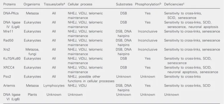
Table 1. Eukaryotic non-homologous end joining proteins.
Proteins Organisms Tissues/cellsa Cellular process Substrates Phosphorylationb Deficienciesc
DNA-PKcs Metazoa All NHEJ, V(D)J, telomeric DSB Yes Sensitivity to cross-links,
maintenance SCID, senescence
DNA ligase Eukaryotes All NHEJ, V(D)J, telomeric DSB Yes Sensitivity to cross-links, SCID,
IV (Lig4) maintenance senescence, neuronal apoptosis
Mre11 Eukaryotes All NHEJ, V(D)J, telomeric DSB, DNA Inconclusive Sensitivity to cross-links, senescence
maintenance hairpins
Rad50 Eukaryotes All NHEJ, V(D)J, telomeric DSB, DNA Inconclusive Sensitivity to cross-links, senescence
maintenance hairpins
Xrs2 Metazoa, All NHEJ, V(D)J, telomeric DSB, DNA Inconclusive Sensitivity to cross-links, senescence
fungi maintenance hairpins
Ku70/Ku80 Eukaryotes All NHEJ, V(D)J, telomeric DSB Yes Sensitivity to cross-links, SCID,
maintenance senescence
XRCC4 Eukaryotes All NHEJ, V(D)J, telomeric DSB Yes Sensitivity to cross-links, SCID,
maintenance neuronal apoptosis, senescence
Pso2 Eukaryotes All NHEJ, possible other Unknown Unknown Sensitivity to cross-links functions in cellular processes
Artemis Metazoa Lymphocytes NHEJ, V(D)J DSB, DNA Yes Sensitivity to cross-links, SCID hairpins
DNA ligase Plants Unknown Unknown Unknown Unknown Unknown
VI (Lig6)
aProteins present in different types of tissues or cells. bIndicates if protein activity is induced or modified by site-specific phosphorylation. cPhysiological deficiencies induced by partially functional or non-functional proteins related to NHEJ, V(D)J recombination, and telomeric
V(D)J, is present in eukaryotes as diverse as yeast, plants, and metazoa (26). The homo-logue of the mammalian gene for DNA ligase IV was isolated from A. thaliana, and its expression profile indicates that this gene is regulated by ionizing radiation-induced DSBs (26). Deletion of mammalian DNA ligase IV results in death during embryogenesis due to massive neuronal apoptosis (Table 1) (14). A highly radiation-sensitive human cell line iso-lated from a leukemia patient was found to express a dysfunctional form of DNA ligase IV (Table 1) (14).
XRCC4
XRCC4 exists in a tight complex with DNA ligase IV (27), which is essential for the ligation step in NHEJ and may also be in-volved in alignment or gap filling prior to ligation (28). In mammalian cells, XRCC4 can interact with DNA, DNA-PKcs, Ku, and DNA polymerase µ, but its precise role in NHEJ is unknown (1). Cells that lack XRCC4 are radio-sensitive, defective in V(D)J re-combination and DSB repair, and disruption of XRCC4 in mice is embryonically lethal due to neuronal apoptosis (Table 1) (14). A plant gene with high homology to mammalian XRCC4, that also interacts with DNA ligase IV and has its expression pattern modulated by DSBs, was identified in A. thaliana (29).
Rad50/Xrs2/Mre11
The Rad50/Xrs2/Mre11 complex is also very well conserved in all eukaryotes studied so far. These three physically interacting gene products were best characterized in yeast, where they participated in Ku-depend-ent end joining in vitro (30). Mammalian homologues for Rad50p and Mre11p have been identified, but due to the lethality of the mutations no mutants exist (Table 1). In human cells the Mre11p, Rad50p, Nbs1p (MRN complex) is involved in DNA damage signaling, possibly by holding opposing ends
of a DSB in proximity, or participating, via its exonuclease activity, in processing DNA ends prior to ligation (30). It is interesting to note that many proteins participating in NHEJ or V(D)J recombination share a high homol-ogy from yeasts to plants and animals, indi-cating the essentiality of these mechanism to cellular well-being. One protein that partici-pates in NHEJ and V(D)J recombination, and whose function is still largely unknown, is Pso2p/Artemis, which belongs to the metallo-ß-lactamase associated CPSF Artemis SNM1/PSO2 (ß-CASP) family.
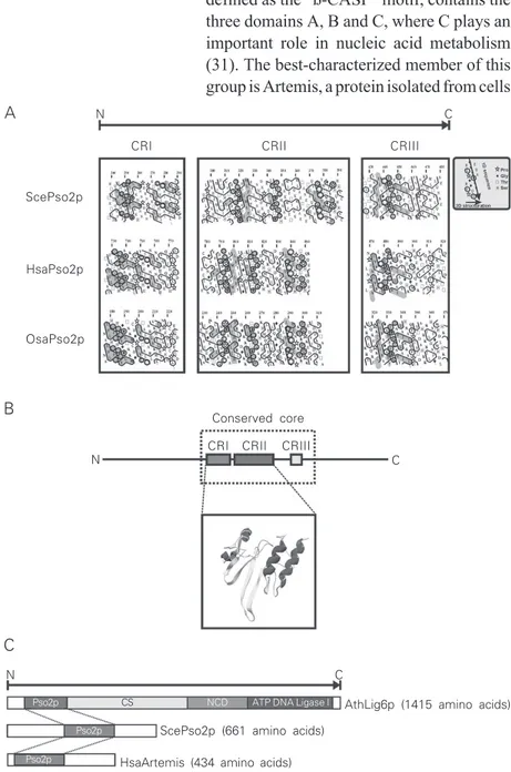
The ß-CASP family
fam-ily, a conserved carboxy-terminal region, defined as the “ß-CASP” motif, contains the three domains A, B and C, where C plays an important role in nucleic acid metabolism (31). The best-characterized member of this group is Artemis, a protein isolated from cells
of patients suffering from a special type of SCID associated with radiosensitivity (RS-SCID) (32). This disease was found in a group of Athabascan-speaking American Indians and has been genetically character-ized (33). An Artemis/DNA-PKcs complex, with endonucleolytic activity on DSBs or hairpins generated by the Rag1/Rag2 pro-teins, might act on NHEJ and V(D)J recom-bination, respectively (34,35). Preliminary protein sequence analyses, including the Artemis/Pso2 sequences, Elac1, Elac2, Cpsf 73-, and Cpsf 100-kDa proteins, indicate similar functions (31). The activity of Elac1/ Elac2 proteins is unknown, but sequence analysis suggests a hydrolase function (36,37). Elac1/Elac2 mutant variants have been associated with human prostate cancer (36). Cpsf 100 kDa and Cpsf 73 kDa hydro-lyze mRNA, and this protein group has con-served domains in eukaryotes as well as in archaea (38). They are important compo-nents of the eukaryotic machinery that pro-cesses the 3' end of mRNAs, acting together with two other Cpsf proteins (30/160 kDa), as well as with the cleavage stimulation factor, poly(ADP-ribose) polymerase, two additional cleavage factors (Im and IIm), and poly(A)-binding protein II (38). Of the three motif domains A, B and C of ß-CASP, domain C, according to HCA, has a con-served hydrophobic residue typical of pro-teins that use DNA as substrate and a histi-dine residue conserved in proteins that bind RNA (31). Our phylogenetic analysis indi-cates that Elac1/Elac2, Cpsf 73/Cpsf 100 and Artemis/Pso2 proteins are paraphyletic, not sharing a recent common ancestor. More-over, the phylogeny of these proteins shows only a functional homology, based on nucleic acid phosphodiesterase activity (Bonatto D, Revers LF, Brendel M and Henriques JAP, unpublished results).
The Pso2/Snm1 protein
Experimental data accumulated over the
N C
CRI CRII CRIII
ScePso2p
HsaPso2p
OsaPso2p
Conserved core
CRI CRII CRIII
N C
N C
AthLig6p (1415 amino acids)
ScePso2p (661 amino acids)
HsaArtemis (434 amino acids) C
B A
Pso2p CS NCD ATP DNA Ligase I
Pso2p
Pso2p
20 years since the isolation and characteriza-tion of pso2/snm1 mutants of S. cerevisiae (39-43; for reviews, see 44,45) so far give no clue to the function of the Pso2p/Snm1p in ICL repair (Table 1). Clearly, pso2/snm1 mutants are extremely sensitive to ICL-in-ducing agents, irrespective of their chemical composition (e.g., ICL induced by 8-MOP + UVA, nitrogen or sulfur mustards, cisplatin, and many others; 39-42); however, they are only mildly sensitive to UVC and not sensitive to ionizing radiation (41,42). Furthermore, S. cerevisiaepso2/snm1 mutants, though inca-pable of forming high molecular weight DNA (data from neutral sucrose gradient assays) during repair of ICL, are not defective in repair of DSBs (40,43). Stability of the mito-chondrial DNA is also affected in these mutants, as they have a higher-than-wild-type phenohigher-than-wild-type frequency of spontaneous “petit” mutations (46). This suggests a pos-sible function for Pso2p/Snm1p in mtDNA recombination or repair in yeast. Pso2p/ Snm1p mutants also have lower induced mutagenesis when compared to the wild-type strain (41).
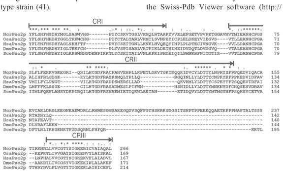
In order to better understand the possible functions of Pso2p in DNA repair of S. cerevisiae, we have used an in silico analysis combining a phylogenetic approach and HCA to characterize the conserved regions (CRs) found between Pso2p and its orthologues. All sequences were obtained directly from GenBank in the National Center for Biotech-nological Information web page [http:// www.ncbi.nlm.nih.gov/] followed by global pair-wise multiple-alignments. The results of the alignments were then used for HCA (DRAWHCA program, available as a freeware at http://www.lmcp.jussieu.fr). Using the closest species of S. cerevisiae, as well as more distant fungal species, we could iden-tify three CRs that are also found in the Artemis/Pso2p/Lig6p sequences of metazoa, protozoa, and plants (Figures 2A-C and 3). These three CRs, which share many con-served amino acid residues (Figures 2A and 3), compose the Pso2p conserved core (CRI, CRII, and CRIII; Figures 2B and 3). It is interesting to note that both CRI and CRII could be three-dimensionally modeled with the Swiss-Pdb Viewer software (http://
Figure 3. Multiple alignment of Pso2p conserved region sequences (CRI to CRIII) from yeast (Saccharomyces cerevisiae, ScePso2p), humans (Homo sapiens, HsaPso2p), filamentous fungi (Neurospora crassa, NcrPso2p), fruit flies (Drosophila melanogaster,DmePso2p), and rice (Oriza sativa, OsaPso2p). Identical amino acid residues are indicated by an asterisk and amino acid residues with similar physico-chemical characteristics by one or two dots in CRs. The positions of the CRs are indicated by arrows.
CRI
CRII
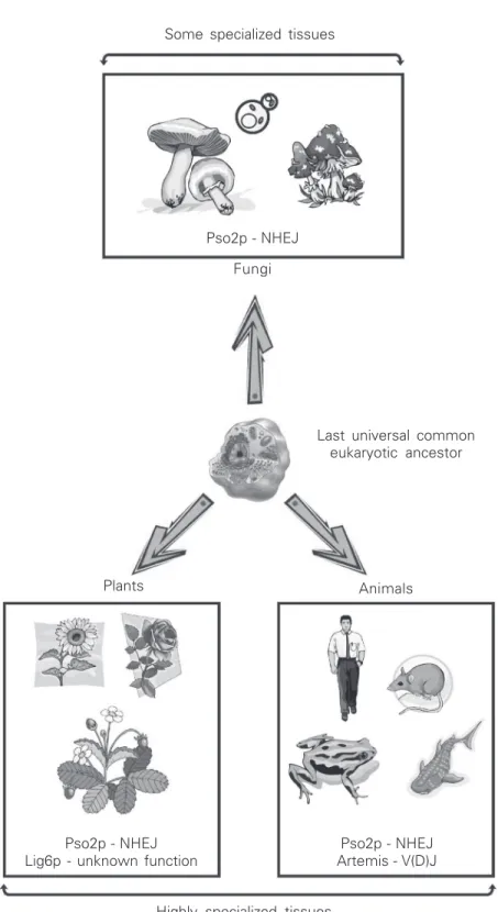
Figure 4. Evolutionary diversification of Pso2p in fungi, animals, and plants from a last universal common eukaryotic ancestor. Artemis and Lig6p are represented within animals and plants, respectively. Animals and plants contain paralogous PSO2 genes, but they are represented by a single sequence for clarity. Fungi contain only one Pso2p sequence. This diversification might be linked to the tissue diversity found in higher eukaryotes (animals and plants). NHEJ = non-homologous end joining.
Some specialized tissues
Highly specialized tissues Pso2p - NHEJ
Fungi
Last universal common eukaryotic ancestor
Animals Plants
Pso2p - NHEJ Lig6p - unknown function
Pso2p - NHEJ Artemis - V(D)J
www.expasy.org/spdbv) (Figure 2B) using as template the penicillinase sequence of Pseudomonas aeruginosa, which belongs to the metallo-ß-lactamase superfamily (Pro-tein Data Bank accession number 1dd6) and exhibited some degree of similarity with Pso2p. All Pso2p sequences analyzed so far show highly divergent N- and C-termini, indicative of different types of enzymatic regulations (Bonatto D, Brendel M and Henriques JAP, unpublished results). More-over, the conserved Pso2p core was found to be associated with other functional do-mains, e.g., plant-specific DNA Lig6p, which contains a DNA ligase I domain in its C-terminus (Figure 2C), and the Pso2p of Aspergillus nidulans, which has a cyto-chrome P450 domain also in its C-terminus (data not shown). The biochemical signifi-cance of these fused domains is still un-known, but we may speculate that these proteins have specific roles in DNA repair or even in chromatin remodeling.
The phylogenetic data indicate the pres-ence of multiple paralogous PSO2 genes that arise from a last universal common eukary-otic ancestor of metazoa and plants. Again we can speculate that the presence of paralo-gous PSO2 genes in multicellular eukaryotes may be associated with the tissue diversity unknown for fungi, suggesting a more spe-cialized function for DNA repair or genome caretaking in plants or metazoa (Figure 4).
In fact, the function of this long 5' UTR may be to maintain hPso2p at low levels since over-expression should be highly toxic to mammalian cells and appears to result in apoptosis (48). Nevertheless, the regulation of hPSO2/hSNM1 during mitosis suggests that this gene may play a role in mitotic progression, particularly in response to ICL-inducing agents, and especially during the G2/M transition. In this regard, it is interest-ing to note that cisplatin-treated cells of the S. cerevisiae pso2 mutant arrest permanently during the G2/M transition (49). The pro-longed arrest in G2/M suggests that the cell is attempting repair or initiating repair in this phase of the cell cycle but cannot complete it without a functional Pso2p (49). Recent data reported by Yu et al. (50) indicate a possible function of Pso2p in DNA repair of hairpins induced by transposition of Ac/Dc elements from Zea mays in S. cerevisiae. In this case, the expression of Ac/Dc elements in S. cerevisiae allows to assay the repair of excision sites in a variety of yeast mutant backgrounds, specifically of DNA hairpins that appear to form in the host DNA during transposition. This indicates that Pso2p may recognize a DNA hairpin as a structure simi-lar to a covalent ICL lesion and may bind to it, as the Artemis protein of vertebrates does during V(D)J recombination (50).
The Artemis protein
The best-characterized member of the ß-CASP family is Artemis (Table 1), which was isolated from cells of patients suffering from a special type of RS-SCID (33). SCID is clinically characterized by opportunistic infections, frequent diarrhea, and failure to thrive. Patients generally die within the first year of life unless treated with, e.g., bone marrow transplantation.
Artemis has 5' to 3' exonucleolytic activ-ity with single-strand DNA specificactiv-ity and, when associated with DNA-PKcs, forms a phosphorylated complex with
endonucle-olytic activity on both 5' and 3' DNA over-hangs; furthermore, it can cleave hairpins generated by the Rag1/Rag2 proteins in V(D)J recombination (34,35). It has been shown that Artemis cooperates with p53 to sup-press chromosomal translocations and tu-mor development in mice. Therefore, it can be considered a tumor suppressor gene. Like other NHEJ/p53 doubly deficient mice, most Artemis-deficient mice succumb to pro-B cell lymphomas by 11-12 weeks of age (10). Despite the striking relationship between NHEJ deficiencies and tumorigenesis in mouse models, potential roles for NHEJ in tumor suppression in humans have remained unclear (10). However, inactivating muta-tions of Ku70, Ku80, DNA-PKcs, XRCC4, and ligase IV have not been observed in the context of human immunodeficiencies, pos-sibly because of a more severe impact of NHEJ mutations on human cells (10). In contrast, mutations in Artemis have been identified in several cohorts of human SCID patients (10). Therefore, the finding that Artemis functions as a tumor suppressor in mice raises the possibility of a similar func-tion in humans. In this regard, hypomorphic alleles of Artemis have been identified in humans and have been associated with a predisposition to lymphomas (18).
Interestingly, the use of a transposition system named Sleeping Beauty in an Artemis-deficient mammalian cell line does not
in-crease the cell’s sensitivity to DSB (51). Sleeping Beauty is a Tc1/mariner-like trans-posable element that, like retroviral integrases
Rad4p-Rad23p complex
Hairpin formation ICL
HR pathways
Mre11p/Rad50p/Xrs2p complex
Cruciform DNA structure ICL
ICL
ICL induction
Exponential growth phase
Artemis/DNA-PK/Ku - Vertebrates Snm1p/Pak1p/yKup - Yeast
Degradation
Pso2p/(Artemis) - phosphorylated Pak1p (DNA-PK)
yKup (Ku)
DNA Pol4p (DNA polymerase lambda) DNA ligase IV
yKup (Ku) PCNA
PCNA
High molecular weight DNA
SSB proteins
Topoisomerase I
Normal DNA replication DNA polymerase
complex
3’ 5’
3’ 5’ 3’
5’
3’ 5’
NER and HR proteins
Figure 5. Non-homologous end joining recombination mediated by Snm1p/Pso2p in growing cells after inter-strand cross-link-ing (ICL) induction durcross-link-ing DNA replication. In the presence of a sister strain, DNA repair may pro-ceed via homologous recombi-nation (HR pathways) mediated by Rad4p-Rad23p and HR pro-teins. Alternatively, the ICL can induce the formation of cruci-form DNA structures, especially when palindromic sequences are present. These cruciform structures are recognized by Mre11p/Rad50p/Xrs2p com-plex that cuts the single-strand DNA regions and induces the formation of DNA hairpins. These DNA hairpins are cleaved by the phosphorylated Artemis (Snm1p-like) DNA-PK/Ku pro-tein complex in metazoa or by phosphorylated Snm1/Pak1p/ yKup in fungi, generating a sub-strate for DNA polymerase λ (Pol4p in yeast) and DNA ligase IV, which perform, together with Ku and PCNA, the rejoining of non-homologous DNA frag-ments (gray DNA chain) and res-titute the DNA replication pro-cess. NER = nucleotide exci-sion repair; PCNA = proliferat-ing cell nuclear antigen; SSB = single-strand binding proteins.
3’ 5’ 3’
5’
and the Rag1 V(D)J recombinase, catalyzes a remarkably similar “overall chemistry” of DNA recombination. However, the structure of Sleeping Beauty transposition intermedi-ates is unknown, and they probably do not comprise DNA hairpins, as was seen in Ac/ Ds elements of maize (51).
Artemis protein was recently used by Poinsignon et al. (52) for site-specific mu-tagenesis in order to dissect the role of the metallo-ß-lactamase and ß-CASP domains of Artemis with regard to V(D)J recombina-tion and DNA repair after ionizing radiarecombina-tion. This study demonstrated that Artemis can be divided into two critical regions, with the COOH-terminal region probably playing an important role in protein stabilization and in DNA repair after ionizing radiation (52). However, the authors concentrated their ef-forts on the study of the CRI and CRII of the Pso2p catalytic core (which encompasses the metallo-ß-lactamase and ß-CASP do-mains), necessary for V(D)J recombination but not for DNA repair. In this case, the CRIII should be required for DNA repair functions induced by ionizing radiation or even by ICLs.
The Pso2p/Snm1p of plants: a special case
In contrast to animals, plants are con-stantly being challenged by sunlight-con-tained UV radiation because of their obliga-tory requirement of sunlight for photosyn-thesis (53). This radiation penetrates plant surface tissues and damages their genome and other cellular targets such as photosys-tem II and plasma membrane ATPase (53). Characteristically, plants also show endo-phytic fungi living asymptomatically within their tissues (54), where they can produce potentially DNA-damaging mycotoxins (55). Moreover, secondary metabolites (e.g., furocoumarin) can be photo-activated by sunlight and induce DNA ICLs in leaves or aerial parts (55). It is thus likely that different
DNA repair systems are required to repair the errors induced by biotic or abiotic factors in a plant’s genome. The NHEJ process in plant tissues is largely unknown, and the DSB repair products have been characterized as excision products of transposable elements, or insertion products of Agrobacterium sp T-DNA (56). Interestingly, the analysis of NHEJ proteins in A. thaliana (e.g., DNA ligase IV, Ku80, and XRCC4) indicates the conservation of basic DSB repair mechan-isms (26).
Using the available genomic information from public databases, we have carried out a phylogenetic study with the aim to find plant-specific Pso2p sequences. Interestingly, we detected paralogous PSO2 genes in the com-plete genomes of A. thaliana and O. sativa, and also a new group of ATP-dependent DNA ligases that contain a Pso2p catalytic core (Table 1, Figure 4) (57). The sequence analyses of these proteins show that the Pso2p catalytic core is localized within the N-terminal part of the protein, while a DNA ligase I domain can be detected in the C-terminal end (Figure 2C), with both domains displaying homology with Pso2p and DNA ligase I of animals and yeasts. Moreover, additional data of microsynteny analysis indi-cate that these genes of the new DNA ligase family are linked to the S and SLL2 loci of Brassica sp and A. thaliana, respectively. It should be noted that the Brassica S and the
Arabidopsis SLL2 loci consist of a gene
func-References
1. Lees-Miller SP & Meek K (2003). Repair of DNA double strand breaks by non-homologous end joining. Biochimie, 85: 1161-1173.
2. Jackson SP (2002). Sensing and repairing DNA double-strand breaks. Carcinogenesis, 23: 687-696.
3. Kuziminov A (2001). Single-strand interruptions in replicating chro-mosomes cause double-strand breaks. Proceedings of the Na-tional Academy of Sciences, USA, 98: 8241-8246.
4. Bernstein C, Bernstein H, Payne CM & Garewal H (2002). DNA repair/pro-apoptotic dual-role proteins in five major DNA repair pathways: fail-safe protection against carcinogenesis. Mutation Research, 511: 145-178.
5. Khanna KK & Jackson SP (2001). DNA double-strand breaks: signaling, repair and the cancer connection. Nature Genetics, 27: 247-254.
6. Rich T, Allen RL & Wyllie RH (2000). Defying death after DNA
tion seems to be essential. Biochemical anal-ysis as well as mutational studies are cur-rently in progress in order to determine the roles of these plant-specific DNA ligases in DNA metabolism.
Unfortunately, little is known about the Pso2 proteins in plants. However, the pres-ence of paralogous PSO2 genes in A. thali-ana and O. sativa is a good indication that, like the tissue diversity found in metazoa, the presence of specialized plant tissues may have specific requirements for repair of DSB or ICL DNA repair.
Concluding remarks
The studies of Pso2p functions in DNA repair or in genome maintenance are just beginning. Since most of the information on putative Pso2p functions comes from its human homologue Artemis, more research is necessary in order to clarify the exact role of Pso2p in DNA metabolism. Since its first genetic studies using mutants of S. cerevi-siae sensitive to photo-addition of bi-func-tional psoralens and to nitrogen mustards (39-44), little information has been obtained by conventional genetical approaches. If Pso2p is necessary for reconstitution of high molecular weight DNA, why do yeast mu-tants, cell lines, or even animal models knocked-out for PSO2 show a wild-type response phenotype to DNA damaging agents, except ICL-generating chemicals? The an-swer to this question may be found in the structure of DNA, more specifically in the secondary structures like DNA hairpins that
can arise from palindromic regions during DNA replication slippage or stalled DNA replication forks (59). Recently, we pro-posed a model where Pso2p would act on DNA hairpin substrates induced by ICLs during DNA replication (46), a feature also shown in the present review (Figure 5). This model proposes that the potential endonucle-olytic function of Pso2p is activated via Pak1p-induced phosphorylation. The spe-cific function in DNA repair of this potential protein kinase of S. cerevisiae is unknown, but when over-expressed, Pak1p acts as a suppressor of thermo-labile DNA polymer-ase α mutations (60). Pak1p was identified in a two-hybrid screening of potential protein partners of Pso2p (Revers LF, Strauss M, Bonatto D, Brendel M and Henriques JAP, unpublished results). Our model helps to explain the specific function of Pso2p in repair of DSB that are generated during repair processing of ICLs. Moreover, it also helps to explain the evolution of Artemis in terms of its function on V(D)J recombination. Since Artemis also binds hairpin-capped DNA ends induced by RAG proteins, it may also have the ability to bind hairpin-intermediates gen-erated during some step(s) of DNA ICL repair.
damage. Nature, 407: 777-783.
7. Richardson C & Jasin M (2000). Frequent chromosomal transloca-tions induced by DNA double-strand breaks. Nature, 405: 697-700.
8. Rooney S, Alt FW, Lombard D, Whitlow S, Eckersdorff M, Fleming J, Fugmann S, Ferguson DO, Schatz DG & Sekiguchi J (2003). Defective DNA repair and increased genomic instability in Artemis-deficient murine cells. Journal of Experimental Medicine, 197: 553-565.
9. Takata M, Sasaki MS, Sonoda E, Morrison C, Hashimoto M, Utsumi H, Yamaguchi-Iwai Y, Shinohara A & Takeda S (1998). Homologous recombination and non-homologous end-joining pathways of DNA double-strand break repair have overlapping roles in the mainte-nance of chromosomal integrity in vertebrate cells. EMBO Journal, 17: 5497-5508.
10. Richardson C & Jasin M (2000). Coupled homologous and non-homologous repair of a double-strand break preserves genomic integrity in mammalian cells. Molecular and Cellular Biology, 20: 9068-9075.
11. Lieber MR, Ma Y, Pannicke U & Schwarz K (2003). Mechanism and regulation of human non-homologous DNA end-joining. Nature Reviews. Molecular Cell Biology, 4: 712-720.
12. Gu Y, Seidl K, Rathbun GA et al. (1997). Growth retardation and leaky SCID phenotype of Ku70-deficient mice. Immunity, 7: 653-665.
13. Frank KM, Sekiguchi JM, Seidl KJ, Swat W, Rathbun GA, Cheng HL, Davidson L, Kangaloo L & Alt FW (1998). Late embryonic lethality and impaired V(D)J recombination in mice lacking DNA ligase IV. Nature, 396: 173-177.
14. Gao Y, Sun Y, Frank K et al. (1998). A critical role for DNA end-joining proteins in both lymphogenesis and neurogenesis. Cell, 95: 891-902.
15. Grawunder U, Zimmer D, Fugmann S, Schwarz K & Lieber MR (1998). DNA ligase IV is essential for V(D)J recombination and DNA double-strand break repair in human precursor lymphocytes. Mo-lecular Cell, 2: 477-484.
16. Li GC, Ouyang H, Li X et al. (1998). Ku70: a candidate tumor suppressor gene for murine T cell lymphoma. Molecular Cell, 2: 1-8.
17. Ma Y, Pannicke U, Schwarz K & Lieber MR (2002). Hairpin end opening and overhang processing by an Artemis/DNA-dependent protein kinase complex in non-homologous end joining and V(D)J recombination. Cell, 108: 781-794.
18. Rooney S, Sekiguchi J, Whitlow S, Eckersdorff M, Manis JP, Lee C, Ferguson DO & Alt FW (2004). Artemis and p53 cooperate to suppress oncogenic N-myc amplification in progenitor B cells.
Proceedings of the National Academy of Sciences, USA, 101: 2410-2415.
19. Labhart P (1999). Nonhomologous DNA end joining in cell-free systems. European Journal of Biochemistry, 265: 849-861. 20. Smith GC & Jackson SP (1999). The DNA-dependent protein
kinase. Genes and Development, 13: 916-934.
21. Hartley KO, Gell D, Smith GC, Zhang H, Divecha N, Connelly MA, Admon A, Lees-Miller SP, Anderson CW & Jackson SP (1995). DNA-dependent protein kinase catalytic subunit: a relative of phos-phatidylinositol 3-kinase and the ataxia telangiectasia gene prod-uct. Cell, 82: 849-856.
22. Poltoratsky VP, Shi X, York JD, Lieber MR & Carter TH (1995). Human DNA-activated protein kinase (DNA-PK) is homologous to phosphatidylinositol kinases. Journal of Immunology, 155: 4529-4533.
23. Dynan WS & Yoo S (1998). Interaction of Ku protein and DNA-dependent protein kinase catalytic subunit with nucleic acids.
Nucleic Acids Research, 26: 1551-1559.
24. West CE, Waterworth WM, Story GW, Sunderland PA, Jiang Q & Bray CM (2002). Disruption of the ArabidopsisAtKu80 gene dem-onstrates an essential role for AtKu80 protein in efficient repair of DNA double-strand breaks in vivo. Plant Journal, 31: 517-528. 25. Tamura K, Adachi Y, Chiba K, Oguchi K & Takahashi H (2002).
Identification of Ku70 and Ku80 homologues in Arabidopsis thalia-na: evidence for a role in the repair of DNA double strand breaks.
Plant Journal, 29: 771-781.
26. van Attikum H, Bundock P, Overmeer RM, Lee L-Y, Gelvin SB & Hooykaas PJJ (2003). The ArabidopsisAtLIG4 gene is required for the repair of DNA damage, but not for the integration of Agrobacte-rium T-DNA. Nucleic Acids Research, 31: 4247-4255.
27. Lee KJ, Huang J, Takeda Y & Dynan WS (2000). DNA ligase IV and XRCC4 form a stable mixed tetramer that functions synergistically with other repair factors in a cell-free end-joining system. Journal of Biological Chemistry, 275: 34787-34796.
28. Lee JW, Yannone SM, Chen DJ & Povirk LF (2003). Requirement for XRCC4 and DNA ligase IV in alignment-based gap filling for nonhomologous DNA end joining in vitro. Cancer Research, 63: 22-24.
29. West CE, Waterworth WM, Jiang Q & Bray CM (2000). Arabidopsis
DNA ligase IV is induced by γ-irradiation and interacts with an
Arabidopsis homologue of the double-strand break repair protein XRCC4. Plant Journal, 24: 67-78.
30. D’Amours D & Jackson SP (2002). The Mre11 complex: at the crossroads of DNA repair and checkpoint signalling. Nature Re-views in Molecular and Cellular Biology, 3: 317-327.
31. Callebaut I, Moshous D, Mornon J-P & de Villartay J-P (2002). Metallo-ß-lactamase fold within nucleic acids processing enzymes: the ß-CASP family. Nucleic Acids Research, 30: 3592-3601. 32. Noordzij JG, Verkaik NS, Van der Burg M et al. (2003).
Radiosensi-tive SCID patients with Artemis gene mutations show a complete B-cell differentiation arrest at the pre-B-cell receptor checkpoint in bone marrow. Blood, 101: 1446-1452.
33. Moshous D, Li L, Chasseval R, Philippe N, Jabado N, Cowan MJ, Fischer A & de Villartay J-P (2000). A new gene involved in DNA double-strand break repair and V(D)J recombination is located on human chromosome 10p. Human Molecular Genetics, 9: 583-588. 34. Jeggo P & O’Neill P (2002). The Greek goddess, Artemis, reveals
the secrets of her cleavage. DNA Repair, 1: 771-777.
35. Schlissel MS (2002). Does Artemis end the hunt for the hairpin-opening activity in V(D)J recombination? Cell, 109: 1-4.
36. Tavtigian SV, Simard J, Teng DHF et al. (2001). A candidate prostate cancer susceptibility gene at chromosome 17p. Nature Genetics, 27: 172-180.
37. Vogel A, Schilling O, Niecke M, Bettmer J & Meyer-Klaucke W (2002). ElaC encodes a novel binuclear zinc phosphodiesterase.
Journal of Biological Chemistry, 277: 29078-29085.
38. Chanfreau G, Noble SM & Guthrie C (1996). Essential yeast protein with unexpected similarity to subunits of mammalian cleavage and polyadenylation specificity factor (CPSF). Science, 274: 1511-1514. 39. Henriques JA & Moustacchi E (1980). Isolation and characterization of pso mutants sensitive to photo-addition of psoralen derivatives in Saccharomyces cerevisiae. Genetics, 95: 273-288.
Sciences, USA, 79: 1722-1726.
41. Cassier C, Chanet R, Henriques JA & Moustacchi E (1980). The effects of three PSO genes on induced mutagenesis: a novel class of mutationally defective yeast. Genetics, 96: 841-857.
42. Ruhland A, Haase E, Siede W & Brendel M (1981). Isolation of yeast mutants sensitive to the bifunctional alkylating agent nitrogen mustard. Molecular and General Genetics, 181: 346-351. 43. Wilborn F & Brendel M (1989). Formation and stability of
inter-strand cross-links induced by cis- and trans-diamminedichloroplati-num (II) in the DNA of Saccharomyces cerevisiae strains differing in repair capacity. Current Genetics, 16: 331-338.
44. Brendel M & Henriques JA (2001). The pso mutants of Saccharo-myces cerevisiae comprise two groups: one deficient in DNA repair and another with altered mutagen metabolism. Mutation Research, 489: 79-96.
45. Brendel M, Bonatto D, Strauss M, Revers LF, Pungartnik C, Saffi J & Henriques JAP (2003). Role of PSO genes in repair of DNA damage of Saccharomyces cerevisiae. Mutation Research, 544: 179-193.
46. Da Silva KVCL & Henriques JAP (1987). Induction of the cytoplas-mic ‘petite’ mutation in pso mutants of Saccharomyces cerevisiae
by photoaddition of furocoumarins or by ultraviolet radiation. Mu-tagenesis, 2: 187-197.
47. Lambert S, Mason SJ, Barber LJ, Hartley JA, Pearce JA, Carr AM & McHugh PJ (2003). Schizosaccharomyces pombe checkpoint re-sponse to DNA inter-strand cross-links. Molecular and Cellular Biology, 23: 4728-4737.
48. Zhang X, Richie C & Legerski RJ (2002). Translation of hSNM1 is mediated by an internal ribosome entry site that upregulates expression during mitosis. DNA Repair, 1: 379-390.
49. Grossmann KF, Ward AM & Moses RE (2000). Saccharomyces cerevisiae lacking Snm1, Rev3 or Rad51 have a normal S-phase but arrest permanently in G2 after cisplatin treatment. Mutation Research, 461: 1-13.
50. Yu J, Marshall K, Yamaguchi M, Haber JE & Weil CF (2004). Microhomology-dependent end joining and repair of transposon-induced DNA hairpins by host factors in Saccharomyces cerevisiae.
Molecular and Cellular Biology, 24: 1351-1364.
51. Izsvák Z, Stüwe EE, Fiedler D, Katzer A, Jeggo PA & Ivics Z (2004). Healing the wounds inflicted by Sleeping Beauty transposition by double-strand break repair in mammalian somatic cells. Molecular Cell, 13: 279-290.
52. Poinsignon C, Moshous D, Callebaut I, de Chasseval R, Villey I & de Villartay J-P (2004). The metallo-ß-lactamase/ß-CASP domain of Artemis constitutes the catalytic core for V(D)J recombination.
Journal of Experimental Medicine, 199: 315-321.
53. Tuteja N, Singh MB, Misra MK, Bhalla PL & Tuteja R (2001). Molecular mechanisms of DNA damage and repair: progress in plants. Critical Reviews in Biochemistry and Molecular Biology, 36: 337-397.
54. Faeth SH (2002). Are endophytic fungi defensive plant mutualists.
OIKOS, 98: 25-36.
55. Choi JJ, Klosterman SJ & Hadwiger LA (2001). A comparison of the effects of DNA-damaging agents and biotic elicitors on the induc-tion of plant defense genes, nuclear distorinduc-tion, and cell death.
Plant Physiology, 125: 752-762.
56. Scott L, LaFoe D & Weil CF (1996). Adjacent sequences influence DNA repair accompanying transposon excision in maize. Genetics, 142: 237-246.
57. Bonatto D, Brendel M & Henriques JAP (2005). A new group of plant specific ATP-dependent DNA ligases identified by protein phylogeny, hydrophobic cluster analysis, and three-dimensional modeling. Functional Plant Biology (in press).
58. Matton DP, Nass N, Clarke AE & Newbigin E (1994). Self-incompat-ibility: how plants avoid illegimate offspring. Proceedings of the National Academy of Sciences, USA, 91: 1992-1997.
59. Lobachev KS, Gordenin DA & Resnick MA (2002). The Mre11 complex is required for repair of hairpin-capped double-strand breaks and prevention of chromosome rearrangements. Cell, 108: 183-193.