Drug Delivery Systems for Bone Regeneration
Joana Vicente Nave
M
ASTERT
HESISI
NTEGRATEDM
ASTERS INB
IOENGINEERINGSupervisor: Inês Alencastre (PhD)
Co-Supervisor: Ana Paula Pêgo (PhD)
ii
iii
R
ESUMO
A remodelação óssea é regulada por um equilíbrio entre as actividades osteoblástica e osteoclástica. No entanto, em condições clínicas complexas, como sejam as fracturas críticas, a regeneração óssea fica comprometida, sendo requerido um auxílio externo. Deste modo, novos métodos que permitam regular a homeostasia do osso têm sido estudados.
O Neuropéptido Y (NPY) exerce um papel importante na regulação do metabolismo e massa óssea. Estudos realizados no receptor Y1 (um dos 5 receptores do sistema NPY) demonstraram que o seu bloqueio tem um impacto positivo na massa óssea. No entanto, o receptor Y1, que se encontra nas membranas celulares, tem uma ampla distribuição ao longo do organismo, com impacto em outros sistemas fisiológicos para além do osso. Deste modo, o objectivo geral deste projecto consiste na concepção de um sistema de entrega local de fármacos para a entrega no meio extracelular do BIBP3226, um potente e selectivo antagonista do receptor Y1. Com este objectivo, sistemas baseados em partículas e nanofibras foram preparados e optimizados.
Estudos revelaram que para tamanhos abaixo de 200 nm e acima de 500 nm, as partículas têm tendência a ser internalizadas por, respectivamente, células não-fagocíticas e fagocíticas, limitando assim a pretendida distribuição extracelular do fármaco. Deste modo, através da aplicação dos métodos de Salting-out e Nanoprecipitação, foram preparadas partículas de Poli(ácido lático-co-ácido glicólico) com tamanhos compreendidos entre 100 e 1000 nm, de modo a testar a hipótese formulada sobre a internalização em células osteoclásticas derivadas da medula óssea. Resultados mostraram que se uma célula estiver capacitada de realizar fagocitose, ela irá internalizar partículas com tamanhos acima de 500 nm numa escala superior quando comparado a partículas de tamanhos inferiores a 500 nm. Em adição, foi ainda notável que células com mais de 8 núcleos, internalizam menos que células com 2 ou 3 núcleos, levando a concluir que à medida que a célula é mais matura em relação ao fenótipo osteoclástico, menor é a sua capacidade de internalizar partículas. Num estudo realizado em paralelo, os volumes utilizados no método de Salting-out foram variados. Os resultados desta variação mostraram diferenças significativas na eficiência de encapsulação das partículas, mas não para nos seus tamanhos.
Relativamente às nanofibras, estas foram preparadas por Electrospinning, usando Policaprolactona. Estes sistemas têm não só a capacidade de realizar uma entrega de fármacos eficiente, como também fornecem suporte como scaffold. Diferentes formulações, no que diz respeito à concentração de polímero e tipo de solvente orgânico, foram testadas. No entanto, devido à degradação do polímero, o procedimento não foi optimizado. Deste modo, novas experiências neste tópico devem ser realizadas.
v
A
BSTRACT
Bone remodeling is tight regulated by an equilibrium between the osteoblastic and osteoclastic activity. However, in complex clinical conditions, such as critical fractures, bone regeneration is impaired and external help is required. Therefore, new methods of controlling bone homeostasis have been studied.
Neuropeptide Y (NPY) was found to have an important role in the regulation of bone metabolism and mass. In effect, studies have shown that blocking Y1 receptor (one of the 5 receptors from the NPY system) has a positive impact in bone mass. However, Y1 receptor, which is located at cell membranes, has a wide distribution throughout the body, with impact in other physiological systems. Therefore, the overall goal of the project is the design of a local drug delivery system for the extracellular delivery of BIBP3226, a potent and selective Y1 antagonist. Therefore, particles and nanofibers based systems were optimized and prepared.
Studies have revealed that for sizes below 200 nm and above 500 nm, particles have a tendency to be internalized by non-phagocytic and phagocytic cells, respectively, thereby hampering the aimed extracellular delivery. Therefore, the Salting-out and Nanoprecipitation methods were applied in the preparation of Poly(lactide-co-glycolide) particles with sizes ranging from 100 nm to 1000 nm, in order to test the abovementioned internalization hypothesis in Bone Marrow derived Osteoclast Lineage cells. Results have shown that if a cell is capable of internalize particles with sizes above 500 nm (i.e., if a cell is capable of performing phagocytosis), it will internalize them in a larger scale than particles with sizes below 500 nm, as Large particles were, in a general way, more internalized than the other differently sized particles. Furthermore it was noticeable a tendency for a lower internalization in cells with more than 8 nuclei, leading to the conclusion that as the cell is more mature towards osteoclasts phenotype, it has less capacity to internalize particles. In a parallel study, the volumes used in the preparation of particles by Salting-out were varied in order to assess if this variability had influence on the particles’ size and drug loading efficiency. The obtained results have shown significant differences in loading, but not in size.
Regarding nanofibers, these were prepared by Electrospinning, using Polycaprolactone. Nanofibers have not only the capacity of performing an efficient drug delivery, but they can also provide support as a scaffold. Different formulations regarding polymer concentration and the organic solvent were tested. However, due to the polymers’ degradation, this procedure could not be optimized. Therefore, further experiments on this topic should be performed.
vii
A
KNOWLEDGMENTS
Em primeiro lugar, quero agradecer à minha orientadora, Inês Alencastre, por todo o apoio, carinho e pela paciente orientação que me disponibilizou ao longo deste percurso, exigindo sempre mais e melhor. Estou também verdadeiramente grata à minha co-orientadora, Ana Paula Pêgo, por todos os conselhos, apoio incondicional e ensinamentos que contribuíram para o meu crescimento intelectual e como pessoa. Agradeço ainda a toda a equipa NOG que me acolheu no desenvolver deste projecto: à Meriem Lamghari por toda a motivação e ânimo no trabalho, à Dani e ao Francisco que tanto batalharam comigo para que tudo corresse bem, à Juliana pela simpatia, preocupação e auxílio, à Estrelinha que tantas vezes abdicou do seu tempo para me ensinar e orientar sempre no melhor caminho, com toda a simpatia, carinho e alegria e, porque os últimos são sempre os primeiros, ao Luís Campeão, o meu maior apoio destes meses, a quem um bilião de agradecimentos seria muito pouco. Quero também expressar a minha gratidão a todos os que de uma maneira ou outra ajudaram e disponibilizaram o seu tempo no desenvolvimento deste projecto, em especial à Rute Nunes, à Vicky, à Juliana Dias, à Joana Furtado, à Maria Lazaro e ao João Martins.
Aos meus pais quero agradecer com todo o meu coração por sempre me terem dado do pouco que tinham para eu conseguir alcançar uma vida melhor. Ao meu irmão agradeço pelo companheirismo, amizade e por toda a educação que me deu como segundo pai que sempre foi. Aos meus primos, tios e avó, por toda a amizade, alegria e apoio ao longo desta caminhada, deixo-lhes um brinde ao Monsieur Conan Correia!
A todos os grandes (enormes!!) amigos que fiz durante estes 5 anos, agradeço por todo este percurso de jantaradas, risadas e boas (ou más acabadas em boas) histórias. Aos Sal(sich)inhas por todos os dias de trabalho que se tornaram hilários. Ao conjunto das 8 Bacocas mais os Faustos, de quem guardo as melhores recordações (mas decerto que o futuro ainda nos guarda muitas mais!). To Sarah, Iza, Ula and Laura, the funniest and happiest girls I’ve ever met! À minha Clarinha e Bruna, por todas as vezes em que pensaram no meu colesterol e ainda assim acharam por bem fazer-me massa com atum para jantar, por todas as conversas incluídas nos capítulos “um dia, quando formos velhinhas, vamo-nos lembrar destas conversas de sofá” e por todo o apoio e amizade incondicionais, 23 horas por dia (na outra hora eu estava a lavar a louça… sozinha). Ao António, por sempre acreditares no meu sucesso, por toda a paciência, amizade e carinho que partilhámos ao longo deste tempo.
ix
“I have not failed. I’ve just found 10,000 ways that won’t work.”
xi
C
ONTENTS
CHAPTER 1 ... 19 Literature Review ... 19 1.1. BONE ... 19 1.1.1. General Considerations ... 19
1.1.2. Formation and Remodeling ... 23
1.2. NPY SYSTEM IN BONE REMODELING ... 26
1.2.1. NPY Y1 Receptor ... 26
1.2.2. Anti Y1 Receptor Therapeutic Strategies ... 28
1.3. DRUG DELIVERY SYSTEMS TO BONE ... 33
1.3.1. Current Therapeutic Approaches for Bone Regeneration ... 33
1.3.2. Nanoparticles Drug Delivery Systems ... 35
1.3.2.1. Polycaprolactone, Poly(lactide-co-glycolide) and Monoethoxy poly(ethylene glycol)-poly(trimethylene carbonate) Polymers ... 36
1.3.2.2. Nanoparticle Delivery Systems for the Extracellular Space ... 38
1.3.3. Nanofibrous Drug Delivery Systems ... 39
1.3.3.1. Electrospinning Process and Therapeutic Applications ... 40
CHAPTER 2 ... 43
Aim of the Thesis ... 43
CHAPTER 3 ... 45
Materials and Methods ... 45
3.1. Preparation and Characterization of the Polymeric Particles ... 45
3.1.1. Particles Preparation ... 45
xii
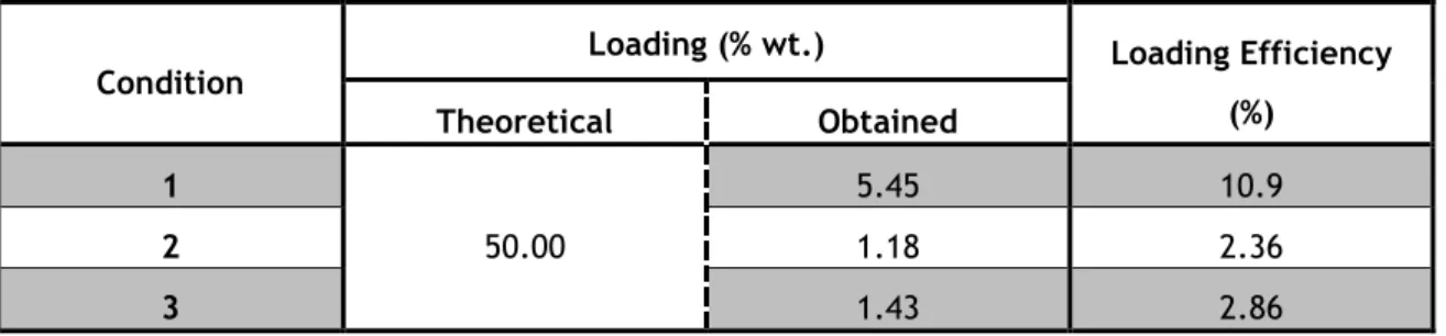
3.1.5. Loading Efficiency of Dexamethasone in the Particles ... 47
3.2. Biocompatibility and Internalization assays ... 48
3.2.1. Cell Culture ... 48
3.2.2. Resazurin Assay ... 48
3.2.3. Live/Dead Assay ... 48
3.2.4. Immunocytochemistry for F-Actin assay ... 49
3.3. Electrospun Fibers ... 49
3.4. Statistical Analysis ... 49
CHAPTER 4 ... 51
Results and Discussion ... 51
4.1. Optimization of the Particles Preparation Procedure ... 51
4.1.1. Polymer Effect ... 53
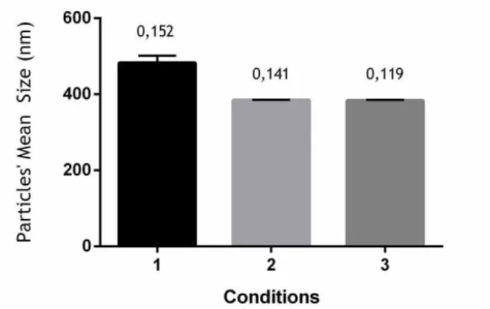
4.1.2. Effect of Volumes’ Variations on both Particles Size and Drug Loading Efficiency . ... .54
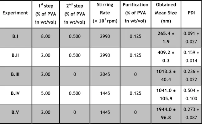
4.1.3. Particles’ Size Optimization ... 56
4.2. Particles Cytotoxicity ... 63
4.3. Evaluation of the Internalization ... 65
4.4. Nanofibers ... 76
CHAPTER 5 ... 79
Conclusion and Future Perspectives ... 79
xiii
L
IST OF
S
YMBOLS AND
A
BBREVIATIONS
α – MEM Minimum Essential Medium Eagle – Alpha Modification ACN Acetonitrile
AR Aspect-Ratio
BMC Bone Marrow derived Osteoclast Lineage Cells BMPs Bone Morphonegenetic Proteins
BSA Bovine Serum Albumin C Conditional Knockout
d Diameter
DAPI 4′,6-Diamidino-2-phenylindole dihydrochloride DCM Dichloromethane
DLS Dynamic Light Scattering DMF N,N- Dimethylformamide DMSO Dimethyl Sulfoxide ECM Extracellular Matrix
EDTA Ethylenediamine Tetraacetic Acid FBS Fetal Bovine Serum
G Germline Knockout
h Height
Hyp Hypothalamus Knockout
M-CSF
Macrophage Colony-Stimulating Factor MgCl2.6H2O Magnesium Chloride HexahydratemPEG-PTMC Monoethoxy poly(ethylene glycol)-poly(trimethylene carbonate) MPS Mononuclear Phagocyte System
MSCs Mesenchymal Stem Cells MSNs Mesoporous Silica Nanoparticles NPY Neuropeptide Y
PBS Phosphate Buffered Saline PCL Polycaprolactone
PDI Polydispersity Index PEG Polyethylene Glycol PFA Paraformaldehyde PLA Poly Lactid Acid PGA Poly Glycolic Acid
xiv PTMC Poly(trimethylene carbonate) PVA Poly(vinyl alcohol)
PVN Paraventricular Nucleus
RANKL Receptor Activator of Nuclear Factor Kappa-B L
igand
rhBMP-2 Recombinant Human Morphogenetic Protein-2 rhBMP-7 Recombinant Human Bone Morphogenetic Protein-7 RNA Ribonucleic Acid
Rpm Rotations per Minute RT Room Temperature
RT-PCR Real Time – Polymerase Chain Reaction TRAP Tartrate Resistant Acid Phosphatase TGF-β Transforming Growth Factor β THF Tetrahydrofuran
Wt Weight
xv
L
IST OF
F
IGURES
Figure 1. Schematic view of human long and cortical bone. ... 21
Figure 2. Overview of the different levels on bone structure.. ... 22
Figure 3. Schematic view of the electrospinning process. ... 41
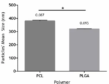
Figure 4. Mean Size of the particles prepared with PCL and PLGA. ... 53
Figure 5. Mean size of the particles prepared with different volumes. ... 55
Figure 6. Size of the particles prepared with the nanoprecipitation method.. ... 60
Figure 7. Particles Cytotoxicity results for BMC after 3 days of differentiation and upon 3 hours of incubation with Coumarin-6 loaded particles. ... 64
Figure 8. Particles Cytotoxicity results for BMC after 7 days of differentiation and upon 3 hours of incubation with Coumarin-6 loaded particles. ... 64
Figure 9. Representative fluorescent micrographs showing Bone Marrow derived Osteoclast Lineage Cells (BMC) after 3 days of culture and upon 3 hours of incubation with Coumarin-6 loaded particles. ... 67
Figure 10. Representative fluorescent micrographs showing Bone Marrow derived Osteoclast Lineage Cells (BMC) after 7 days of culture and upon 3 hours of incubation with Coumarin-6 loaded particles. ... 69
Figure 11. Percentage of Internalization of Coumarin-6 loaded particles, upon 3 hours of incubation, in mono and multinucleated cells cultured for 3 days (A) and 7 days (B). ... 69
Figure 12. Representative fluorescent micrographs showing Bone Marrow derived Osteoclast Lineage Cells (BMC) after 3 days of culture and upon 3 hours of incubation with Coumarin-6 loaded particles.. ... 72
Figure 13. Representative fluorescent micrographs showing Bone Marrow derived Osteoclast Lineage Cells (BMC) after 7 days of culture and upon 3 hours of incubation with Coumarin-6 loaded particles.. ... 73
Figure 14. Average number of internalized Coumarin-6 loaded particles, upon 3 hours of incubation, per mono and multinucleated cell after 3 days (A) and 7 days (B) of incubation.. . 74
Figure 15. Nanofibers produced by electrospinning at 1Kv/cm, with a distance of 14 cm between the spinneret and the collector.. ... 78
xvii
L
IST OF
T
ABLES
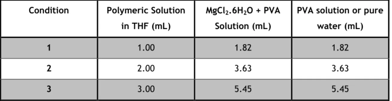
Table I. Summary of bone phenotypes from mice after deletion of Y receptors. ... 27 Table II. Summary of the peptide and non-peptide antagonists for Y1 Receptor ... 28 Table III. Volumes of solutions used in the Salting-out method. ... 46 Table IV. Centrifuge forces used in the purification of the prepared particles with different polymers: PCL, mPEG-PTMC and PLGA. ... 46 Table V. Drug loading efficiency of the prepared Dexamethasone loaded PLGA particles with different volumes. ... 56 Table VI. PLGA particles size and preparation conditions. ... 58 Table VII. PLGA particles size after the optimization of the parameters in the preparation procedure.. ... 58 Table VIII. Sizes and respective PDI results for the particles used in the cytotoxic assessment and internalization studies after 3 and 7 days of cell culture differentiation.. ... 62 Table IX. Percentage of Internalization of Coumarin-6 loaded particles, upon 3 hours of incubation, in mono and multinucleated cells cultured for 3 days.. ... 70 Table X. Percentage of Internalization of Coumarin-6 loaded particles, upon 3 hours of incubation, in mono and multinucleated cells cultured for 7 days. ... 70 Table XI. Average number of internalized Coumarin-6 loaded particles, upon 3 hours of incubation, per mono and multinucleated cell after 3 days of culture. ... 74 Table XII. Average number of internalized Coumarin-6 loaded particles, upon 3 hours of incubation, per mono and multinucleated cell after 7 days of culture. ... 75
19
C
HAPTER 1
Literature Review
1.1. BONE
1.1.1. General Considerations
Bone is a complex and dynamic tissue with the ability to adapt to its functional demands and repair itself [1]. It is considered to be a connective tissue that functionally interacts with several other organs and tissues [2]. This tissue has an important role in mechanical (it supports the whole body and allows locomotion), protective (it shields vital organs and bone marrow) and metabolic (it acts in the regulation of calcium and phosphate homeostasis) functions [2]. In addition, bone is responsible for the storage of minerals and production of blood cells [3].
In terms of classification, bones can be divided into flat, which corresponds to flat, slender and usually curved bones (e.g. skull), long, which are classified as being longer than wider (e.g. femur), and short or cuboid, which are bones that are nearly as wide and thick as they are long (e.g. carpus) [3, 4].
Bone is essentially made up of cells, extracellular matrix, which is mineralized, water and lipids. Regarding the cellular component, bone is formed by four different cellular interveners: the osteoblasts, with osteogenic functions, the osteocytes, that play a role as mechanosensors in bone remodeling, the bone-lining cells, that cover the bones’ surface, and the osteoclasts, with resorption functions [2, 5]. More details about the bone cells will be described in the next section. In what concerns the bone matrix, it is composed by 35% of organic components (mainly formed by fibers of collagen and proteoglicans) and 65% of inorganic component (mainly formed by hydroxyapatite) [2, 3]. Collagen is responsible for the flexible resistance, while the mineral components confer compression resistance, which allows the weight support [3].
Structurally, it can be identified two types of bone: cancellous or trabecular and cortical or compact bone (Figure 1-A). Cancellous bone is the most active bone part in terms of growth, calcium homeostasis and hematopoiesis [4]. It is constituted by interconnected bone
20
plates nominated trabeculae, between which there is free space that is filled with bone marrow and vessels. The trabeculae are constituted by several lamellae, being the osteocytes placed between them. As, usually, blood vessels cannot cross the trabeculae, the osteocytes can only reach nutrients from the canaliculi, which are the channels that associate the osteocytes in bone. Regarding the trabeculae surface, it is constituted by a cell layer, which is mainly composed by osteoblasts and few osteoclasts [3]. Its supportive functions occur mainly in locations where compression type of loading is predominant (e.g. in vertebral bodies) [4]. Cortical bone (Figure 1-B) is stronger, more static, thicker and with less free space than the cancellous one [3, 4]. The osteocytes and lamellae present in this type of bone are positioned around the blood vessels, which penetrate the bone mass. In fact, in cortical bone, there are two main channels where blood vessels penetrate: the channels parallel to the bone axis, which are called Havers or central channels, and the channels perpendicular to the bigger bone axis, which are called Volkmann or perforating channels. Furthermore, it can also be found in cortical bone the osteon or Havers system, which is constituted by a central channel and its content (which includes blood vessels, nerves and loose connective tissue), concentric lamellae and osteocytes. In fact, it can be distinguished three different types of lamellae present in cortical bone: the concentric lamellae, previously referred as being part of the osteon, which are concentric circular layers of bone matrix that round a common center (center channel); the circumferential lamellae, which are considered to be flat plates that form the external surface of cortical bone; and the interstitial lamellae, which are present between the osteons and consist of remaining of concentric or circumferential lamellae [3]. The main locations of cortical bone include the shafts of long bones and peripheral lining of flat bones [4].
Microscopically, it can also be distinguished two types of bone, accordingly to their collagen fibers organization: the woven and the lamellar bone. In the woven bone the collagen fibers are randomly organized, in different directions. It is present in newborns and in places where fast bone formation occurs (e.g. after a fracture). The lamellar is considered to be the mature bone that organizes itself in thin layers of lamellae. In this case, the collagen fibers of each lamella are organized parallel to one another and angled to the collagen fibers present in the adjacent lamellae. The osteocytes are present in the interior of the gaps, between the lamellae layers [3, 4]. After the formation of woven bone, usually it is organized to become lamellar bone, in a process named remodeling [4]. Figure 2 schematizes the different types of bone at different structural levels, including the cellular level.
21
Figure 1. Schematic view of human long (A) and cortical (B) bone. Adapted from [3].
(A)
22
Figure 2. Overview of the different levels on bone structure. (A) Macroscopic aspect of a right femur,
with special emphasis on the cortical and spongeous (trabecular) bones. (B) Cortical and the inner spongeous bone in detail, showing the trabeculae that compose the latter referred type of bone. (C) Single trabeculae with bone marrow cavities lined with endosteum on each side. (D) Woven type of bone. (E) Lamellar type of bone. (F) An example of osteoblast from woven bone. (G) An example of osteoclast from woven bone. (H) An example of an osteocyte and surrounding structures. Adapted from [3, 4].
(F)
(G)
23
1.1.2. Formation and Remodeling
Bone is continuously being renewed trough a dynamic balance between bone resorption and formation. This constitutes the central basis for the maintenance of normal bone mass and architecture and calcium homeostasis, which is mediated essentially by the osteoclasts (responsible for bone resorption) and osteoblasts (in charge of bone formation) [6].
The bone cells mentioned in the previous section are divided into two main groups: osteoclasts and the osteoblastic family (which includes osteoblasts, osteocytes and bone lining cells) [6].
Osteoblasts derive from mesenchymal stem cells. An example of this type of cell is shown on Figure 2-(F). Osteoblasts have important functions in bone formation, as they are responsible for the deposition of the uncalcified bone matrix (named osteoid), and subsequent mineralization. This mineralization is reached in two steps: firstly, hydroxyapatite crystals are formed within the matrix vesicles; secondly, these mineral crystals are elongated into the extracellular space, filling gaps between collagen fibrils [2]. Moreover, another important function attributed to osteoblasts is the regulation of osteoclast differentiation, as osteoblasts produce important cytokines for this process [2]. At the end of the bone formation phase, osteoblasts have different fates: they can be subjected to apoptosis, become inactive osteoblasts or bone lining cells, or be trapped in the bone matrix as osteocytes [2]. Regarding the bone lining cells, they form a thin continuous layer that covers all bones’ surfaces, enabling a controlled movement of ions between the body and the bone. It is considered that the layer of cells located outside the bone is named periosteum (this term often includes the collagenous sheet that covers the outer surface); the cells’ layer placed on the inside of the bone, it is termed as endosteum (Figure 1-(A)) [5]. In what concerns the osteocytes (Figure 2-(H)), they correspond to the cells present in the body of the bone [5]. Although it is possible for them to produce essential components to maintain the bone matrix, compared to the osteoblasts, osteocytes are relatively inactive [3]. Osteocytes are trapped in the hard bone tissue, occupying free spaces called lacunae and connecting with neighboring osteocytes and bone lining cells through cellular prolongations in the canaliculi [3, 5]. Gases and nutrients can circulate through a small quantity of liquid that surrounds cells in the lacunae and canaliculi or by moving from cell to cell, through the gap junctions that join the cellular prolongations [3]. Osteocytes have different functions, from which can be highlighted their role as mechanosensors (they perceive mechanical strain variations and translate it into biochemical signals that will affect bone formation and/or resorption), the regulation of phosphate homeostasis and mineralization, and, in early studies, it was also suggested that osteocytes have the ability to regulate calcium homeostasis [2].
Osteoclasts are big and multinucleated cells that derive from the monocyte-macrophage line. An exemplificative image of these cells is represented on Figure 2-(G). Osteoclasts are responsible for bone resorption or destruction, which is conducted through two different
24
processes: the acidification to dissolve the inorganic matrix and the secretion of proteolytic enzymes that digest the organic compounds of the bone [2, 3, 5]. The first step occurs when the cellular membrane contacts with the bone matrix, leading to the formation of several projections that will give rise to a ruffled border. Hydrogen ions are pumped through this border, producing an acidic medium that decalcifies the bone matrix. In the second phase, osteoclasts release enzymes that digest the matrix protein components. Some of the products that result from this procedure are conducted to the osteoclast interior through an endocytosis process. Besides this procedure, osteoblasts can also interfere in the resorption process by degrading the non-mineralized organic matrix part. This step performed by osteoblasts allows the direct contact between the osteoclasts and the mineralized bone matrix, enabling a better degradation process [3].
Bone cells mediate not only the formation, but also the bone remodeling. This process occurs in order to substitute the old bone by a new one. Therefore, osteoclasts remove the old bone through resorption processes, and later the osteoblasts deposit the new bone. During the remodeling, the woven bone will be substituted by the lamellar one, making it possible to accomplish different functions, including bone growing, changings in bone configuration, development of new osteons, adaptation to mechanical stress, calcium ions regulation and bone repair [3].
Bone repair can be divided in two basic types: primary and secondary bone healing. Primary healing it is rare and corresponds to the attempt of cells in cortical bone to re-establish the disrupted continuity [7]. In the case of secondary bone healing, it has a characteristic sequence of events that include hematoma, inflammation, formation of soft and then hard callus and remodeling [7]. In the case of, for example, a fracture, there is a disruption of the local tissue integrity, interruption of normal vascular function and distortion of the marrow architecture [3, 7]. This usually leads to the formation of a hematoma with sequential release of inflammatory mediators. This response is characterized by the same steps that occur in other injured tissues: increased blood flow, vascular permeability, migration of inflammatory cells and release of cytokines, and activation of the complement cascade [7]. Consequently, there is a formation of a soft callus, which is a cartilaginous callus. Although it is initially avascular, its subsequent replacement with woven bone involves vascular invasion [3, 7]. The hard callus formation is characterized by an active stage of osteogenesis, osteoblastic activity and formation of mineralized bone matrix. During the formation of the hard callus, the soft callus is gradually removed as revascularization occurs [7] and woven bone is formed [3]. These steps are similar to the ones that occur in endochondral and intramembranous bone formation, which are the processes responsible for skeletogenesis [4, 8]. In fact, the formation of the hard callus can be associated to endochondral bone formation, while the intramembranous bone formation is responsible for the formation of compact or more spongeous cancellous bone [4]. The final process in bone repair will correspond to bone remodeling, which was previously described.
25
In the clinical setting, the most common form of bone regeneration occurs in fracture healing, which is a specific case of bone repair [8]. However, in some cases, like in large bone defects, the normal biological procedure of regeneration is not enough, what leads to the need of an external help.
26
1.2. NPY SYSTEM IN BONE REMODELING
1.2.1. NPY Y1 Receptor
Neuropeptide Y (NPY) is a 36-amino acid neurotransmitter [9-12]. that was first isolated from pig brain by Tatemoto et al. [13]. NPY is abundantly expressed in several brain regions [9, 10, 12] with the highest expression occurring in the hypothalamic arcuate nucleus [9, 12, 14]. In the periphery, the adrenal medulla is the primary source of circulating NPY but this neuropeptide is also expressed in liver, heart, spleen and endothelial cells of blood vessels [10], as well as on vascular smooth muscle cells, pancreatic cells, periosteum, bone marrow (particularly in megakaryocytes) [15] and in osteoblasts, osteocytes [9, 12, 15], chondrocites [15], and osteoclasts (unpublished data) [9, 12]. It is also present in the sympathetic nervous system, being co-stored and co-released with noradrenaline upon nerve stimulation [9, 10]. In what concerns the function, NPY is known to play major roles in food consumption regulation, blood pressure, anxiolysis induction, memory retention, circadian rhythms and energy homeostasis [1, 11, 16]. Besides all of these, the discovery of NPY-immunoreactive fibers in bone tissue, associated to blood vessels, bone lining and bone marrow cells [17], indicates that NPY has a central role in regulating bone homeostasis, by performing neuroendocrine actions on bone cells [1, 12].
NPY functions are exerted throughout 5 NPY receptors, termed as Y receptors Y1, Y2, Y4, Y5 and Y6 [1, 9, 12, 18]. From these, so far only Y1 and Y2 were found to play a role in the regulation of bone homeostasis (Table I).
Y2 receptors are expressed in several areas of the brain, such as the hypothalamus, hippocampus, amygdala and brain stem [9, 12, 16]. Peripherally, the Y2 receptors are expressed in white and brown adipose tissue, liver, muscle, spleen and intestine, but not in osteoblasts [9, 12]. The definitive evidence that NPY plays a role in the regulation of bone homeostasis was first revealed by Baldock et al. [19] after germline deletion of Y2 receptors in mice [20]. The results obtained from this deletion shown an increase in cancellous bone volume, which was associated with an amplified osteoblastic activity [19]. Likewise, selective deletion of hypothalamic Y2 receptors in mice resulted in an identical increase in trabecular bone volume within 5 weeks [19]. As no changes of circulating hormones were detected during the experiments, the modulation of bone formation was attributed to the hypothalamic Y2 receptors through neural mechanisms. In addition to this, it was observed that whereas in wild type bone marrow stromal cells Y1 expression was detected (but Y2, Y4, Y5 and Y6 expression was absent), in Y2 knockout bone marrow stromal cells it wasn’t detected any Y receptors expression [21]. This was further evidence that the resulting observable effects on bone were related to Y2 central mechanisms, which were probably associated to changings in the Y1 receptor, and not by direct mechanisms in bone cells [19, 21].
27
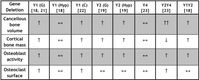
Table I. Summary of bone phenotypes from mice after deletion of Y receptors. For Y5 and Y6 receptors there aren’t reported conclusions.
Gene Deletion [18, 21] Y1 (G) Y1 (Hyp) [18] Y1 (C) [22] Y2 (G) [19] Y2 (Hyp) [19] Y4 [23] Y2Y4 [23] Y1Y2 [18] Cancellous bone volume
↑
↔
↑
↑
↑
↔
↑↑
↑
Cortical bone mass↑
↔
↑
↑
↑
↔
↓
↑
Osteoblast activity↑
↔
↑
↑
↑
↔
↑
↑
Osteoclast surface↑
↔
↑
↔
↔
↔
↑
↔
G – Germline Knockout; C- Conditional Knockout; Hyp – Hypothalamus Knockout; ↑ - Increased; ↔ - Unaltered; ↓ - Decreased.
Regarding Y1 receptors, they have the widest distribution in the brain, being expressed in cerebral cortex, thalamus, amygdala [11, 16] and particularly in the paraventricular nucleus (PVN) of the hypothalamus [9, 12]. Also, Y1 receptors have a broad distribution in peripheral tissues, including colon, pancreas, adipose tissues, kidney adrenal gland, heart, placenta and, regarding the bone, Y1 receptors are so far the only ones found to be expressed in the osteoblastic lineage and bone marrow stromal cells [12, 15]. In order to evaluate how Y1 receptors knockout would directly influence bone homeostasis, Lundberg et al. [21] performed a study using germline Y1 receptor knockout mice and it was concluded that the alteration of the Y1 receptor signaling leads to a modification in bone formation, as the germline deletion of Y1 receptors resulted in greater bone mineral content and bone mineral density. The same results were obtained by Baldock et al. [18], who demonstrated that the resulting greater bone volume in germline Y1 receptor knockout mice was associated with both osteoblastic and osteoclastic activity. In fact, the results showed that, besides bone formation and unlike what happened in the Y2 receptor knockout studies, bone resorption was also altered in germline Y1 knockout mice, with significant greater osteoclast surface. However, when a hypothalamus-specific deletion of Y1 receptors was performed, the achieved results exhibited no alterations in bone volume neither in bone cell activity when compared to the control group. This led to the conclusion that the Y1 receptor regulates bone homeostasis by a peripheral, non-hypothalamic, pathway. Therefore, with this new insight, new studies have been made in order to evaluate the potential of peripheral Y1 receptors knockout as a new therapeutic strategy on bone metabolism. Lee et al. [24] experiment demonstrated that osteoblastic-specific Y1 deletion resulted in elevated osteoblast activity in male mice, increasing cancellous and cortical bone mass. This suggested that Y1 receptors would be a potential target to treat bone associated diseases. In keeping with this, Sousa et al. [22] showed that the oral administration of a Y1 receptor antagonist to wild type mice resulted in increased bone mass, with high
28
mineral apposition rate in both cortical and cancellous bone of mice. However, besides this, it was observed an increase in bone resorption indices, which was a behavior only observed in germline deletion of Y1 receptors. This result suggested that Y1 receptor blockage would not only lead to elevate bone mass, but could also lead to an adequate bone turnover, thereby possibly avoiding hypocalcaemia and related complications [22].
As previously referred, there is an Y2-dependent inhibition of Y1 expression in osteoblastic lineage cells that suggests the existence of a mechanism where central Y2 signaling moderates tissue homeostasis [18, 21]. Likewise, the osteoblastic effects from Y1 knockout and Y2 knockout were similar. Therefore, there was the hypothesis that these two receptors were linked in the regulation of bone homeostasis [18]. In order to evaluate this supposition, in the same study previously referred, Baldock et al. [18] performed a germline Y1Y2 double knockout. However, the results showed that deletion of both Y1 and Y2 receptors did not result in additive effects on bone mass when compared to Y1 knockout and Y2 knockout mice. Thus, it was concluded that, although Y1 and Y2 appear to share common pathways in the regulation of bone tissue, there are discrete actions of individual Y receptors, with probable central Y2 and peripheral Y1 effects on bone tissue [15, 18].
Therefore, in order to explore the potential of Y1 receptors, several Y1 receptor antagonist strategies have been investigated in Y1 receptor targeting therapies for diseases such as anxiety [25-31], obesity [32-40], epilepsy [41, 42], between several others. Thus, several effective Y1 receptor antagonists are commercially available in order to determine the pharmacological role of Y1 receptors in targeted diseases.
1.2.2. Anti Y1 Receptor Therapeutic Strategies
The Y1 receptor has a great range of peptide and non-peptide antagonists available. These antagonists have been applied in several experiments, with different purposes. In Table II a summary of the available Y1 receptor antagonists, therapeutic applications and obtained results with the respective administration methods is presented.
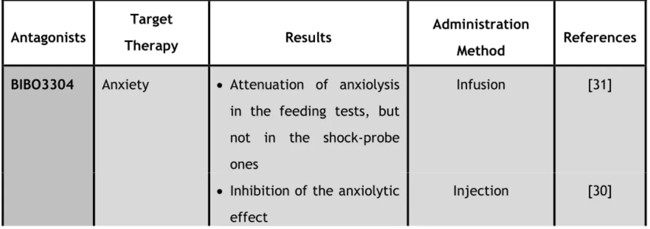
Table II. Summary of the peptide and non-peptide antagonists for Y1 Receptor and some of the respective studied applications and results.
Antagonists
Target
Therapy Results
Administration
Method References BIBO3304 Anxiety Attenuation of anxiolysis
in the feeding tests, but not in the shock-probe ones
Infusion [31]
Inhibition of the anxiolytic effect
29
Bone Mass Dose-dependently increase in bone mass
Oral [22]
Depression Attenuation of the antidepressant-like effects
Injection, Infusion [43, 44]
Inflammatory Response
Abolition of the pro-inflammatory action
Injection [45]
Food Intake Inhibition of feeding behavior
Injection, Injection [39, 40]
Pain Modulation
Prevention of the anti-allodynic actions Injection [46] Post-Traumatic Stress Disorder Prevention of the inhibition of fear responses Infusion [47]
BIBP3226 Anxiety Potentiation of the anxiogenic and depressive effects of Cholecystokinin tetrapeptide Injection [25] Induction of an anxiogenic-like effect Injection, Injection, - [26-28]
Atherosclerosis Increased atherosclerotic lesion areas
Injection [48]
Depression Attenuation of the antidepressant-like effects
Injection [43]
Epilepsy Reduction of the number and time of seizures
Infusion [41]
Heart Associated Functions
Dose-dependent inhibition of the increase in blood pressure
- , Infusion [49, 50]
No significant change in heart rate
Smaller infarct volume
Injection [51] Inhibition of the vasoconstriction -, In vitro, Infusion, In vitro, In vitro [49, 52-55] Hypothalamic- Pituitary-Adrenal Regulation
The increase of plasma corticosterone levels was contradicted
30 Microglial
Reactivity
Suppression of the reduction of interleukin-1β and tumor necrosis factor-α protein generation
In vitro [57]
Food Intake Inhibition of food intake Injection, Injection [36, 38] Renal Function No influence in renal
functions in ischaemic heart failure rats
Infusion [50]
Reproductive Function
Inhibition of the increase of Gonadotropin-Releasing Hormone-1 mRNA
In vitro [58]
GI264879A Food Intake Decrease in food intake and body weight
Injection [32]
GR231118 Anxiety Decrease in time spent in social interaction (increased anxiolysis)
- [27]
Epilepsy Reversion of the anticonvulsant effect of intrahippocampal ghrelin
Microinjection [42]
Depression Increased depression-like behavior
Injection [59]
Heart Associated Functions
Inhibition of both cardiac slowing and decrease in arterial pressure
Increase in ventilation
Injection [60]
Food Intake Decrease in food intake stimulated by orexin Injection [34] Vascular Secretory Function of Salivary Glands Inhibition of the attenuation of vasodilation
Inhibition of the pressor response
Injection [61]
H409/22 Anxiety Increase the anxiolytic-effect
Injection [29] Colitis Attenuation of the clinical
manifestation of the disease
31
Food Intake Suppress of food intake Infusion [37] Heart Associated Functions Inhibition of the vasoconstriction Infusion [63]
J-104870 Food Intake Suppress of food intake Injection [35]
J-115814 Food Intake Suppress of food intake and body weight
Oral [64]
LY357897 Food Intake Suppress of food intake Injection [33]
SR120819A Food Intake Dose-dependent reduction of food intake Injection [39] Heart Associated Functions Attenuation of vasoconstriction on the kidney - [65] Lipolysis and Leptin Secretion Blockage of the antilipolytic efect evoked by hPYY
Prevention of adipocyte leptin secretion
In vitro [66]
(-) Corresponds to administration methods with unknown information.
BIBO 3304 is the abbreviation for N-[[4-(Aminocarponylaminomethyl)-phenyl] methyl]-N2-(diphenylacetyl)-argininamide trifluoroacetate. This non-peptide compound exhibits selective binding to the Y1 receptor subtype, with more than 1000 to 10000-fold lower affinity for human Y2, human and rat Y4 and human and rat Y5 receptors. Likewise In what concerns its properties to inhibit the NPY mediated signals, no agonistic properties were found to BIBO3304. Thus, it is considered that BIBO 3304 is a potent Y1 receptor antagonist [40] and, when compared to BIBP 3226, it has a 10-20 fold higher affinity for both human and rat Y1 receptors [67]. However, this compound isn’t available in radiolabelled form [68]. Chemically, it presents a molecular weight of 757.69.
BIBP 3226 corresponds to the short name for R-N2-(Diphenylacetyl)-N-(4-hydroxyphenyl)-methyl argininamide. It corresponds to a non-peptide Y1 receptor antagonist, exhibiting competitive and excellent Y1 receptor selectivity [49, 69], as it doesn’t bind to human Y2 receptor subtype nor cross-react to 60 other experimented receptor types and 15 enzyme systems [70]. BIBP3226 doesn’t reveal agonistic properties towards Y1 receptors [69, 70] and therefore it is considered to be the first true non-peptide Y1 receptor antagonist, with characteristics that make it a suitable tool to study the role of NPY in pathophysiological
32
conditions [70]. This compound has shown to be a fully antagonistic for both neural cells and peripheral tissues [71]. Chemically, BIBP3226 is a hydrophobic compound with a molecular weight of 473.57.
Regarding GI264879A, it is an abbreviation for 1-Substituted-4-methylbenzimidazole (413), N--[3,3-bis(1-naphthyl)propionyl]-D-arginine N-[(S)-1-benzyl-2-methoxyethyl] amide. It is considered to be a non-selective and weak NPY receptor ligand, with decreasing affinity for Y1 (which is equal for the Y4), Y5 and Y2 receptors. Regarding its behavior towards NPY mediated signals, GI264879A was tested only for the Y1 receptor and it showed antagonistic properties [32].
GR23118, also named GW1229 or 1229U91, is the abbreviated name for Homodimeric Ile-Glu-Pro-Dpr-Tyr-Arg-Leu-Arg-Tyr-CONH2. Although this peptide compound reveals potent Y1 receptor antagonistic properties, it was discovered that GR23118 is also an agonist for Y4 receptors [72]. Regarding the compound chemistry, it has a molecular weight of 2352.77.
In what concerns H409/22, it corresponds to the abbreviation of the (2R)-5-([Amino(imino)methyl]amino)-2-[(2,2-diphenylacetyl)amino]-N-[(1R)-1-(4-hydroxyphenyl)ethyl ]-pentanamide compound. H409/22 is considered to be a potent and selective ligand to Y1 receptors, with antagonistic properties and very low affinity for Y2 and Y5 receptors. However, this compound reveals 5 times lower potential than BIBO 3204 [37]. The molecular weight of this non-peptide compound is approximately 487.61.
J-104870 is the abbreviation for 2-[(4-Chlorophenoxy)methyl]benzimidazoles (411), 6(5ethyl1,3thiazol2ylthiomethyl)2[3methoxy5(2propenyloxycarbonylamino) benzylamino] -4-morpholinopyridine. This compound corresponds to a potent and selective antagonist for the Y1 receptor, revealing low affinities for Y2, Y4 and Y5 receptors. Besides this, it is considered that this non-peptide compound has oral bioavailability and brain penetrability [35]. Its molecular weight is approximately 555.72.
In what concerns J115814, it is the abbreviation for (-)-2-[1-(3-chloro-5-isopropyl oxycarbonyl aminophenyl) ethylamino] -6-[2-(5-ethyl-4-methyl-1,3-thiazol-2-yl)ethyl]-4-morpholinopyridine. This compound has a high affinity to Y1 receptors, but low affinity for Y2, Y4 and Y5 receptors, what reveals its potent and selective Y1 receptor antagonistic properties [73].
LY357897 corresponds to the 1-((1-[3-((3s)(3-Piperidyl))-propyl]-2-[(4-chlorophenoxy)-methyl]indol-3-yl]-2-(4-piperidylpiperidyl)ethan-1-one compound. At the time of its discover, it was classified as being the first selective, subnanomolar Y1 antagonist [33], as it doesn’t show appreciable binding affinity for Y2, Y4 and Y5 receptors [71].
Regarding SR120819A, it corresponds to the (2R)-N-[(2R)-3-[4-[N'-[[4-(dimethylaminomethyl) cyclohexyl] methyl] carbamimidoyl] phenyl]-1-oxo-1-pyrrolidin-1-ylpropan-2-yl]-2-(naphthalen-2-ylsulfonylamino)-3-phenylpropanamide compound. It is considered to be a non-peptide, orally active and selective Y1 receptor antagonist [74, 75], as it did not reveal affinity for Y2, Y4 or Y5 receptors [71]. SR120819A has a molecular weight of 750.39.
33
1.3. DRUG DELIVERY SYSTEMS TO BONE
1.3.1. Current
Therapeutic
Approaches
for
Bone
Regeneration
For situations where normal bone regeneration is impaired or insufficient, there are currently several therapeutic approaches that, when used alone or in combination, aim the enhancement or management of these complex clinical situations [8].
Bone grafting is a common surgical procedure used to enhance bone regeneration [8]. Bone grafting can be defined as being a strategy where bone from somewhere else is applied in the place where it is needed a stimulation of bone formation. There are three possibilities of bone grafting, being the preferable one the autologous bone graft (the used bone is the patient’s own, being usually harvested from locations with relative excess of bone, like the pelvis). The other two options of bone grafting correspond to the allogenic bone graft (the harvested bone belongs to other humans) and the xenogenic bone graft (the used bone graft belongs to other animals from other species) [4]. The autologous bone graft is considered to be the gold standard bone grafting material, as it comprises several advantageous properties: osteoinduction (the extracellular matrix contains bone morphogenetic proteins and other growth factors), osteogenesis (the graft contains osteoprogenitor cells that will contribute to bone formation after the graft is vascularized) and osteoconduction (it works as a scaffold that allows the deposition of bone and integration by the surrounding bone) [4, 8, 76]. Besides this, the autologous bone graft is histocompatible and non-immunogenic, reducing the probabilities of immunoreactions and transmission of infections [8]. Likewise, it can be completely resorbed and remodeled, not interfering with physiologic bone adaptation [4]. However, it presents some disadvantages, as for example limited mechanical strength, quantity restrictions, diverse physical injuries that arise from surgical complications and substantial costs [4, 8, 77]. As an alternative to autologous bone graft, allografts and xenografts were also thought as possible options. The allografts are obtained from human cadavers or living donors. This type of graft surpasses the problems of quantity, as allogeneic bone is available in many preparations [8]. However, as they are devitalized and sometimes demineralized, they do not possess any cellular component, what leads to reduced osteogenic and osteoinductive properties and, consequently, to a delay in the regeneration process [4]. Furthermore, this type of grafts is commonly associated to immunogenicity and rejection reactions and cost problems [4, 8, 78]. In what concerns the xenografts, they are obtained from animals of other species. However, several problems associated to ethics and graft rejection are attributed to this type of graft.
As grafts present several disadvantages, bone-graft substitutes have been developed in the field of bone tissue engineering. These substitutes, named scaffolds, are made of natural or
34
synthetic biomaterials and aim to promote migration, proliferation and differentiation of bone cells [8]. In order to achieve these goals, bone tissue engineering usually combines scaffolds with biologically active factors, which can be cells, proteins or a combination of both. By doing this strategy, osteoconduction, osteoinduction and osteogenesis are reached. Thus, it becomes necessary to define three different components: scaffolds, growth factors and cells [4].
Scaffolds serve as a delivery vehicle for osteoinductive molecules and/or osteogenic cells. It must fill a gap in a bone defect, facilitating, at the same time, the healing process. However, to perform its purposes, scaffolds for bone regeneration must be biocompatible, osteoconductive, porous (the material must have an interconnected porous architecture in order to facilitate the bone growth inside of the material), biodegradable (this characteristic depends on the final aim of the scaffold, but usually, as bone is regenerated, the material must gradually degrade) and intrinsically osteoinductive [4]. Likewise, scaffolds must have good mechanical properties [4, 8]. In what concerns growth factors, they correspond to signaling molecules that influence certain cellular functions as they bind to specific receptors in cell membranes [4]. The most extensively studied molecules are the bone morphonegenetic proteins (BMPs), which correspond to potent osteoinductive factors [4, 8]. Other growth factors have also been used in different strategies, with different functions regarding cell proliferation, chemotaxis and angiogenesis. These include platelet-derived growth factors, transforming growth factor-β, insulin-like growth factor-1, vascular endothelial growth factor and fibroblast growth factor, among others [8]. Regarding the cellular component, an adequate supply of cells, such as mesenchymal stem cells (MSCs) and osteoprogenitors, is of major importance in bone regeneration strategies. Usually, these cells are obtained from the bone-marrow, which also contains growth factors, in a minimally invasive procedure [8].
Besides the aforementioned strategies, gene therapies are rising as potential procedures to improve bone regeneration. This method involves the transfer of genetic material into the genome of the target cells, in order to promote the expression of bioactive factors for a prolonged time. However, to apply gene therapies, issues of cost, biological safety and efficacy problems must be solved [8].
In some cases, drugs can also improve bone regeneration. An ideal drug for bone regeneration should restrict its pharmacological activity specifically to bone sites, with minimal effects at a systemic level [79]. However, these drugs are usually administrated alone, orally or parenterally. As bone is a peripheral tissue with limited blood supply, during their pathway, drugs are exposed to various phsycochemical and biological factors, which will affect the drugs bioavailabilty and, thus, their efficient delivery to the required sites. In order to overcome this problem, larger drugs doses are administrated, leading to toxicity problems [80, 81]. Other complications may also arise from the fact that some drugs need to be taken for prolonged duration, resulting in problems associated with the patient therapy compliance [81]. Therefore, new agents and efforts should be made in order to develop new strategies that overcome the
35
abovementioned problems, enabling improvements in drug efficiency. An example of strategy that has been developed is the controlled drug delivery systems, which make use of different materials with different configurations (e.g. microspheres and nanoparticles). These new developed systems, to be considered as a drug carrier, must be non-toxic (i.e. bioinert or biodegradable), biocompatible, able to incorporate a drug physically or chemically and retain it until it reaches a specific target site, where the drug carrier will gradually be degraded and deliver the drug in a controlled way over time [82]. The materials from which drug carriers are made of, will be chosen accordingly to the desired characteristics, which include chemical characteristics (hydrophobicity or hydrophilicity, molecular size, pi, between others), desired delivery rate, drug release mode, target bone/bones, injury size, whether materials degradation is desired, between other parameters, such as carrier size and structure and drug loading [83, 84]. Therefore, through the combination of different parameters and materials, it is possible to develop new ways of drug delivering, enabling improvements in the field of bone regeneration.
Drug delivery systems can be divided into systemic or local drug delivery. The systemic drug delivery systems are less invasive and usually associated to an oral or parenteral administration. However, these systems can provoke some side effects on non-skeletal tissues [85]. Also, the drugs used in this type of delivery systems should have prolonged circulation time, be able to distribute and accumulate on the target tissues and also they should be protected from enzymatic and chemical degradation [86]. Although it is a more invasive procedure, local delivery of drugs has some advantages towards the systemic drug delivery, as the needed drug quantity is reduced, the side effects are lower, drugs can be retained in the specific local for increased periods and it allows a time-controlled deliver. Ideally, this local drug delivery process should deliver the drug through minimally invasive procedures (e.g. injection to the local site) and possess the ability for in situ matrix formation, allowing an accelerated wound healing, ease in the recognition of requirements of irregular shape defects and augmented patients’ comfort and compliance [87, 88].
Therefore, several approaches can be studied in order to improve the actual solutions used to bone regeneration, with special attention to the drug delivery systems.
1.3.2. Nanoparticles Drug Delivery Systems
Nanoparticles have become an important area in the drug delivery systems field as they have the ability to deliver a wide range of drugs to different areas of the body for sustained periods of time, while their small size makes them suitable for systemic circulation [89]. Recent advances in synthesis of polymeric materials with enhanced and advantageous characteristics (biocompatibility, controlled biodegradation profile and responsiveness to biologically-relevant stimuli) are leading to the development of new drug delivery systems [90]. These nanocarriers are divided into two categories: nanocapsules (vesicular system with a
36
polymeric shell and inner core) and nanospheres (polymeric matrices) [84]. Nanocapsules differ from nanospheres in their constitution: nanocapsules are used in a reservoir form, in which a solid material surrounds a core which is liquid or semisolid at room temperature; nanospheres are matrix particles, which means that their entire mass is solid [91]. In both cases, nanoparticles can be loaded with a wide variety of drugs, including hydrophilic and hydrophobic drugs, proteins, vaccine and biological macromolecules [89]. Drugs can be entrapped inside the nanoparticles or adsorbed on their surface. The first option can be performed by an association of the drug and the nanocarrier during the preparation of the nanoparticles; for drugs that are highly susceptible to degradation, the adsorption on the surface of the nanoparticles becomes a more suitable procedure [91].
There are several methods for the preparation of nanoparticles that allow the production of polymeric nanoparticles with different properties that ensure a proper drug delivery and targeting. These methods can be divided in procedures that include two main steps and the ones that include only one main step. The two steps methods comprise the preparation of an emulsified system (first step), followed by the nanoparticles formation (second step). Usually the second step is achieved either by precipitation or gelation of a polymer or polymerization of monomers. The methods that only require one step are based on the precipitation of a polymer in conditions of spontaneous dispersion formation or due to the self-assembly of macromolecules to form nanogels or polyelectrolyte complexes from a polymeric solution [91].
As nanoparticles have the same size range as parts of natural bone, they are considered as potential candidates for local applications as bone-specific carriers [82]. Furthermore, nanoparticles offer other several advantages, including controlled release properties, protection to the compound of interest and possibility of changing the nanoparticles surface for actively targeting them to specific desired tissues [84, 90]. In order to achieve the desired results, as previously mentioned in section 1.3.1, nanoparticles should comprise a set of characteristics: be nontoxic for cells, effectively carry the molecule of interest and exert their actions specifically on the target, therefore reducing the side effects systemically [82].
As also mentioned in the previous section, the main criteria in choosing a material for drug delivery include not only the chemical characteristics of the drug to be delivered, but also bioavailability, biocompatibility, straightforward production and degradation rate, which enables a sustained release of drugs encapsulated in nanoparticles [84]. Therefore, it becomes essential to perform a previous study on the required delivery parameters and available materials and correspondent characteristics.
1.3.2.1. Polycaprolactone, Poly(lactide-co-glycolide) and
Monoethoxy poly(ethylene glycol)-poly(trimethylene
carbonate) Polymers
Polymers are versatile materials that can be useful in several applications. For example, hydrophilic polymeric nanoparticles can be applied and used for hydrophilic drugs, gene,
37
oligonucleotides and small interfering RNA; on the other hand, hydrophobic polymers can be used for water-insoluble drugs [90]. Usually, biodegradable polymeric nanoparticles are preferred as they enable controlled/sustained release, subcellular size and biocompatibility with tissues and cells. Furthermore, these compounds are stable in blood, non-toxic, biodegradable, nonthrombogenic, nonimmunogenic, noninflammatory, do not activate neutrophils and avoid reticuloendothelial system [92]. Polycaprolactone (PCL), Poly(lactide-co-glycolide) (PLGA) and Monoethoxy poly(ethylene glycol)-poly(trimethylene carbonate) (mPEG-PTMC) were previously tested in our group in nanoparticles’ preparation procedures [93]. Therefore, a revision on these three polymers was performed in order to understand their characteristics and suitability for the proposed theme.
PCL has a hydrophobic nature [90]. In physiological conditions, this polymer degradation occurs by hydrolysis of its ester linkages. PCL receives a special attention for the preparation of long-term implantable devices [92], as it has a degradation rate of 2 to 3 years, low tensile strength and very high elongation ate breakage [94]. It is also considered to be a semi-crystalline polyester with great organic solvent solubility [94]. Regarding its applicability, PCL has been used as a drug carrier in several studies, including anticancer, diabetes (insulin), antifungal (amphotericin B) and clonazepam drugs [92]. However, so far, no PCL nanoparticle delivery system to bone has been reported.
PLGA corresponds to a copolymer of poly lactic acid (PLA) and poly glycolic acid (PGA). PLA is more hydrophobic than PGA and, consequently, lactide rich PLGA copolymers are less hydrophilic, adsorb less water and degrade more slowly. In fact, the 50:50 ratio of PLA/PGA exhibits the highest rate of degradation; for other ratios, as the content of PGA is higher, the degradation rate also augments [95]. After hydrolysis, PLGA produces metabolite monomers (lactic and glycolic acid), which are two monomers with which the body usually deals. However, the acidic nature of this copolymer makes it not suitable for drugs or bioactive molecules. This problem can be overcome by blending the PLGA with other compounds [92]. In what concerns the mechanical strength, swelling behavior, capacity to undergo hydrolysis and biodegradation rate are dependent on the PLGA physical properties (molecular weight, polydispersity index and cristallinity) [95]. This polymer has been widely used to encapsulate and deliver several types of drugs, including anticancer, diabetes (insulin), psychotic, hormonal (estradiol) and tetanus drugs [92]. Regarding specifically the targeting to bone, drug delivery systems based on PLGA micro and nanoparticles have been developed and used as carriers for recombinant human morphogenetic protein-2 (rhBMP-2) [96, 97], recombinant human bone morphogenetic protein-7 (rhBMP-7) [98], transforming growth factor β (TGF-β) [99], nafcillin [100], estrogen [101] and dexamethasone [102].
mPEG-PTMC is a diblock copolymer and it is considered to be an amphiphilic compound: the mPEG component constitutes the hydrophilic part, while the PTMC segments are the hydrophobic ones [103, 104]. It degrades very slowly in water and in acidic environments, although in lipase solutions it can be degraded by an enzymatic surface erosion process [104]. In fact, block copolymers containing PEG have been widely used for the development of
38
controlled delivery systems of drugs. On the other hand, as PTMC doesn’t form acidic compounds during its degradation by surface erosion, PTMC-based polymers are also very attractive for the delivery of drugs [104]. The use of this copolymer has been widely described by Zhang et al [104] in several studies regarding the compound characteristics and its use in the preparation on micro and nanoparticles. However, besided the aforementioned studies, mPEG-PTMC has not been described in literature.
1.3.2.2. Nanoparticle Delivery Systems for the Extracellular
Space
The cell membrane acts as a barrier or biomembrane that separates the external environment from the inside of the cell. Understanding interactions between particles and this membrane becomes important to predict subsequent biological effects [105].
Internalization of nanoparticles in cells is influenced by several physical and chemical properties of the materials, as well as by the medium characteristics in the moment of the exposure. There are several nanoparticle characteristics that influence their uptake: size, shape and geometry, surface charge and hydrophilicity. The bulk characteristics of the nanoparticle do not play an important role in the particles internalization, as the surface characteristics are the ones that determine the protein corona and therefore biological impact [106].
Several studies have been made with the aim of evaluating how the particles size can influence their uptake by cells. Accordingly to Sylvester et al. [107], nanoparticles of 100 and 200 nm were easily internalized by cells, while particles with 500 nm remained in the extracellular space. Thus, it was concluded that 350 nm would be the ideal size to retain the nanoparticles in the extracellular space. Same results were obtained by Sivaraman and Ramamurthi [108], who also concluded that, although 500 nm size particles are completely excluded by cells, in certain cases they can be phagocyted. These results are in agreement with several others that demonstrate that nonphagocytic cells favor the uptake of smaller particles while larger particles aren’t internalized by these same cells [109-114]. However, there isn’t agreement in which is the exact size that leads to the exclusion of the particles by nonphagocytic cells [114]. Regarding the phagocytic cells, although some results have shown that there is a particle size-dependent uptake [115], there isn’t clear evidences that this procedure is unequivocally related to particle size [106].
Regarding the shape and geometry, Gratton et al. [114] studied the uptake of monodisperse cross-linked PEG hydrogel particles by HeLa cells. The results showed that cylindrical nanoparticles with high-aspect-ratio (diameter (d)=150nm, height (h)=450nm) were internalized faster than the symmetric (aspect-ratio (AR)=1, d=h=200 nm) cylindrical nanoparticles, although they have the same volume. When comparing cylindrical nanoparticles with similar aspect ratio but different diameter (d=100nm vs d=150nm), the results indicated that the particles with smaller diameter were less internalized than the larger ones. The same happened
![Figure 1. Schematic view of human long (A) and cortical (B) bone. Adapted from [3].](https://thumb-eu.123doks.com/thumbv2/123dok_br/15840372.1084239/21.892.201.707.106.801/figure-schematic-view-human-long-cortical-bone-adapted.webp)