Universidade de Trás-os-Montes e Alto Douro
Cranial Cruciate Ligament Rupture
Surgical Treatment by Tibial Plateau Leveling Osteotomy
Dissertation of the Integrated Master in Veterinary Medicine
Tiago Avelino Rolo Viana
Supervisor: Professor Doutor Luis Miguel Viana Maltez da Costa
iii
Universidade de Trás-os-Montes e Alto Douro
Cranial Cruciate Ligament Rupture
Surgical Treatment by Tibial Plateau Leveling Osteotomy
Dissertation of the Integrated Master in Veterinary Medicine
Tiago Avelino Rolo Viana
Supervisor: Professor Doutor Luis Miguel Viana Maltez da Costa
Jury composition:
___________________________
___________________________
___________________________
iv
Declaração
Nome: Tiago Avelino Rolo Viana
Correio electrónico: viana.tiago@hotmail.com
Designação do mestrado: Mestrado Integrado em Medicina Veterinária
Título da dissertação de Mestrado em Medicina Veterinária: Cranial Cruciate Ligament Rupture - Surgical Treatment by Tibial Plateau Leveling Osteotomy
Orientadores: Professor Doutor Luis Miguel Maltez Viana da Costa Ano de conclusão: 2016
Declaro que esta dissertação de mestrado é resultado da minha pesquisa e trabalho pessoal e das indicações do meu orientador. O seu conteúdo é original e todas as fontes consultadas estão devidamente mencionadas no texto e na bibliografia final. Declaro ainda que este trabalho não foi apresentado em nenhuma outra instituição para obtenção de qualquer grau académico.
Vila Real, Fevereiro de 2016
_____________________________________
v
vii
Acknowledgements
My entire academic career culminates in this work. Certainly, I would not have achieved it alone, hence I express here my thanks to all those who directly or indirectly contributed so I could turn a Veterinary Surgeon.
I would like to thank to Rector Magnificus of the Universidade de Trás-os-Montes e Alto Douro, Professor Doutor António Fontainhas Fernandes for the granted opportunities.
I naturally thank to Professor Doutor Luis Maltez, supervisor for this dissertation, for all the help provided in its elaboration but above all, for all the knowledge that he has conveyed to me throughout my academic career. Undoubtedly, a master who awakened in me the passion for the small animal surgery.
Also to all the teachers from the Integrated Master in Veterinary Medicine that I had the honour to meet and be a student of, my many thanks.
I thank all the HVTM team for all the granted opportunities and for the kindness they have shown to me during my volunteering period. A special thanks to Dr. Paulo Pimenta and Dra. Maria João for all the attention they gave me.
To Dr. Jorge Leite, it is hard to express all the gratitude I keep for providing me an exceptional traineeship at Bonematrix, full of valuable knowledge. A model, both by the technical skills as by the philosophy with which he faces the challenges and surgeries. For all the opportunities, for the friendship, many thanks.
I thank to all the team from Hospital ARS in Barcelona for the kindness they received me with, especially to Dr. Artur Font and Dr. Joan Mascort. To Dr. Luís Feo and the Interns Marta, Felix, Ana, Mario, Jorge and Natalia my sincere thanks for all the patience and availability to taught me but above all, for making me feel at home.
I also would like to thank to all the QVSH team in Cambridge, for all the trust they have putted on me and by all the granted chances. To Matt McMillan for all the attention and extreme care he had with me, but also to Jon Hall, Gareth Arthurs, to the interns and the nursing team.
viii
I could not fail to thank to all my friends and colleagues. Impossible to mention everyone’s name unfortunately. To Diogo Marques, Diogo Brito, Tomás Basto e Pedro Meireles, a big hug. Truly, friendships that shaped my personality and made me grow up as a person. Also to Anocky, Sté, Frias, Gb, Yuri and Sapo, many thanks. To my “non-veterinarian” friends, especially to the “Ferreiros do Monte & Companhia” and to the “Internationals” thank you all for the good moments we lived together.
And of course, to my family for all the support they always have given me. To my parents, my grandmother and my brothers, true role models.
I couldn’t help but also thank all the people who I had the fortune to meet recently, who make the HVBV team for all of the kindness and support they've shown upon welcoming me.
ix
Resumo
A rotura de ligamento cruzado cranial é uma das mais importantes doenças ortopédicas que afectam o cão e a causa mais frequente de osteoartrite do joelho e claudicação do membro posterior. Muito se tem vindo a descobrir desde a primeira publicação em 1926, no entanto a patogenia exacta da doença permanece desconhecida.
A rotura pode ser traumática ou degenerativa, representando a ultima a maioria dos casos. Pode ocorrer em cães de qualquer tamanho, idade ou raça embora seja mais frequente em cães jovens adultos de raças grandes.
O diagnóstico baseia-se na história de claudicação, anamnese e outras ferramentas específicas de diagnóstico como radiografias ou artroscopia. A rutura do ligamento cruzado cranial pode ser tratada medicamente ou cirurgicamente. O tratamento cirurgico é frequentemente o recomendado pois atinge-se uma estabilização do joelho mais rápida bem como um retorno mais precoce à sua função.
Actualmente, os procedimentos cirúrgicos mais utilizados no tratamento da da rotura de ligamento cruzado cranial são a Tibial Plateau Leveling Osteotomy (TPLO), Tibial Tuberosity Advancement (TTA) e Extracapsular Stabilization (ES). Contudo, o grande número de técnicas existentes sugere que não há uma técnica que seja unanimemente aceite. Esta escolha deverá ser baseada em vários critérios como tamanho, idade, cronicidade da lesão, aceitação do proprietário, preferência do cirurgião e nível de experiência com um ou outro procedimento.
Este estudo retrospectivo tem por objetivo analisar os resultados da Tibial Plateau Levelling
Osteotomy (TPLO) avaliando os resultados clínicos e cirúrgicos bem como as complicações
associadas à técnica.
xi
Abstract
Cranial cruciate ligament disease is one of the most important orthopaedic conditions of the dog and the most common cause of stifle osteoarthritis and pelvic limb lameness. Much has been learned since the first report in 1926, however the exact aetiopathogenesis of this condition remains unknown. The rupture can be traumatic or degenerative, the last one represents the majority of the cases. It can occur in dogs of any size, age or breed, though it is more frequent in young adult dogs belonging to large breeds.
Diagnosis is based in the history of lameness, anamnesis and specific diagnostic tools (radiographs, arthroscopy). Cranial cruciate disease may be treated either medically or surgically. Surgical treatment is frequently recommended to achieve a more rapid stifle stabilization, meniscal treatment and earlier return to clinical function.
Currently, the most commonly performed surgical procedures include the Tibial Plateau Levelling Osteotomy (TPLO), the Tibial Tuberosity Advancement (TTA) and the Extracapsular Stabilization (ES). Nevertheless, the large number of techniques performed suggests that no single technique is unanimously accepted. This selection should be based upon numerous criteria including size, age, chronicity of the injury, acceptance of the owner, surgeon’s preference and experience level with the procedure.
The objective of this retrospective study is to analyse the results of the Tibial Plateau Levelling Osteotomy by evaluating the clinical and surgical outcomes and eventual complications associated with the technique.
xiii
Table of Contents
Declaração ... iv Acknowledgements ... vii Resumo ... ix Abstract ... xiTable of Contents ... xiii
Figure Index ... xv
Table Index ... xvi
List of Abbreviations and Acronyms... xvii
Introduction ... xix
Chapter I ... 1
1. Anatomy of the Stifle ... 3
1.1. The cruciate ligaments ... 7
2. Epidemiology and Pathogenesis ... 9
3. Diagnosis ... 16 3.1. Clinical Presentation... 16 3.1.1. Signalment ... 16 3.1.2. History ... 16 3.1.3. Physical Examination ... 17 3.2. Diagnostic Imaging ... 21 3.2.1. Radiography ... 21 3.2.2. Ultrasonography ... 23 3.2.3. Computed Tomography ... 24
3.2.4. Magnetic Resonance Imaging ... 25
3.2.5. Scintigraphy ... 26
3.2.6. Thermography ... 27
3.3. Arthroscopy ... 28
3.4. Meniscal Injury ... 29
3.4.1. Classification of Meniscal Tears ... 30
3.4.2. Meniscal Injury Treatment ... 31
3.5. Arthrotomy ... 32
xiv
3.7. Differential Diagnosis ... 33
4. Cranial Cruciate Ligament Rupture Treatment ... 34
4.1. Medical Management ... 34 4.2. Surgical Treatment ... 36 4.2.1. Intracapsular Techniques ... 37 4.2.1.1. Paatsama Technique ... 37 4.2.1.2. Over-the-Top Technique ... 38 4.2.2. Extracapsular Techniques ... 39 4.2.2.1. Imbrication Techniques ... 40 4.2.2.2. TightRope ... 41
4.2.2.3. Fibular Head Transposition ... 41
4.2.3. Osteotomy Techniques ... 43
4.2.3.1. Cranial Tibial Wedge Osteotomy (CTWO) ... 45
4.2.3.2. Tibial Plateau Leveling osteotomy (TPLO) ... 46
4.2.3.3. Tibial Tuberosity Advancement (TTA) ... 55
4.2.3.4. Modified Maquet Procedure (MMP) ... 57
4.2.3.5. Triple Tibial Osteotomy (TTO) ... 58
Chapter II ... 61
1. Objectives ... 63
2. Materials and Methods ... 63
3. Results ... 68
4. Discussion ... 69
5. Conclusions ... 73
xv
Figure Index
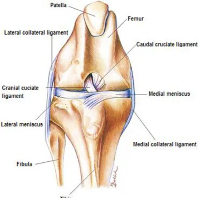
Figure 1 Normal Stifle Joint ... 3
Figure 2 Joint Capsule of the left stifle ... 4
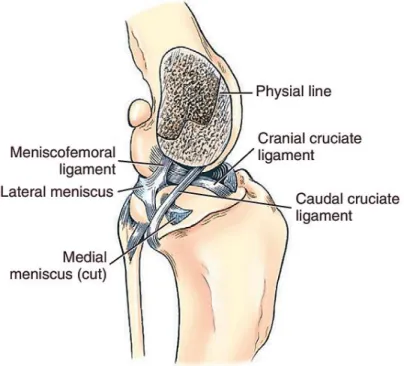
Figure 3 Menisci and ligaments of left stifle joint, dorsal aspect ... 5
Figure 4 Ligaments of the Stifle Joint ... 6
Figure 5 Cruciate and meniscal ligaments of the stifle, medial aspect. ... 8
Figure 6 Ruptured Cranial Cruciate Ligament ... 9
Figure 7 Schematic representation of the forces acting on the tibia during weigh bearing ... 12
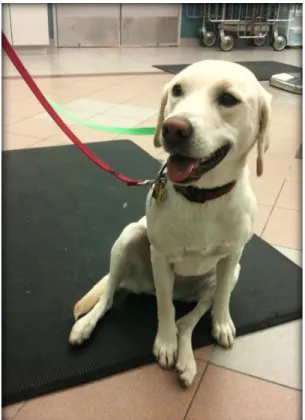
Figure 8 Dog with positive "sit test" ... 18
Figure 9 Tibial compression test ... 20
Figure 10 Cranial drawer test... 20
Figure 11 Mediolateral radiographic image of a stifle joint with chronic CrCL rupture. ... 22
Figure 12 Tibial compression radiographs of a dog with CrCL rupture ... 23
Figure 13 Ultra sonographic image of a ruptured CrCL ... 24
Figure 14 Transverse CT image of an avulsion fracture at the insertion of the CrCL. ... 25
Figure 15 Magnetic resonance imaging of a normal stifle and a stifle with CrCL rupture ... 26
Figure 16 Bone scintigraphy of stifle ... 26
Figure 17 Thermal image of a stifle with a ruptured cranial cruciate ligament ... 27
Figure 18 Arthroscopic examination of the Cruciate Ligaments ... 28
Figure 19 The "wedge-effect" ... 29
Figure 20 Classification of meniscal tears ... 30
Figure 21 Caudal meniscotibial release ... 32
Figure 22 The A-TraC Dynamic stifle brace for dogs with CrCL insufficiency ... 35
Figure 23 Fascia lata substitution (Paatsama) ... 37
Figure 24 Over-the-Top Procedure ... 38
Figure 25 Isometric points in the lateral aspect of the canine stifle ... 39
Figure 26 Modified Retinacular Imbrication techniqu ... 40
Figure 27 Three-in-One Technique ... 40
Figure 28 TightRope (A); Corkscrew/FasTak Anchor (B); and Knotless SwiveLock Anchor (C) 41 Figure 29 Fibular Head Transposition ... 42
Figure 30 Joint reaction force in the CrCL-deficient stifle according to Slocum ... 43
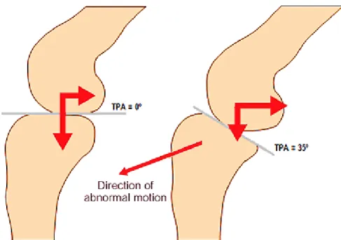
Figure 31 Method of measuring the tibial plateau angle in the stifle joint of the dog ... 44
xvi
Figure 33 Representation of the tibiofemoral forces in the stifle joint, according to Slocum, before
(left) and after (right) TPLO ... 46
Figure 34 TPLO rotation - Quick Reference Chart ... 47
Figure 35 Osteotomy position planning ... 48
Figure 36 Insertion of the Jig pins ... 49
Figure 37 Partial osteotomy using a saw guide ... 50
Figure 38 Postoperative representation of the TPLO ... 50
Figure 39 Lateral radiograph view showing thickening of the patellar tendon ... 53
Figure 40 Fixation failure resulting in ‘rock back’ of the tibial plateau ... 54
Figure 41 Representation of the tibiofemoral forces in the stifle joint, according to Montavon and Tepic, before (A) and after (B) TTA ... 55
Figure 42 Postoperative representation of the TTA ... 56
Figure 43 TTA rapid implants... 57
Figure 44 Modified Maquet Procedure ... 58
Figure 45 Position of the osteotomies and postoperative illustration of TTO ... 59
Figure 46 TPLO Surgical procedure. ... 65
Figure 47 Post-operative radiographs of TPLO. ... 66
Table Index
Table 1 Potential risk factors for CrCL rupture ... 15Table 2 Classification of surgical procedures for CrCL insufficiency ... 36
xvii
List of Abbreviations and Acronyms
BCS - Body Condition Score CaCL - Caudal Cruciate Ligament CrCL - Cranial Cruciate Ligament CT - Computed Tomography CTT - Cranial Tibial Thrust
CTWO - Cranial Tibial Wedge Osteotomy DJD - Degenerative Joint Disease
ECVS - European College of Veterinary Surgeons
ECVDI - European College of Veterinary Diagnostic Imaging ES - Extra-capsular Stabilization
FHT - Fibular Head Transposition FI - Fascial Imbrication
GAG - Glycosaminoglycans LFS - Lateral Fabellar Suture LSA - Lateral Suture Anchors
MMP - Modified Maquet Procedure (MMP) MMP-2 - Matrix Metalloproteinase-2 MRI - Magnetic Resonance Imaging
MRIT - Modified Retinacular Imbrication Technique NSAID’s - Nonsteroidal anti-inflammatory drugs OA - Osteoarthritis
QVSH – The Queen’s Veterinary School Hospital TPA - Tibial Plateau Angle
TPLO - Tibial Plateau Leveling Osteotomy TR - TightRope
TTA - Tibial Tuberosity Advancement TTO - Triple Tibial Osteotomy
xix
Introduction
Cranial cruciate ligament (CrCL) disease is one of the most important orthopaedic conditions of the dog and the most common cause of stifle osteoarthritis and pelvic limb lameness. Much has been learned since the first report in 1926; however the exact aetiopathogenesis of this condition remains unknown. Unlike humans, normally, the degeneration of the ligament first leads to a partial rupture which progresses to a complete rupture following an unspectacular trauma. Although cruciate disease is particularly common in large- and giant-breed dogs, dogs of all breeds and ages may be affected. Its socioeconomic impact is substantial: it was estimated that 1.7 million dogs were treated for CrCL disease in 2003 in the United States where pet-owners spend more than a thousand million dollars annually with the disease. Partial or complete CrCL disruption causes stifle joint instability, leading to a cascade of inflammatory and pathologic changes resulting in synovitis, osteoarthritis, meniscal injury, and altered stifle kinematics. Animals with chronic history of pain and lameness have, predictably, an impaired quality of life. Therefore, the intervention of the veterinarian surgeons is indispensable to return the limb to its normal function.
Cranial cruciate ligament disease may be treated either medically or surgically. Surgical treatment is frequently recommended to achieve more rapid stifle stabilization, meniscal treatment and earlier return to clinical function. The first report of surgical correction in a dog with CrCL rupture was described in the 1950’s, since then numerous surgical techniques to stabilize the stifle joint have been described. These techniques are broadly classified as extra-articular, intra-articular and tibial osteotomy techniques. Unlike extra- and intra-articular techniques that provide static stabilization through the use of stabilizing sutures or addition of bioscaffolds, tibial osteotomy techniques provide dynamic stability through changes in the stifle joint kinematics. Currently, the most commonly performed surgical procedures include the Tibial Plateau Levelling Osteotomy, the Tibial Tuberosity Advancement and the Extracapsular Stabilization. Nevertheless, the large number of techniques performed suggests that no single technique was unanimously accepted. This selection should be based upon numerous criteria including size, age, chronicity of the injury, acceptance of the owner, surgeon preference and experience level with one or another procedure. The objectives of this work are to perform a literature review about the CrCL rupture, its epidemiology and pathogeny, diagnosis as well as the surgical techniques for its treatment including the most recent ones. It is also intended to compare the clinical and surgical outcomes
xx
of the Tibial Plateau Levelling Osteotomy. The data collection, part of the practical component for this dissertation, was done during the traineeship over the academic year of 2014/2015 in The Queen’s Veterinary School Hospital – University of Cambridge (Cambridge, UK). In this traineeship, among others clinical procedures I had the opportunity to attend to referral consults, diagnostic imaging, treatments, surgical preparation, surgeries and re-checks or routine consults.
1
Chapter I
3
1. Anatomy of the Stifle
The anatomy of the canine stifle joint is complex. Changes in any of its anatomical components will not only lead to dysfunction of the joint but it will also lead to damage of the other structures involved (Carpenter & Cooper 2000). Thus, understanding of normal anatomy and function of each component it is essential for proper diagnosis and treatment of any stifle joint disease.
Defined as a hinge synovial joint, the stifle comprises in fact, two distinct but interdependent joints: the femorotibial joint (formed by the femur and the tibia) and the femoropatellar joint (formed by the femur and the patella) (Evans & de Lahunta 2013). As any synovial joint, the stifle is characterized by having a joint cavity, a joint capsule, synovial fluid and articular cartilages (Vasseur 2002; Konig 2007).
Figure 1 Normal Stifle Joint (Hill´s Pet Nutrition 2004)
The synovial fluid (synovia) serves mainly to lubricate the contact surfaces of the joints but in addition transports nutrients to the hyaline cartilage and removes products of the metabolism from it (Evans & de Lahunta 2013).
4
The joint capsule of the stifle joint is the largest in the dog and it has three different cavities, all of which freely intercommunicate (Carpenter & Cooper 2000). The largest of those cavities is located between the patella and femur and the other two (lateral and medial) are present between the femoral and tibial condyles. The joint capsule (Figure 2) is composed by an outer fibrous layer and an inner layer or synovial membrane which is a specialized, vascular, connective tissue that produces synovia. It is structurally reinforced by a fibrous retinaculum and by the tendons and ligaments that encompasses the joint. Distal to the patella, the synovial and fibrous layers are separated by the infrapatellar fat body (Vasseur 2002; Evans & de Lahunta 2013; Konig 2007).
Figure 2 Joint Capsule of the left stifle (Evans & de Lahunta 2013)
The femorotibial articulation is formed between the condyles of the femur and the proximal end of the tibia and is the primary responsible for weight bearing. The incongruence that exists within this articulation is compensated by two semilunar fibrocartilages, the menisci, which are interposed between each femoral condyle and the tibia (Figure 3) (Evans & de Lahunta 2013; Vasseur 2002). The menisci are primarily compose of water (>64%) but also contain large amounts of collagen, proteoglycans and glycosaminoglycans (GAG) (Muir 2010; Hayashi et al. 2004). Their primary function is transmission of load but also act as shock-absorbers protecting the articular surfaces of the femur and the tibia and lubricating the joint (Evans & de Lahunta 2013). It was shown in humans that they can absorb up to 50% of the total load that crosses the knee joint (Fukuda et al. 2000). Menisci have a small blood supply and are capable of regeneration but they must rely on the synovial fluid to nourish them (Vasseur 2002; Muir 2010).
5
Figure 3 Menisci and ligaments of left stifle joint, dorsal aspect (Konig 2007).
In cross-section, they are wedge-shaped (Figure 5) with the thicker border peripherally attached to the joint capsule, and the axial borders thin and concave (Konig 2007). The proximal surface has a concave shape to provide better coaptation with the femoral condyles and the distal surface is flattened and faces towards the tibia. The lateral meniscus is larger than the medial and forms a slightly greater arc (Vasseur 2002; Carpenter & Cooper 2000).
Each meniscus is attached to the tibia by a cranial and a caudal meniscotibial ligament. The lateral meniscus has an additional ligament to the distal femur called meniscofemoral ligament (Figure 3). The two menisci are united by the intermeniscal or transverse ligament. This is an important landmark because it lies just cranial to the CrCL. The medial meniscus is firmly attached to the joint capsule and to the medial collateral ligament which makes the lateral meniscus significantly more mobile when compared to the medial (Barone 2000; Evans & de Lahunta 2013). These ligaments are richly innervated and also irrigated by blood vessels suggesting the menisci may have a sensory function in joint proprioception and protection from excessive loading (Muir 2010). The femoropatellar articulation is formed between the trochlea of the femur and the patella. The patella is the largest of four sesamoids bones present in the canine stifle, being the others the medial fabella, the lateral fabella and the popliteal sesamoid. It has an oval shape and is intercalated in the tendon of insertion of the quadriceps muscle. The portion of the tendon that extends from the patella to the tibial crest is called patellar ligament (Carpenter & Cooper 2000; Barone 2000). The patellar ligament is separated from the joint capsule by a large quantity of fat,
6
the infrapatellar fat pad. A small synovia bursa is frequently located between the distal part of the patellar ligament and the tibial tuberosity. The patella provides a greater surface bearing area for the tendon, altering the direction of pull of the quadriceps, and providing protection for the tendon (Evans & de Lahunta 2013; Barone 2000). The patella is held in the trochlea of the femur mainly by the thick fascia lata and the medial femoral fascia. Aiding in this function are two delicate structures: the medial and lateral femoropatellar ligaments (Figure 3). They are narrow bands of loose fibers that prolong from the patellar border until the respective fabella and are partially blended with the femoral fascia. The borders of the patella are continued into the femoral fascia by the medial and lateral parapatellar fibrocartilages which usually meet dorsally (Muir 2010; Konig 2007).
The primary ligamentous support is provided by the femorotibial ligaments: the medial and lateral collateral ligaments (Figure 4), the cranial cruciate ligament and the caudal cruciate ligament (CaCL). The collateral ligaments develop in the fibrous layer of the joint capsule on either side of the stifle and provide support to the joint capsule but nonetheless they are exclusively extra-articular. They are primarily responsible to limit the movements of varus (lateral collateral ligament) and valgus (medial collateral ligament). The collateral ligaments function together with the cruciate ligaments to provide rotational stability to the joint both in flexion and extension but their effects are most pronounced in extension (Vasseur 2002; Canapp 2007).
Figure 4 Ligaments of the Stifle Joint (Evans & de Lahunta 2013)
The medial collateral ligament is a thick ligament that extends between the medial epicondyle of the femur to the medial proximal border of the tibia. It blends with the medial meniscus forming a
7
strong attachment (Vasseur 2002; Konig 2007). During the normal range of motion of the stifle, its cranial portion remains always taut but the caudal portion becomes lax in flexion. The lateral collateral ligament origins just proximal to the insertion of the popliteus muscle, on the lateral epicondyle of the femur and extends caudodistally to the head of the fibula with some fibers directing to the lateral condyle of the tibia. The entire ligament is lax in flexion and become taut during extension the stifle. This loosing of the lateral collateral ligament during flexion allows the lateral femoral condyle to move caudally resulting in internal rotation of the tibia during flexion (Canapp 2007; Vasseur 2002).
1.1. The cruciate ligaments
The cruciate ligaments are multifascicular structures compose of numerous bundles of collagen fibers (Hayashi et al. 2004). They are mainly situated in the intercondylar fossa of the femur between the two synovial sacs of the femorotibial joint. They spiral around one another as they course distally. The CaCL arises in the intercondylar fossa from the lateral surface of the medial femoral condyle and passes caudodistally to insert on the lateral aspect of the popliteal notch of the tibia. In contrast, the CrCL arises from the caudomedial part of the lateral condyle of the femur and extends craniodistally to insert on the central intercondylar area of the tibia (Figure 5). The CaCL is slightly thicker and longer than the CrCL (Barone 2000; Vasseur 2002; Muir 2010). Functionally, each ligament is divided in two bands; the CaCL has a relatively larger cranial band that is taut in flexion and loose in extension and a caudal band that is taut in extension and loose in flexion. The CrCL has a larger caudolateral band that is taut in extension but loose in flexion and a craniomedial band that is taut in flexion and extension (Canapp 2007; Muir 2010; Jerram & Walker 2003).
8
Figure 5 Cruciate and meniscal ligaments of the stifle, medial aspect (Evans & de Lahunta 2013).
When the dog is bearing weight in the hind limbs, the ground reaction forces are counteracted by the contraction of the antigravity muscles (quadriceps and gastrocnemius muscles) (P.B. et al. 1985; Slocum 1993). These collective forces across the stifle compress the femur against the caudal and distal sloped proximal tibial plateau. The slope of the tibial plateau converts the femorotibial compression into a cranially directed shear force, the cranial tibial thrust (Slocum 1993). In healthy canine stifles this shear force is constrained by the intact CrCL. The magnitude of the cranial tibial thrust results from the external ground reaction forces, internal muscular forces and the slope of the tibial plateau, when the cranial tibial thrust exceeds the tensile strength of the healthy CrCL or a weakened, degenerative ligament, completely or partially ruptures occurs (Slocum 1993; Canapp 2007).
The CrCL mainly prevents cranial translation of the tibia relative to the femur but also limits excessive internal rotation during flexion by twisting together with the CaCL and prevents hyperextension of the stifle. The CaCL prevents caudal translation of the tibia relative to the femur and helps limit internal rotation of the tibia along with the CrCL (Vasseur 2002; Evans & de Lahunta 2013; Arnoczky 1981). It is also a secondary restraint to hyperextension and helps to prevent varus and valgus motion during flexion (Canapp 2007).
9
The primary blood supply to the cruciate ligaments comes from the synovial tissues that enfold the ligaments such as the infrapatellar fat body and soft tissues caudal to the joint (Hayashi et al. 2004; Evans & de Lahunta 2013). There are however few vessels located in the central core of the midsection of each ligament, therefore minor injuries and stress due to twisting of the ligaments further reduces the blood supply, which limits the capability of the ligaments to be repaired and consequently results in weakening of the ligaments (Muir 2010).
2. Epidemiology and Pathogenesis
Cruciate ligament disease is one of the most important orthopaedic conditions of the dog and the most common cause of stifle osteoarthritis and pelvic limb lameness and usually leads to rupture of the CrCL (Figure 6) (Hayes et al. 2010; Dillon et al. 2014; Bergh et al. 2014; Adams et al. 2011). The exact aetiopathogenesis of canine CrCL rupture is not defined although several factors have been identified. It is clear that dogs of nearly any age, reproductive status, breed, size, body condition, and intended function can be affected, therefore we should consider CrCL rupture as a disease caused by a spectrum of causes and risk factors that result in a final common pathway of abnormal biomechanics and biology (J. L. Cook 2010b; Kowaleski et al. 2013). It is of much importance to understand these to fully comprehend the cruciate ligament disease.
10
Traditionally, CrCL failure can result from traumatic or degenerative causes. Both are related since ligaments weakened by degenerative causes are more susceptible to trauma (Guthrie et al. 2012). Most dogs with rupture have a more chronic course without history of a distinct trauma (Vasseur 2002). Acute traumatic ligament rupture can have different aetiologies but is most commonly associated with the hyperextension of the stifle and it is usually seen in giant or large breed dogs with up to 4 years of age. This may happen if the dog’s foot becomes trapped in a hole or a fence but can also occur while jumping if the force of the cranial tibial trust exceeds the breaking strength of the ligament. A sharp turn while the foot is weight-bearing produces excessive internal rotation of the tibia, the CrCL becomes very strongly twisted, and fibres are torn loose by rotation against the lateral femoral condyle (Slocum 1993; Muir 2010; Rooster 2001). Rupture of the CrCL can be either complete or partial. High energy traumas usually cause complete rupture while low energy traumas usually cause partial rupture. In most cases, partial rupture evolves to complete rupture due to the subsequent inflammation in the joint, flagging of the ligament and changes in the biomechanical balance (J. L. Cook 2010b). Traumatic events generally involve a multiple ligamentous injury and leads to joint luxation; injuries to both the cranial and the CaCL associated with medial collateral ligament injury are common. Isolated CrCL rupture is more often seen in puppies, classically is unilateral and associated with avulsion of the ligament at its tibial attachment site (Hayashi et al. 2004). Nonetheless, cruciate ligament rupture as a result of a traumatic event is not common, representing only 20% of all affected animals. Generally, there is a history of a more chronic and progressive lameness consistent with a degenerative process associated with other factors (Whitehair et al. 1993; Vasseur 2002).
The peak of incidence of CrCL rupture is usually seen between the age of 2 to 10 years (Adams et al. 2011; Venzin et al. 2004; Grierson 2012). Degenerative changes within the ligament are natural aging processes that develop in all dogs. This means that the strength of a dog’s CrCL declines with aging correlating with histological changes to the matrix that include loss of ligament fibroblasts and chondroid transformation of surviving cells (Vasseur 2002; Muir et al. 2011). Vasseur and colleagues (1985) reported that by the age of five years, there was microscopic evidence of degenerative disease in the CrCL of dogs weighing more than 15 kg and these changes progressed with age. There is a tendency for dogs weighting less than 15 kg to rupture the CrCL later in life (>7 years) compared with other dogs. It has been reported that the most frequent degenerative changes occurred in the central core of the CrCL partly because of its poorer vascularisation, with the caudal ligament being less severely affected (Vasseur 2002; Adams et al. 2011; Kowaleski et al. 2013). Yet, if normal aging processes were a primary and sole
11
cause of spontaneous CrCL rupture, the frequency of bilateral cruciate disease would be higher than the actually observed (Moore & Read 1996; Adams et al. 2011). In the past, development of stifle arthritis was thought to occur secondary to the development of stifle instability associated with progressive CrCL rupture. However, recent studies have shown that stifle arthritis precedes the development of CrCL rupture and the associated stifle instability in the majority of affected dogs (Muir et al. 2011; Muir 2010).
The risk of CrCL rupture is also higher in certain breeds while others almost never experienced the condition which suggests a genetic predisposition (Wilke & Muir 2010). Medium, large and giant breeds such as Rottweiler, Labrador and Chesapeake Bay retriever, Newfoundland, Akita, Neapolitan mastiff and Bullmastiff, Chow Chow, Saint Bernard and Staffordshire bull terrier are clearly predisposed to suffer from CrCL rupture while smaller, chondrodystrophic breeds as Dachshund, Schnauzer Miniature, Shih Tzu and Pekingese but also Greyhounds, are significantly at lower risk (Hayashi et al. 2004; Jerram & Walker 2003; Harasen 2003; Wilke & Muir 2010; Vasseur 2002; Adams et al. 2011). Breed variations in the properties of the CrCL have been reported. In vitro study compared CrCL from Rottweilers and racing Greyhounds and revealed that the Rottweilers’ ligaments had significantly greater cross-sectional area at their distal attachments and were more vulnerable to damage requiring only half of the load of the Greyhound ligaments to suffer complete rupture (Wingfield et al. 2000).
The relative anatomy of the stifle has been recently extensively studied; skeletal abnormalities such as chronic patellar luxation, angular shaft deformities and straight limb conformation may all contribute to abnormal stress and micro injuries of the CrCL. The mainstream opinion has focused on the conformation of the proximal tibia as the main contributor to abnormal biomechanics leading to degenerative joint disease and excessive stress within the ligament, instigating chronic deterioration and eventual rupture (Selmi & Padilha Filho 2001). Although one study has found a significant difference in tibial plateau angle (TPA) between dogs with and without CrCL rupture (Morris & Lipowitz 2001), not all studies have identified this correlation and some studies in fact contradict it (Zeltzman et al. 2005). Moreover, many dogs with a steep TPA do not develop cruciate disease and in most dogs, the functional TPA is parallel to the ground. Also, muscular strength, body size, obesity, rapid weight gain, relative inactivity, and exercise can all modify the amount of stress sustained by CrCL in addition to the TPA. Thus, there is no conclusive evidence that either the TPA or Patellar Tendon-Tibial Plateau Angle are a significant risk factor for cruciate ligament disease. Theoretic considerations and ex vivo research advocate that an abnormal slope of the
12
tibia, increases CrCL strain (Figure 7) and shear component of total joint force, which may originate early ligament failure but not as a primary cause.
Figure 7 Schematic representation of the forces acting on the tibia during weigh bearing. An increased
TPA results in a greater cranial force (Cranial Tibial Thrust) (Schulz 2013).
However, a recent study might change completely the way we see the acting forces in the canine stifle: Using uni-planar fluoroscopic kinematography, Rey et al. showed that the cranio-caudal stifle instability following CrCL rupture is characterized by caudal slippage of the femur, instead of cranial tibial thrust (CTT), which changes the common understanding of cranial tibial subluxation. “The historical focus on tibial instability following CrCL rupture might have limited our understanding of stifle kinematics in the past and might have to be revised in the future” (Rey et al. 2014).
Other theories revolve around the intercondylar notch dimensions, limb alignment and proximal tibial conformation. Because there is a contact between the intercondylar fossa and the CrCL, a narrowing of the intercondylar notch might also contribute to ligament fibers impingement and subsequent CrCL failure, as reported in humans (Lewis et al. 2008; Comerford et al. 2011). Smaller tibial tuberosity wideness is thought to increase the CTT and promote CrCL degeneration in younger animals. Inherent instability is another potential abnormality that could be involved in the development of cruciate ligament disease; this can result from a dysfunction of the canine stifle stabilizers (i.e. ligaments, quadriceps and hamstring muscles, tendons…). Collectively, all of the stabilizers work to help maintain stifle joint kinematics (Canapp 2007; de Medeiros et al. 2011).
13
No previous study has objectively compared affected population with a control population to assess obesity or sedentary lifestyle as a risk factor for CrCL rupture. Some studies propose that heavier dogs are at increased risk whereas others state that it doesn’t play a significant role (Guthrie et al. 2012; Cook 2010). Nonetheless, a high percentage of affected dogs are overweight and inactive, “obese” dogs are almost 4 times as likely to suffer from CrCL rupture as those of “normal” dogs (Adams et al. 2011). A sedentary life style weakens the collagen of the CrCL itself but also the other stabilizing mechanisms (Johnson & Johnson 1993; Rooster 2001). Theoretically, in obese animals, there will be increased loading of the limbs and increased tension on the ligaments within the joints which will submit repeatedly the CrCL to higher stress that could accelerate the degenerative process (Hilde de Rooster, Tanya de Bruin 2010; Adams et al. 2011). In obese humans, there is an increased risk of osteoarthritis (OA) in certain non-weight bearing joints which suggests that metabolic factors are likely to play a role in the process of OA (Gualillo 2007). Additionally, due to overweight, the stifle angulation increases and the leg assumes a more straight position. Probably, in an obese animal, maintaining a closer stifle angulation requires additional muscular effort, although keeping the leg in a straight position alleviates the muscular effort. This increases the TPA and consequently the CTT. Thus, obesity associated with muscular loss is a risk factor to CrCL rupture (Vezzoni 2004). So far, body condition score (BCS), body mass index or bone, muscle or fat contents have not been studied comprehensively as they relate to CrCL rupture. These features warrant further investigation into the role of obesity in cruciate ligament disease (Guthrie et al. 2012).
A significant factor to consider in the epidemiology of CrCL rupture in dogs is the occurrence of bilateral rupture. According to the most recent literature, the incidence of dogs with bilateral rupture at the time of initial clinic presentation range between 11 and 31% (Kiefer et al. 2015; Chuang et al. 2014; J. L. Cook 2010b). Among dogs presented with unilateral CrCL rupture, a large proportion will develop contralateral CrCL rupture within 12 to 24 months of initial diagnosis. The risk of developing contralateral ligament rupture was reported to be as high as 37% but this would increase to 59% if radiographic changes were visible in the contralateral stifle (Doverspike et al. 1993; Chuang et al. 2014). One study with Labradors found that approximately half of the animals affected by unilateral CrCL rupture will have contralateral rupture within 6 months and the same study revealed that gender, age, weight or TPA are not good predictors (Chuang et al. 2014). Bilateral disease is likely to be due to degenerative changes and can also be due to increased loading in the contralateral limb following unilateral failure, rather than trauma or athletic injury.
14
This has an importance that cannot be unappreciated when talking to the owners regarding their expectations and commitment to treatment (J. L. Cook 2010b; Guthrie et al. 2012).
Several studies stated that hormonal influences might have a direct or indirect role of on the mechanisms of the disease. Although the exact mechanism which correlates oestrogen levels with the CCL metabolism still remains unknown, oestrogen receptors were found on the surface of the CrCL, suggesting that oestrogen might play a role in decreasing the incidence of CrCL rupture (Muir 2010). One study performed in rats found that ovariectomy decreases the elastine content and fiber diameter in the coxofemoral joint capsule and another study conducted in humans showed that in women, the incidence of Anterior Cruciate Ligament rupture was associated with elevated oestrogen concentrations in pre-ovulatory phase of the menstrual cycle (Renstrom et al. 2008) proposing the possibility of sexual hormones playing a role in CrCL rupture. Neutered males and spayed females have considerably increased probabilities (up to two times) of having CrCL rupture when compared with intact dogs of both genders, not only because of direct hormonal influences but also because of the tendency to gain weight (Hayashi et al. 2004; Adams et al. 2011; Whitehair 1993; Guthrie et al. 2012). Additionally, it was reported that early neutering (before 6 months of age) was a risk factor for development of excessive tibial plateau angles in large breed dogs (J. L. Cook 2010b).
Evidences that immune-mediated response has an influence in several types of joint pathologies have been reported. Elevation of collagenase, anti-collagen antibodies and immune complexes have been detected in synovial fluid of dogs with CrCL rupture (Rooster et al. 2010; Muir 2010; Comerford 2010). However, the presence of these anti-collagen antibodies does not necessarily imply that the initiation of CrCL rupture is immune-mediated (Rooster et al. 2010; J. L. Cook 2010a). A key factor to this disease mechanism is that the CrCL is intra-articular but extra-synovial, and for this reason the ligament is “protected” from the synovial environment. Several structures such as the articular cartilage, synovial intima or the meniscus continuously communicate with the CrCL but this communication is filtered by the synovial sheath. When this sheath is damaged the Collagen type I, one of the main constituents of the cruciate ligaments, has the potential to act as an antigen. Whether the exposure is of biological or biomechanical causes or both is not known to date (J. L. Cook 2010a; Knebel & Meyer-Lindenberg 2014). Further research in this field is highly required to explain all unanswered questions (Comerford 2010; Bleedorn & Muir 2012). It is clear though that this process has severe consequences for both the ligament and the joint, with
15
the release of inflammatory mediators and degradative enzymes, recruitment of inflammatory and immune system cells an production of anti-collagen antibodies in some cases (J. L. Cook 2010a). Vascular mechanisms may also play a role in CrCL rupture. Cranial cruciate ligament blood supply is predominantly of soft tissue origin. The intra-ligamentous network is relatively limited whereas the core of the middle third is even less vascularized. Taking this into account, it was hypothesised that hypoxia states may weaken the ligament (Rooster et al. 2010).
Concluding, prevalence of CrCL rupture in dogs has increased over the past few decades. To the date, relatively little is known about this disease but with the current knowledge, we know cruciate disease includes a spectrum of aetiopathogenesis, risk factors, clinical presentations and treatment approaches (Knebel & Meyer-Lindenberg 2014). A inclusive understanding of epidemiology and disease mechanisms is vital to pursue the best treatment options, accurate and comprehensive communication with our clients (J. L. Cook 2010b; Hayashi et al. 2004).
Table 1 Potential risk factors for CrCL rupture (Harasen 2003; J. L. Cook 2010b)
Category Potential risk factors
Age > 4 years Breed Newfoundland Rottweiler Labrador retriever Saint Bernard Boxer Chow Chow
American Staffordshire Terrier
Weight >22 kg
Reproductive
Status Neutered (before 6 months may increase further)
Stifle Anatomy
Narrow intercondylar notch
Excessive tibial plateau angle: > 28° (degree varies among studies) Relatively small proximal tibial width
Cranial angulation of the proximal tibia Distal femoral torsion
16
3. Diagnosis
The diagnosis of CrCL rupture is based on a history of lameness simultaneously with physical and radiographic examination (Vasseur 2002; Carobbi & Ness 2009). Sometimes, the clinician will suspect of cruciate ligament disease based on the history reported by the owner. However, clinical signs can vary greatly between affected individuals (Rooster 2001).
3.1. Clinical Presentation
3.1.1. Signalment
Dogs of any age or breed can be affect, yet, we can divide them in four broad groups (Jerram & Walker 2003):
1. Middle-aged, medium- to large-breed dogs that have a history of chronic pelvic-limb lameness that become acutely lame following only mild exercise (Sandman & Harari 1995). 2. Young large-breed dogs with conformational abnormalities that develop acute lameness
after mild or moderate activity;
3. Small miniature or toy-breed, middle-aged dogs that present with CrCL injury following mild to moderate exercise, which is secondary to medial patellar luxation (Moore & Read 1996);
4. Large to medium-sized active, athletic dogs that suffer an acute traumatic injury to the CrCL during vigorous exercise or work;
3.1.2. History
In most cases, dogs became acutely lame following rupture of the CrCL (Rooster 2001). Owners may provide a history suggestive of trauma, but more careful analysis will often reveal that the onset of the lameness was insidious or that the lameness was observed after a minor trauma during normal daily activity (Muir 2005).
Acute injury, chronic injury and partial tears are the three clinical presentations associated with CrCL injury. A small percentage of dogs have acute traumatic rupture and are severely lame and occasionally do not bear any weight on the affected limb. The lameness gradually diminishes without treatment (particularly in animals weighting less than 15 kg) if no meniscal injury is present, and by 3 to 5 weeks the dog will have a mild to moderate limp and some muscle atrophy will develop with time (Vasseur 2002; Kim et al. 2008). A large majority of dogs do not have a history
17
of obvious trauma, presenting a more deceptive lameness, often intermittent that worsen with exercise. These animals may have a history of difficulty rising and sitting, owners may also tell the dog sits with the affected limb out to the side of the body. (Vasseur 2002; Muir 2005). Partial tears are difficult to diagnose in the early stages of injury. Dogs have a mild lameness associated with exercise that resolves with rest. As the ligament continues to tear and the stifle becomes more unstable, degenerative changes worsen and the lameness becomes more obvious. Subsequent instability from CrCL rupture invariably leads to development of progressive stifle osteoarthritis and often results in secondary meniscal damage (Kim et al. 2008; Schulz 2013).
3.1.3. Physical Examination
A systematic approach is necessary to ensure that all points of the orthopaedic examinations are covered and no abnormality is missed. We should always obtain a full and comprehensive history, perform a general clinical examination and then an orthopaedic examination to observe the dog sitting and walking, evaluate the gait and carry out with the orthopaedic physical examination. A list of differential diagnosis should be formulated and further diagnostic tests should be carried as appropriate to establish a definitive diagnosis (Arthurs 2011).
Orthopaedic examination generally begins with the dog in a standing position to allow comparison with the contralateral limb; this will also facilitate the examination because dogs are usually less stressed when standing compared with being restrained in lateral recumbency. The dog has to be examined from behind and from the side at rest to assess the degree of weight-bearing, the angle and position of the stifle joint. Plus, the patient should be inspected for skeletal abnormalities which may predispose to CrCL rupture (Jerram & Walker 2003). The gait evaluation must be performed in a location where adequate footing and room for trotting and running is provided (Arthurs 2011; Canapp 2007). Loss of CrCL alters the joint motion over the entire gait cycle therefore, closely observing the dog standing still, walking, trotting, running, sitting and rising gives much information regarding CrCL function and whether the problem is bilateral or unilateral. If the lameness is bilateral, dogs will usually lean forward and adjust their stance while sitting and rising to unload the pelvic limbs (Muir 2005; Vasseur 2002). The degree of lameness seen during gait evaluation varies from no lameness to non-weight bearing. Often, lameness that is absent during walking speed is evident when the dog is trotting or running. In dogs with unilateral lameness, external rotation of the affected limb may be evident when walking. Similarly, some affected dogs are not able to sit in a full squat. They hold the affected limb in a non-physiological way beside the body
18
to avoid the discomfort of the full flexion of the stifle necessary for normal sitting, this is referred to as the “sit-test” (Figure 8) (Muir 2010; Canapp 2007).
Figure 8 Dog with positive "sit test": the leg is abducted and stifle flexion is reduced (Benjamino 2012) After determining which limb is primarily affected, the examination of that limb should be saved for last and should start with the least stressful manipulations and gradually proceeds to tests that may provoke some discomfort or pain. Classically, diagnosis of CrCL injury is made by palpation of the affected limb (Vasseur 2002; Jerram & Walker 2003). On palpation, atrophy of the gluteal and quadriceps musculature of the affected limb is usually evident, particularly in cases of chronic lameness. Examination of the stifle usually reveals joint effusion with the lateral and medial margins of the patellar tendon feeling indistinct on palpation (in a normal joint, the edges of the patellar tendon are sharp and distinct) (Arthurs 2011; Canapp 2007). Depending on the chronicity of the injury, variations in the normal range of motion may also be noticed as crepitation, meniscal “click” or pain that suggests meniscal damage. The absence of a clicking sound does not exclude meniscal damage because there is low correlation between physical examination findings and meniscal lesions detected by arthrotomy/arthroscopy (Vasseur 2002). A firm swelling along the medial joint surface (medial buttress) can be felt in more chronic cases as a consequence of osteophyte formation along the trochlear ridges and fibrous formation along the medial condyle
19
and proximal tibia (Schulz 2013; Vasseur 2002). This pathologic change is almost always indicative of CrCL rupture. Rupture of the CrCL may also lead to excessive internal rotation of the tibia relative to the femur, which may be apparent on physical examination (Muir 2005).
Craniocaudal instability can be detected by two different tests: cranial drawer test and tibial compression test (Arthurs 2011; Vasseur 2002; Dillon et al. 2014). Although both of these can be performed in the conscious dog, they are more reliably when performed with the animal sedated or anaesthetized, and placed in lateral recumbency since lack of adequate patient relaxation is a common cause of failure to elicit cranial drawer movement. Some authors consider the cranial drawer movement a diagnostic sign for CrCL rupture (Slocum & Devine 1983). During application of these tests to the stifle, it is important to place the examiner fingers directly on the bony landmarks, to avoid interpreting movement of the skin and overlying soft tissues as indicative of translation of the tibia relative to the femur (Carobbi & Ness 2009; Vasseur 2002).
The Cranial Drawer Test (Figure 10) involves grasping the stifle in two hands. The clinician should be positioned behind the dog and to examine the right stifle, the dog should be placed in left lateral recumbency. The proximal tibia should be taken in the right hand with the thumb on the fibular head and the index finger on the tibial tuberosity. The distal femur should be taken in the left hand with the thumb over the lateral fabella and the index finger on the patella. For the opposite stifle, these positions are inverted. The proximal tibia should then be pushed cranially relative to the distal femur. The stifle should be stable and movement should not be possible. A dog that has CrCL instability will show cranial movement of the proximal tibia relative to the distal femur (Arthurs 2011; Rooster 2001; Vasseur 2002; Canapp 2007; Harasen 2002). With partial tears, early instability is difficult to detect because a portion of the ligament is intact and inhibits craniocaudal movement. To aid in detection of partial tears, the cranial drawer test can be performed with the stifle in extension and in about 30° of flexion (Vasseur 2002; Troy & Bergh 2015). In immature dogs, a small degree of cranial translation of the tibia relative to the femur is consider normal and is indicative of slight laxity of the ligament. However, in these cases, the movement won’t exceed a few millimetres (< 5 mm) and will come to an abrupt stop. Also, there is no associated joint effusion or pain and the dog should have the same degree of ‘instability’ in both limbs. Contrarily, in dogs with incipient CrCL rupture, the movement will end softly and indistinctly (Muir 2005; de Rooster et al. 1998). Another false positive can occur in some adult dogs due to mild internal rotational ‘instability’ of the tibia. Similarly, in these cases, there is no associated joint effusion or pain and the dog should have the same degree of movement in both limbs. Thus, the tibia must
20
be held in a neutral position to avoid internal rotation. Because this is a test of technique and not strength, performing this test successfully requires practice (Arthurs 2011).
The Tibial Compression Test (Figure 9) mimics the muscular forces acting through the stifle during weight-bearing that generate cranial tibial thrust (Jerram & Walker 2003; Arthurs 2011). This test can be performed with the dog standing or in lateral recumbency with the affected limb uppermost (Vasseur 2002). For the right limb, the stifle should be cupped by the palm of the left hand and the index finger extended distally so that the tip of the finger exerts gentle caudal pressure on the tibial tuberosity. The foot should be taken in the right hand with the calcaneus cupped by the palm and the fingers extended distally towards the digits (Arthurs 2011; Troy & Bergh 2015). By flexing the hock, the gastrocnemius muscle tenses, creating cranial tibial thrust force (Slocum & Devine 1983; Canapp 2007; Vasseur 2002). Displacement of the tibial tuberosity is monitored by observing it and by using the third finger of the left hand to feel it. With CrCL rupture, the tibial crest will be advanced forward as the hock is flexed. This can occur at different
Figure 10 Cranial drawer test: With the stifle
flexed and then extended, the tibia is moved cranially and distally relatively to the femur (Canapp 2007)
Figure 9 Tibial compression test: With the limb
in moderate extension, the hock is flexed with the lower hand. Cranial movement of the tibia is an indication of CrCL damage (Canapp 2007)
21
angles of stifle flexion/extension, so the test should be repeated throughout the stifle’s full range of movement (Arthurs 2011; Vasseur 2002). The tibial compression test has the advantage of being easier to perform in larger dogs (Troy & Bergh 2015).
Similar results on instability are found by the classical cranial drawer test and the tibial compression test. In chronic situations of cruciate injury, both tests are less reliable (Carobbi & Ness 2009). During physical examination, the clinician should also palpate the stifle carefully for caudal drawer motion since a small proportion of dogs will also have CaCL rupture (Muir 2005).
3.2. Diagnostic Imaging
For decades, radiography has been the most often used medical imaging technique for diagnose and follow-up of stifle disorders and has been proven to be very useful. Over the years though, more advanced diagnostic imaging methods such as computed tomography (CT) or magnetic resonance imaging (MRI) became more accessible in the veterinary profession. Also arthroscopy has gain popularity among veterinary orthopaedic surgeons for diagnosis and treatment of several stifle diseases and has become a routine procedure in several orthopaedic clinics (I. Gielen, B. Van Ryssen 2009).
3.2.1. Radiography
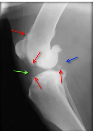
Radiographs of the stifle are not always essential to obtain a diagnosis of CrCL injury, but may be helpful to verify the presence of osteoarthritis (OA) in routine cases, to confirm stifle pathology in challenging cases of partial CrCL tear and to rule out other disorders such as fractures, neoplasia or erosive arthritis (Kowaleski et al. 2013; Canapp 2007). The most common changes related with OA include narrowing of the joint space; subchondral sclerosis of the tibial plateau; osteophytosis which occur typically along the trochlear ridges, patella, fabella, ligament attachments, and caudal tibial plateau; intraarticular mineralization; bone remodelling; and joint capsule distention identified as proximal displacement of the infrapatellar fat pad and caudal displacement of the capsule. Typical radiographic findings in CrCL disease are consistent with OA of the stifle in chronic cases, muscular atrophy of the thigh, joint effusion, periarticular swelling and displacement of the infra-patellar fat pad shadow (Figure 11) (Marino & Loughin 2010; Jerram & Walker 2003). Cranial displacement of the proximal tibia may be visible on a standard lateral radiograph without any special stress radiographs. This specific sign is called “Cazieux positive” and is diagnostic of CrCL rupture (Bree et al. 2010). Radiographs of the contralateral stifle can be useful for comparison and
22
may be of prognostic value if there is evidence of radiographic synovial effusion and osteophytosis in the contra-lateral stifle. These are clinically relevant markers for risk of contra-lateral CrCL rupture (Chuang et al. 2014; Doverspike et al. 1993; Vasseur 2002; Canapp 2007). Most of the abnormalities are seen on standard radiographic views, but stress views may be necessary to solidify the diagnosis (Marino & Loughin 2010). The most reliable method of diagnosing cruciate disease is through the use of tibial compression radiography (Harasen 2002).
Figure 11 Mediolateral radiographic image of a stifle joint with chronic CrCL rupture. The loss of fat pad
definition (green arrow) and distention of the caudal joint capsule (blue arrow) is evident. Also note the osteophyte formation (red arrows) along the trochlear ridge and the subchondral bone sclerosis of the tibial plateau (Schulz 2013).
To perform a tibial compression radiography (Figure 12), a standard lateral radiographic view of the stifle joint is obtained with the joint at 90° of flexion (neutral position). Then, a second radiograph is taken with the stifle in the same position while performing the tibial compression test (de Rooster et al. 1998). The proximal tibia will move cranially in relation to the distal femur when stress is applied. Another parameter in the interpretation of tibial compression radiography is the displacement of the sesamoid bone of the popliteous muscle. Tibial compression radiography is therefore a reliable diagnostic method for both complete and partial CrCL ruptures with a sensibility of 99% and a specificity of 100% especially when there is a lack of cranial drawer sign on clinical examination (Carobbi & Ness 2009; de Rooster et al. 1998; Bree et al. 2010).
23
Figure 12 Tibial compression radiographs of a dog with CrCL rupture: Neutral (left) and tibial compression
(right). By flexing the hock joint, a displacement between the distal femur and the tibial plateau becomes noticeable (black arrows). Also note the ventral displacement of the sesamoid bone of the popliteous muscle (white arrow). (Bree et al. 2010)
Radiography may be sufficient to make a diagnosis when clinical signs and physical examination findings are accordant in the evaluation. Nonetheless, if radiographic assessment is inconclusive, more advanced imaging is necessary.
3.2.2. Ultrasonography
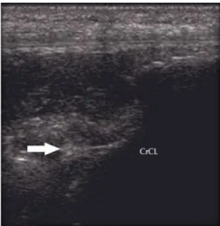
Ultrasonography (US) is a potential valuable imaging technique of the musculoskeletal system in small animals (M. Kramer, M. Gerwing, U. Michele, A. Flöck, M. Risselada, P. van Bree 2004; Nayseh et al. 2015). It can be used to demonstrate pathological changes in soft tissue but is relatively inaccurate to confirm rupture of the CrCL (Gnudi & Bertoni 2001; I. Gielen, B. Van Ryssen 2009). Ultrasonography may be useful for assessing cartilage abnormalities, meniscal tears, muscle, tendon and ligament abnormalities or neoplasia. The cruciate ligaments, being deep intra-articular structures, are the most difficult ligaments of the stifle joint to examine (C. R. Cook 2010). Therefore, accurate examination of joints requires considerable ultra-sonographic experience and a standardised examination procedure (I. Gielen, B. Van Ryssen 2009). Regarding the evaluation of meniscal tears, although ultrasonography grants a high specificity (99,3%), the sensitivity is poor (48,9%) (Mahn et al. 2005). Diagnosis of CrCL rupture can be made by demonstration of the fluttering edges of the ruptured ligament (Figure 13) (C. R. Cook 2010; Nayseh et al. 2015).
24
Figure 13 Ultra sonographic image of a ruptured CrCL: The ligament presents with irregular ends (arrow)
consistent with a complete rupture (C. R. Cook 2010).
Diagnosis of CrCL rupture in dogs is generally cost-effective and readily available but it can be very difficult to reproduce sonographic findings in the canine stifle. There are significant limitations to the use of ultrasound to evaluate the stifle joint which make other diagnostic imaging modalities more useful (Soler et al. 2007; M. Kramer, M. Gerwing, U. Michele, A. Flöck, M. Risselada, P. van Bree 2004; Seong et al. 2005; Gnudi & Bertoni 2001).
3.2.3. Computed Tomography
Better soft-tissue differentiation and absence of superimposition are the major advantages of cross-sectional imaging such as CT over conventional x-ray techniques. Modern scanners allow the reproduction and display of a 3D model which can be coloured according with the anatomic structure (i.e. bone, muscle, cartilage) (Soler et al. 2007; Marino & Loughin 2010). CT has been proven to be extremely sensitive in demonstrating avulsion fractures of intra-articular ligaments like the CrCL (Figure 14) and the tendons of the extensor digitorum longus and the popliteus muscles (Marino & Loughin 2010; I. Gielen, B. Van Ryssen 2009). CT evaluation of the intercondylar notch of canine stifles can be easily obtained and is more reliable when compared with conventional radiographs (Lewis et al. 2008; Soler et al. 2007). Additionally, degenerative changes can be identified in an earlier stage. CT scan is therefore a valuable asset in the diagnosis of several stifle pathologies including partial CrCL rupture (Han et al. 2008).
25
Figure 14 Transverse CT image of an avulsion fracture at the insertion of the CrCL (red arrow) (I. Gielen,
B. Van Ryssen 2009).
3.2.4. Magnetic Resonance Imaging
Magnetic resonance imaging (MRI) is an emerging superior imaging technique for demonstration of soft tissue lesions. In the dog, as in humans, MRI has promising capabilities for imaging stifle pathology (I. Gielen, B. Van Ryssen 2009; Schmohl et al. 2014). Consequently, there is still minimal evidence in veterinary medicine available to show that MRI is better than other types of examinations justified for the cost and risk once general anaesthesia is required (Scrivani 2010). The major advantages of MRI are its excellent image resolution (Figure 15), superior soft tissue contrast, acquisition of images in any plane, and use of a magnetic field rather than ionizing radiation (Soler et al. 2007). In addition, MRI appears to be a very reliable method for diagnosing meniscal tears pre-operatively, with reported sensitivity of 100% and specificity of 94% (Olive et al. 2013; Barrett et al. 2009). A multiplicity of meniscal tears have been described with MRI which wouldn’t have been recognised with other imaging modalities (I. Gielen, B. Van Ryssen 2009). However, the high cost of this procedure limits its use in diagnosing CrCL rupture (Jerram & Walker 2003; Schulz 2013).
26
Figure 15 Left: T1 sagittal magnetic resonance imaging of a normal stifle; Right: T2 sagittal oblique MRI
of a stifle with CrCL rupture (black arrow); The CaCL is marked with the white arrow. (Marino & Loughin 2010)
Both CT and MRI provide excellent images of the stifle joint structures, the most obvious difference comparing both techniques is the greater soft-tissue contrast demonstrated and the exceptional definition in MRI images (Soler et al. 2007).
3.2.5. Scintigraphy
Scintigraphy provides information regarding the metabolic function of the skeleton. It provides an indication of many joint disorders and has been successfully used for staging dogs with OA but its major application is in lameness investigation if pain is not easily localizable to any one joint. Although it is a very sensitive technique, it is not very specific (Figure 16) (Innes et al. 1995; Comerford 2012; I. Gielen, B. Van Ryssen 2009).
Figure 16 Bone scintigraphy of stifle bone uptake 12 weeks after left cranial cruciate ligament transection
27
3.2.6. Thermography
Thermography is a non-invasive diagnostic imaging technique that records cutaneous thermal patterns generated by the infrared emission of surface body heat (Figure 17). Thermography in dogs is now limited to research applications but early accurate detection of stifle diseases may be possible as technology improves (Marino & Loughin 2010; Infernuso et al. 2010).
Figure 17 Thermal image of a stifle with a ruptured cranial cruciate ligament. The patella (black solid
arrow) is cooler than the inflamed joint (black open arrow). (Infernuso et al. 2010)
Regardless of the imaging technique, progresses in computing technology have accelerated advances in diagnostic imaging. Eventually, a multimodality approach will likely provide a complete assessment of complex structures in the canine stifle using the advantages of each technique.
28
3.3. Arthroscopy
The use of arthroscopy as a method of joint exploration in small animals was firstly described in 1978 by Siemering, who reported findings of stifle arthroscopies and concluded the instrumentation was useful in the diagnosis of diseases of the canine stifle joint (Hoelzler et al. 2004). At about the same time, Bennett and Kivumbi from of the United Kingdom reported the utility of arthroscopy for the canine stifle joint (Bardet 2006). Since then, major advances in veterinary arthroscopy have revolutionized both the diagnosis and treatment of joint pathology. Its use has now become common for canine joint disease (Bardet 2006; Beale & Hulse 2011).
Figure 18 Arthroscopic examination of the Cruciate Ligaments: A - normal appearance of the CrCL [crcl]
with the two distinct bands (cranio-medial band [crmb] and caudo-lateral band [cdlb]) and CaCL [cdcl]. The lateral femoral condyle [lfc] is seen. B - normal insertion of the CrCL. Medial meniscus [mm] and medial femoral condyle [mfc] are seen. C - complete chronic tear of the CrCL with a nodular formation on the end of the torn ligament (Beale et al. 2003)