Research Article
Apocynin Dietary Supplementation Delays Mouse Ovarian Ageing
F. Timóteo-Ferreira,
1,2S. Mendes
,
1,2N. A. Rocha,
2L. Matos
,
1,2,3,4A. R. Rodrigues
,
1,2H. Almeida
,
1,2,5and E. Silva
1,21Instituto de Biologia Molecular e Celular (IBMC) and Instituto de Inovação e Investigação em Saúde (I3S),
Universidade do Porto, Portugal
2Unit of Experimental Biology, Department of Biomedicine, Faculty of Medicine of Porto University, Portugal 3Faculty of Dental Medicine of Porto University, Portugal
4Faculty of Nutrition and Food Sciences of Porto University, Portugal 5Obstetrics-Gynecology, Hospital-CUF Porto, 4100 180 Porto, Portugal
Correspondence should be addressed to E. Silva; elisabete.silva@i3s.up.pt Received 26 April 2019; Accepted 10 September 2019; Published 20 October 2019 Academic Editor: Rosalba Parenti
Copyright © 2019 F. Timóteo-Ferreira et al. This is an open access article distributed under the Creative Commons Attribution License, which permits unrestricted use, distribution, and reproduction in any medium, provided the original work is properly cited. Advanced maternal age is associated with higher infertility rates, pregnancy-associated complications, and progeny health issues. The ovary is considered the main responsible for these consequences due to a continuous decay in follicle number and oocyte quality. Intracellular imbalance between oxidant molecules and antioxidant mechanisms, in favour of the former, results in oxidative stress (OS) that is believed to contribute to ovarian ageing. This work is aimed at evaluating whether an age-related increase in ovarian OS, inflammation, and fibrosis may contribute to tissue dysfunction and whether specific antioxidant supplementation with a NADPH oxidase inhibitor (apocynin) could ameliorate them. Mice aged 8–12 weeks (reproductively young) or 38-42 weeks (reproductively aged) were employed. Aged mice were divided into two groups, with one receiving apocynin (5 mM) in the drinking water, for 7 weeks, upon which animals were sacrificed and their ovaries collected. Ovarian structure was similar at both ages, but the ovaries from reproductively aged mice exhibited lipofuscin deposition, enhanced fibrosis, and a significant age-related reduction in primordial and primary follicle number when compared to younger animals. Protein carbonylation and nitration, and markers of OS were significantly increased with age. Moreover, mRNA levels of inflammation markers, collagens, metalloproteinases (MMPs), and tissue inhibitor MMPs (TIMPs) were upregulated. Expression of the antifibrotic miRNA29c-3p was significantly reduced. Apocynin supplementation ameliorated most of the age-related observed changes, sometimes to values similar to those observed in young females. These findings indicate that there is an age-related increase in OS that plays an important role in enhancing inflammation and collagen deposition, contributing to a decline in female fertility. Apocynin supplementation suggests that the imbalance can be ameliorated and thus delay ovarian ageing harmful effects.
1. Introduction
During the last decades, developed and developing countries have experienced economic and educational changes that gave women the opportunity to reach higher professional and decision levels. As a consequence, childbearing has been postponed into a period of life when fertility success decreases and pregnancy-associated disorders increase
sig-nificantly [1].
Human female fertility peaks in the early 20s and gradu-ally declines until the mid-30s. Thereafter, reproductive
potential falls sharply, until it virtually ends at menopause around the age of 50 [2]. The ovary is believed to be the main fertility regulator due to the continuous age-related decay in follicle number and oocyte quality [3].
A theory for ovarian ageing holds that age-related disrup-tion of redox homeostasis affects oocyte quality [4]. The con-tinued generation of reactive oxygen species (ROS) together with an age-related decline in activity and expression of important follicle antioxidant enzymes results in an imbal-ance between ROS production and antioxidant defences. The condition leads to oxidative stress (OS) responsible for
Volume 2019, Article ID 5316984, 11 pages https://doi.org/10.1155/2019/5316984
protein, amino acid, lipid, and DNA damage that underlie ovarian ageing and fertility loss [5]. As a matter of fact,
in follicular fluid of older women, the expression of
important antioxidant enzymes, as catalase and specific
glutathione S-transferases, is significantly lower when
com-pared with younger women [6]. Similarly, superoxide dis-mutase 1, superoxide disdis-mutase 2, and catalase gene expression in granulosa [7] and cumulus oophorus cells [8] are also downregulated during reproductive ageing. In this process, the use of specific antioxidant molecules has shown beneficial effects in delaying follicle depletion and fertility impairment [9–12].
Studies on ovarian ageing have expanded from the oocyte immediate surroundings to the ovarian stroma, mainly com-posed of an arrangement of extracellular matrix components
(ECM) andfibroblasts and smooth muscle, endothelial, and
immune cells. This microenvironment has an important impact on follicle development and oocyte quality. In fact,
Briley et al. [13] verified ovarian microenvironment changes
with age, specifically an increase in fibrosis and inflamma-tion, and suggested its contributory role to the coincident decrease in oocyte quality. ROS are also believed to contrib-ute to the synthesis and activation of various cytokines and growth factors, hence creating common feedforward and
feedback mechanisms that promote tissuefibrosis [14].
Inflammation and ROS formation appear to be key
factors in the pathogenesis of ovarian fibrosis [15], which
reflects a disturbance of the synthesis and degradation of
extracellular matrix (ECM) favouring excessive collagen deposition. Important regulators of ECM homeostasis are metalloproteinases (MMPs), a group of enzymes capable of degrading all types of ECM components [16], and tissue inhibitor metalloproteinases (TIMPs), both affected by age-ing [17]. More recent studies have also identified specific
microRNAs (miRNAs) as important mediators in fibrosis;
they act either by regulating target genes involved in the process of ECM remodelling or by signalling pathways associated with it [18]. Reduction of ROS production by inhibiting NADPH oxidase (NOX) activity with apocynin
has shown beneficial effects on renal [19, 20], cardiac [21],
skeletal muscle [22], and pulmonary [23] fibrosis. Despite
our previous results showing beneficial effects of apocynin supplementation on uterine ageing and fertility [24], its effect on ovarian redox imbalance, inflammation, and fibrosis during reproductive ageing is scarce.
In sum, an age-related low-grade inflammatory state may associate with ovarian collagen deposition and a progressive reduction in follicle number and oocyte quality. In this set-ting, it was hypothesized that an age-related increase in
ovar-ianfibrosis and ROS underlies fertility reduction. To verify it,
markers of oxidative stress, tissuefibrosis, and inflammation,
along with the possibility to ameliorate those features by a specific antioxidant supplementation, were evaluated in vivo.
2. Material and Methods
2.1. Animal Handling and Ovarian Tissue Collection. All the experiments were performed according to the Portuguese law on animal welfare and according to the guidelines issued
by Federation of European Laboratory Animal Science Associations (FELASA). Female mice (C57BL/6J strain) obtained from Harlan were kept under controlled conditions
(12 h light/dark cycle and room temperature at 22°C) and
had free access to tap water and standard mouse chow. Young (8-12 weeks old) and reproductively aged (38-42 weeks old) female mice were employed. Reproductively aged mice were divided into two groups, with one receiving the antioxidant, apocynin, 5 mM, in drinking water 7 weeks
prior to sacrifice. Water bottles were protected from light
and changed twice a week. At the selected ages, female mice were anesthetized with isoflurane and euthanized by cervical dislocation, and the ovaries were excised. One ovary was immediately frozen in liquid nitrogen and subsequently kept
at -80°C for molecular studies and the other was fixed
overnight, in 4% paraformaldehyde, for structural studies. 2.2. Tissue Processing for Histological Techniques. Fixed ovaries were dehydrated with the aid of increasing concentra-tions of ethanol and diaphanized using benzol. Impregnation
and inclusion were carried out in paraffin, and 5 μm thick
sequential sections were mounted on poly-L-lysine-coated
slides and dried overnight at 37°C. They were stored in plastic
boxes to be used for all histological applications throughout this study.
2.3. Morphological Analysis of Ovarian Tissues. Ovarian sections for morphological examination were stained with hematoxylin & eosin (H&E) according to the following protocol. Slides were dewaxed twice with xylol and hydrated with decreasing concentrations of ethanol and water. Subse-quently, slides were stained with Harris hematoxylin for 2 minutes and then stained with alcoholic eosin for 5 minutes. Finally, tissues were dehydrated with increasing concentra-tions of ethanol followed by two xylol passages. Slides were mounted in Entellan and air dried. Ovarian morphology was observed under light microscope equipped with a digital camera, and representative images at ovarian midsection were captured. Other ovarian sections were stained with Sudan Black 0.1% for lipofuscin examination. Slides were dewaxed and hydrated as previously described, stained with Sudan Black 0.1% for 20 minutes, dehydrated, and mounted. 2.4. Follicle Counting at Ovarian Midsection. H&E slides were used for follicle counting. The follicles were branded as pri-mordial, primary, secondary, or antral, and corpus luteum.
Follicles were classified as primordial or primary when
oocytes were surrounded, respectively, by a single layer of squamous or cuboidal granulosa cells. Secondary follicles
were identified by having more than one layer of granulosa
cells with no visible antrum. Antral follicles are the ones that
displayed small areas of follicularfluid (antrum) or a single
large antral space. Corpus luteum were identified as intrao-varian bund structures with morphologically homogeneous round cells, showing enhanced cytoplasm/nucleus ratio, when compared with granulosa cells, and deprived of oocyte. The number of follicles at ovarian midsection was obtained by calculating the mean counts of three ovarian midsections, representative of each animal, and a minimum of four ani-mals per group was used.
2.5. Autofluorescence Lipofuscin Detection. Ovarian sections were dewaxed and hydrated as previously described. After washing in PBS, they were mounted in phosphate-buffered
glycerol and observed under afluorescence microscope (Carl
Zeiss AxioImager Z1) equipped with a digital camera.
2.6. Fluorescent Immunohistochemistry. Afluorescent
immu-nohistochemical technique was performed to detect protein carbonylation and nitration (markers of oxidative stress). Ovarian sections were dewaxed and hydrated as previously described. Slides were washed with phosphate-buffered saline
(PBS) solution and permeabilized with 0.5% Triton™ X-100
in PBS for 5 minutes, followed by washing with PBS. For pro-tein carbonylation assessment, an additional derivatization
process was required before nonspecific signal blocking.
The derivatization protocol was performed as previously published [25]. In brief, sections were incubated in 10 mM 2,4-dinitrophenylhydrazine (DNPH; Sigma-Aldrich) in 10% TFA for 10 minutes, the reaction stopped with 2 M Tris base pH 10, and slides were washed with PBS. Background staining was blocked with 2% bovine serum albumin (BSA) in PBS with 0.1% Tween 20 (PBS-T) for 1 hour at room temperature. Slides were then incubated with a rabbit polyclonal antibody recognizing DNP (1 : 250; Sigma-Aldrich) or a mouse monoclonal antibody recognizing nitrotyrosine (1 : 250; Santa Cruz Biotechnology) in
PBS-T, overnight at 4°C. The following day, slides were washed
in PBS-T, incubated with Alexa Fluor 488-conjugated anti-rabbit IgG secondary antibody (1 : 750; Molecular Probes) or in Alexa Fluor 568-conjugated anti-mouse IgG secondary antibody (1 : 750; Molecular Probes) in PBS-T for 1 hour at room temperature. Then, they were washed again, counter-stained with 4′,6-diamidino-2-phenyl-indole (DAPI), and mounted. Slides were examined, and images were recorded
under afluorescence microscope (Carl Zeiss AxioImager Z1)
equipped with a digital camera. Signal extension was quanti-fied using ImageJ software by identification of the threshold cut point, with blind intervention of three operators. A mini-mum of four animals per group was used, and a minimini-mum of two representative sections of the midovary was examined. 2.7. Fibrosis Evaluation in Ovarian Tissue. Picrosirius red histochemical technique was performed for quantification
of tissue fibrosis. Ovarian sections were dewaxed and
hydrated as previously described. Then, slides were stained with sirius red solution for 90 minutes, rapidly passed through 0.5% acidified water, and subsequently dehydrated, with increasing concentrations of ethanol, followed by two xylol passages. Slides were mounted in Entellan for further visualization and analysis. This technique stains collagen in red and cytoplasm in yellow. Red staining (collagen) was
quantified using the ImageJ software by identification of the
threshold cut point, with blind intervention of three opera-tors. A minimum of four animals per group was used, and a minimum of two sections of the midovary was examined.
2.8. Real-Time PCR. Ovarian RNA extraction and puri
fica-tion was performed with TripleXtractor direct RNA kit
(GRiSP) following the manufacturer’s instructions. Total
RNA was quantified by measuring the absorbance at 260 nm in a NanoDrop (Thermo Fisher Scientific), and RNA purity was evaluated by the ratio of absorbance at 260 and 280 nm. RNA samples were reverse transcribed to cDNA with the NZY First-Strand cDNA Synthesis kit (NZYTech). Real-time PCR was carried out using PowerUp SYBR Green
Master Mix (Thermo Fisher Scientific) and specific primers
(Table 1) in a StepOnePlus™ Real-Time PCR System
(Applied Biosystems). Primers were designed using the
online available specific mRNA sequences and Primer3
pro-gram. The derived sequences were submitted for a BLAST search to ensure exclusive alignment to the desired target genes. Amplification reactions were performed, in duplicate, according to conditions stated in Table 1. To check specific-ity, a dissociation curve was derived at the end of each run. Controls lacking reverse transcriptase were included to ensure no genomic DNA contamination during prepara-tions. Results were normalized to 18S expression.
2.9. MicroRNA Quantification. Total RNA extraction was performed using the commercial Recover ALL™ Total Nucleic Acid Isolation Kit (Ambion), according to the
manu-facturer’s instructions. In brief, for each sample, four
paraffin-embedded ovaries were sectioned at 20 μm thickness. Each
sample was dewaxed in 100% xylene, washed in 100% ethanol,
and incubated with protease and digestion buffer for 2 hours
at 50°C, followed by 15 minutes at 75°C. Nucleic acids were
isolated and incubated with DNase mix for 30 min at room
temperature. After a final purification, 7 μL of total RNA
was reverse transcribed to cDNA using the MystiCq micro-RNA cDNA Synthesis Mix (Sigma-Aldrich Co.). In the pro-cess of cDNA synthesis, miRNAs were subjected to polyadenylation by poly(A) polymerase that catalysed the transfer of adenosine deoxynucleotides to the 3′ end. Ready-Script Reverse Transcriptase and other necessary reagents for cDNA synthesis were subsequently added to convert the
poly(A) tailed microRNAs into first-strand cDNA using an
oligo-dT adapter primer. The unique sequence at the 5′ end
of the adapter primer allows amplification of microRNA
cDNAs in real-time qPCR reactions using the MystiCq micro-RNA qPCR Universal Primer. Real-time PCR was carried out as previous described. Controls lacking reverse transcriptase were included to ensure no genomic DNA contamination dur-ing preparations, along with controls lackdur-ing poly(A) poly-merase. Results were normalized to RNU1A (Qiagen) expression. Specific primers used were 21∗_1; miR-29c_1; miR-215_1; and miR-212-3p_1 miScript Primer Assay
(Qiagen), with annealing temperature of 55°C.
2.10. Statistical Analysis. Arithmetic means are given with standard error of the mean (SEM). Statistical analyses were performed with GraphPad Prism 6.01 using one-way analysis of variance (ANOVA), followed by the Tukey posttest. A
P < 0:05 was considered statistically significant.
3. Results
3.1. Age-Related Alterations in Ovarian Morphology. To observe ovarian morphology, H&E-stained midorgan
sections (Figure 1(a)) revealed follicles in various stages of development, occupying most areas of the ovary, but pre-dominantly in the periphery. Central areas were occupied by a heterogeneous stroma that included vessels, bundles of extracellular connective tissue, and respective cells.
A unique population of stroma cells was detected only in the ovaries of reproductively aged mice. They were large and multinucleated, with pale cytoplasm containing a yellow-brown pigment (Figure 1(a)), which displayed autofluores-cence (Figure 1(b)). Fluoresautofluores-cence quenching by staining with Sudan Black indicated that these were deposits of lipofuscin (oxidized lipids and proteins) (Figure 1(b)). Ovarian stromal cells with the abovementioned characteristics were consis-tently observed in reproductively aged mice and correspond to multinucleated giant macrophages [13, 26]. Follicle num-ber was evaluated and, as expected, in reproductively aged mice, a significant reduction was noticed. This was due to a
decrease in the number of primordial and primary follicles. Apocynin had no effect (Figure 1(c)).
3.2. Age and Antioxidant Supplementation Effect on Ovarian
Oxidative Stress and Fibrosis. Tyrosine nitration and protein carbonylation were used to characterize ovarian oxidative stress (Figures 2 and 3). As shown in Figure 2, protein tyro-sine nitration staining was found on the medullary stromal cells and partially colocalized with lipofuscin deposits. DNP immunoreactivity was observed in stromal cells, theca cells, oocytes, and corpus luteum (Figure 3). Reproductively aged females had increased expression of lipofuscin deposits
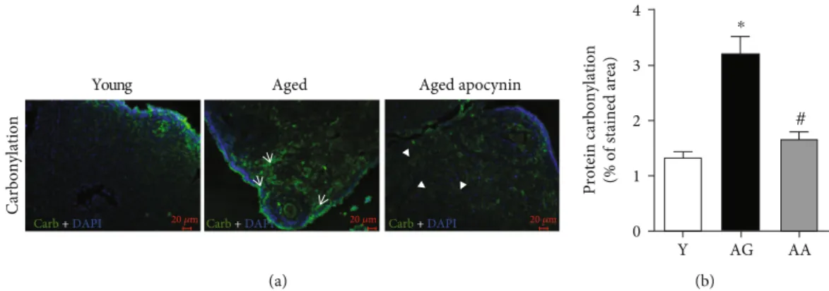
(1:00 ± 0:41 vs. 8:53 ± 2:95), carbonylated (1:34 ± 0:09 vs.
3:17 ± 0:35), and nitrated proteins (1:00 ± 0:20 vs. 2:60 ± 0:26) (Figures 2(b) and 3(b)). The use of apocynin, that inhibits NOX-mediated superoxide production, resulted in
a significant reduction in both carbonylated and nitrated
Table 1: List of primers used in RT-PCR reactions.
Primers Sequence Annealing temperature Fragment (bp)
Col1α1 Fwd 5′-GACGCATGGCCAAGAAGACA-3′ 60°C 85 Rev 5′-CTCGGGTTTCCACGTCTCAC-3′ Col3α1 Fwd 5′-AGCTTTGTGCAAAGTGGAACC-3′ 58°C 114 Rev 5′-ATAGGACTGACCAAGGTGGC-3′ Col5α1 Fwd 5′-CCTGGTTCAGTGAATTCAAGCG-3′ 60°C 81 Rev 5′-TCATTTGTACCACGCCCACG-3′ TGF-β1 Fwd 5′-ATTCCTGGCGTTACCTTGG-3′ 60°C 120 Rev 5′-AGCCCTGTATTCCGTCTCCT-3′ TNF-α Fwd 5′-CCCTCACACTCAGATCATCTTCT-3′ 60°C 61 Rev 5′-GCTACGACGTGGGCTACAG-3′ IL-1β Fwd 5′-TGACGGACCCCAAAAGATGA-3′ 60°C 87 Rev 5′-TGCTGCGAGATTTGAAGCTG-3′ CCL5 Fwd 5′-TGCCCACGTCAAGGAGTATT-3′ 60°C 84 Rev 5′-ACTTGGCGGTTCCTTCGAG-3′ MMP2 Fwd 5′-TGTCGCCCCTAAAACAGACA-3′ 58°C 65 Rev 5′-TGGGGCAGCCATAGAAAGTG-3′ MMP9 Fwd 5′-CCTGGAACTCACACGACATCT-3′ 62°C 72 Rev 5′-CACGCCAGAAGAATTTGCCAT-3′ MMP12 Fwd 5′-GGGCTGCTCCCATGAATGAC-3′ 56°C 85 Rev 5′-GTCATTGGAATTCTGTCCTTTCCA-3′ TIMP1 Fwd 5′-GGTGTGCACAGTGTTTCCCTGTTT-3′ 60°C 72 Rev 5′-TCCGTCCACAAACAGTGAGTGTCA-3′ TIMP2 Fwd 5′-GGATTCAGTATGAGATCAAGC-3′ 55°C 145 Rev 5′-GCCTTTCCTGCAATTAGATAC-3′ 18S Fwd 5′-CGCCGCTAGAGGTGAAATTC-3′ 60°C 67 Rev 5′-CATTCTTGGCAAATGCTTTCG-3′
proteins, to levels similar to those observed in the younger group (Figures 2 and 3).
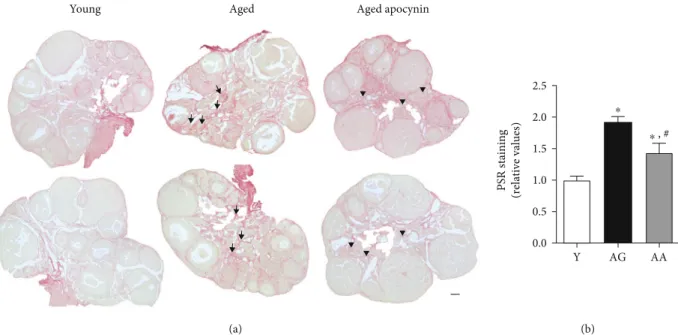
Oxidative stress appears to play a crucial role in the
ethology of tissuefibrosis. Next, picrosirius red (PSR)
histo-chemical technique was used to evaluate ovary collagenfibril
deposition. Slight PSR staining was observed around folli-cles, blood vessels, and epithelium of the ovarian surface (Figure 4(a)). However, a network-like, intense PSR staining,
characteristic offibrotic foci, was only seen on the ovarian
stroma and increased significantly with age (1:00 ± 0:12 vs.
1:92 ± 0:09) (Figure 4). In contrast, reproductively aged
females treated with apocynin exhibited significantly
reduced PSR staining (1:43 ± 0:15) (Figure 4(b)).
3.3. Age and Treatment Effect on Inflammation Factors and Collagen Expression. As previously mentioned, synthesis and secretion of various growth factors and cytokines are interlinked with OS in feedforward and feedback cycle
mechanisms that might contribute tofibrosis via enhanced
inflammation. Reproductive ageing was accompanied by a
significant increase in several cytokines [(CCL5 (1:00 ± 0:92
vs. 18:93 ± 5:96), TNF-α (1:00 ± 0:66 vs. 6:82 ± 1:73),
IL-1β (1:00 ± 0:40 vs. 15:55 ± 5:13)], including TGF-β1
(1:00 ± 0:90 vs. 12:85 ± 3:81) that is considered the most
important cytokine modulatingfibrosis signalling (Figure 5).
All those molecules also regulate specific miRNA expression
involved in fibrosis. Interestingly, as shown in Figure 6,
miRNA29c-3p, a “master fibromiRNA,” was found to be
downregulated in the ovaries of reproductively aged
females (1:00 ± 0:13 vs. 0:57 ± 0:06), unlike miRNA212-3p
(1:00 ± 0:11 vs. 0:86 ± 0:13) and 21a-3p (1:00 ± 0:25 vs.
0:76 ± 0:14) that were not affected by ageing. Expression of miRNA 29c-3p was inversely correlated with collagen
expression. Both collagen types Col1a1 (1:00 ± 0:88 vs.
11:48 ± 3:88) and Col5a1 (1:00 ± 0:91 vs. 10:51 ± 3:10) were significantly increased in aged females. Apocynin treatment normalized cytokine and collagen expression to levels similar to those observed in younger females (Figure 5).
3.4. MMP/TIMP Expression. MMPs regulate not only ECM degradation but also bioavailability and activity of cyto-kines, chemocyto-kines, and growth factors. MMP activity is,
in turn, regulated by its specific TIMPs. During
reproduc-tive ageing expression of MMP9, TIMP1 and TIMP2 were
significantly increased [(1:00 ± 0:95 vs. 16:80 ± 5:63),
(1:00 ± 0:97 vs. 51:62 ± 16:61), and (1:00 ± 0:96 vs. 33:09 Young Aged P P S S SA SA CL CL CL CL SA SA S PM Aged apocynin CL CL CL SA S P (a) Aut ofluor escence Sudan-black (b) Y AG AA * PM P S SA CL ⁎ ⁎ ⁎ ⁎ ⁎ 30 20 10 N u m b er o f f o llic les 0 ⁎ ⁎ (c)
Figure 1: Ovarian structure and follicle number. (a) H&E-stained representative midovarian sections from young and aged mice. Note yellow-brown palely stained multinucleated cells, located in stroma of reproductively aged mice (white arrowheads). (b) Representative images of Sudan Black staining and autofluorescence in the ovaries of aged mice. Multinucleated cells stain intensely with Sudan Black and emit strong autofluorescence, indicating age-related deposition of oxidized proteins and lipids. Average quantification of autofluorescence area (n = 4‐5 per group). (c) Follicle distribution at midovarian sections of young and reproductively aged mice (n = 4 per group), showing a significant decrease in primary and primordial follicle number of older animals. Y: young; AG: aged; AA: aged apocynin; PM: primordial follicles; P: primary follicles; S: secondary follicles; SA: secondary antral follicles; CL: corpus luteum. Bars = 100 μm. Data are presented as mean ± SEM.∗P < 0:05, compared with young female mice.
± 10:88), respectively] (Figure 6). Once more, apocynin dietary supplementation reversed age-related observed changes (Figure 6).
4. Discussion
The present study revealed that, during reproductive ageing, the ovary undergoes morphological and molecular changes related with a disturbance of the redox homeostasis, shown by increased lipofuscin content, protein carbonylation and
nitration, and collagen deposition. Along with those changes, there was an age-related increase of inflammation and fibro-sis, evidenced by higher relative expression of inflammation markers, MMPs, TIMPs, and a specific miRNA. Antioxidant supplementation with apocynin was capable of ameliorating features of the ovarian ageing process.
Over time, cells and tissues display structural changes
that reflect age-related modifications in biomolecules and
signalling pathways, eventually leading to tissue dysfunction [27]. Beyond time-related structural changes, the ovary is
Young Aged Lipofuscin Nitr otyr osine Aged apocynin Mer ge 20 𝜇m 20 𝜇m 20 𝜇m (a) Lipofuscin (relative values) Nitrotyrosine staining (relative values) Y AG AA 0 5 10 15 Y AG AA 0 1 2 3 4 ⁎ ⁎ # (b)
Figure 2: Protein nitration in the mouse ovaries evidenced by fluorescence immunohistochemistry, using a specific antibody. (a) Representative images of young, reproductively aged and apocynin-treated aged mice. Note tyrosine nitration staining and lipofuscin colocalization in the tissue. (b) Average quantification of lipofuscin and nitrated proteins stained area assuming young mice equals 1 (n = 4‐5 per group). Y: young; AG: aged; AA: aged apocynin. Data are presented as mean ± SEM. Data are presented as mean ± SEM.
∗P < 0:05, compared with young female mice;#P < 0:05, compared with reproductively aged female mice.
Young Aged
Carbonylation
Aged apocynin
Carb + DAPI 20 𝜇m Carb + DAPI Carb 20 𝜇m + DAPI 20 𝜇m
(a)
Protein carbonylation (% of stained area)
Y AG AA 0 1 2 3 4 # ⁎ (b)
Figure 3: Protein carbonylation in the mouse ovaries evidenced by fluorescence immunohistochemistry, using a specific antibody for DNP. (a) Representative images of young, reproductively aged and apocynin-treated aged mice. Note the substantial carbonyl labeling increase in ovarian stroma cells (white arrows) and antioxidant-mediated amelioration (arrowheads). (b) Average quantification of stained area (n = 4‐5 per group). Y: young; AG: aged; AA: aged apocynin. Data are presented as mean ± SEM.∗P < 0:05, compared with young female mice;#P < 0:05, compared with reproductively aged female mice.
distinctly affected by additional cyclic ones, tightly controlled by hormonal variations. The mild, albeit continued, inflam-matory nature of such alterations subjects the ovary to
recur-rent OS, inflammatory cell invasion, and fibrosis, especially
intensified during ageing, when a decrease in antioxidant
defences deepens the cellular redox imbalance [6–8, 28, 29].
The regulatory mechanisms contributing to age-related ovarian changes are still poorly known. Previously, we dem-onstrated that dietary supplementation with apocynin enhanced pregnancy outcomes at a latter reproductive age by improving the observed age-related decrease in mouse litter size and restoring decidua layer thickness [24]. In this
work, apocynin was used to study its antioxidant properties in mitigating ovarian ageing effects.
Two features that distinguished young and reproduc-tively aged ovaries were a unique population of multinucle-ated giant cells containing lipofuscin deposits and the
number of follicles. In aged mice, a significant reduction in
the number of primordial and primary follicles was observed, which agrees with the well-known time-related decline of the follicle resting pool [2, 30]. At this age, as expected, dietary supplementation with apocynin did not prevent the decline, because natural oocyte attrition had occurred. However, female fertility is not only dependent on the follicle pool
Young Aged Aged apocynin
(a) PSR staining (relative values) Y AG AA 0.0 0.5 1.0 1.5 2.0 2.5 ⁎ ⁎ , # (b)
Figure 4: Fibrosis in mouse ovarian midsections stained by picrosirius red. (a) Representative images of young, reproductively aged and apocynin-treated aged mice. Note increased deposition of collagen fibers in the stroma of reproductively aged mice (arrows) and antioxidant-mediated amelioration (arrowheads). (b) Relative quantification of stained area assuming young mice equals 1 (n = 4 per group). Y: young; AG: aged; AA: aged apocynin. Bar = 100μm. Data are presented as mean ± SEM. ∗P < 0:05, compared with young female mice;#P < 0:05, compared with reproductively aged female mice.
mRNA (relative expression) CCL5 TGF-𝛽 TNF-𝛼 IL-1𝛽 0 10 20 30 Y AG AA ⁎ ⁎ ⁎ ⁎ # # # # mRNA (relative expression)
mi212-3p mi21a-3p mi29c-3p 0.0 0.5 1.0 1.5 2.0 ⁎ mRNA (relative expression)
Col1a1 Col3a1 Col5a1
0 10 20 30 40 50 # # ⁎ ⁎
Figure 5: Expression of inflammation/fibrotic factors and extracellular matrix proteins. Relative mRNA and miRNA expression was calculated using the 2ΔCT (ΔCT = CTreference RNA− CTtarget), and values were expressed as fold change over young group (n = 4‐7 per
group). Reproductive ageing is associated with an inflammatory and profibrotic ovarian microenvironment, increased levels of cytokines and collagens, and decreased level of the antifibrotic mi29c-3p RNA. Apocynin treatment reverts the expression of inflammation and fibrosis factors. Y: young; AG: aged; AA: aged apocynin. Data are presented as mean ± SEM.∗P < 0:05, compared with young female mice; #P < 0:05, compared with reproductively aged female mice.
but also on follicle and oocyte quality [31, 32], itself
influenced by the ovarian oxidative microenvironment [13].
The ovarian stromal giant multinucleated cells present in reproductively aged mice were previously shown to be posi-tive for a cell surface membrane protein—F4/80—highly expressed in macrophages [13, 20, 22]. Such observation sup-ports the perception that they result from macrophage fusion. In the ovaries, macrophages are important contribu-tors to the regulation of folliculogenesis and corpora lutea establishment and regression [33]. The unique presence of giant multinucleated cells in aged ovaries indicates that they
associate closely to long-term effects on the tissue.
Macro-phage fusion is a hallmark of a low-grade chronic in
flam-matory condition, a recurrent oxidative challenge that potentiates phagocytic function for the disposal of cell debris [34, 35]. Such phagocytic cells are a source of factors that promote OS. Besides lipofuscin, itself a deposit of oxi-dized protein and lipid, our results suggest they also accu-mulate nitrated proteins. Probably, due to lipofuscin undegradable nature and inability to be removed from cells, apocynin treatment did not reduce its deposition, unlike protein carbonylation and nitration content.
The low-grade chronic inflammatory condition favours
overproliferation of fibroblasts, excessive ECM deposition,
andfibrosis [36]. In accordance with a previous study using
two different strains of mice [13], the current study displays a significant age-related increase of fibrosis around follicles, blood vessels, and in the ovarian stroma. The observation
that treatment with apocynin results in less ovarianfibrosis
highlights the role of oxidative imbalance in its formation and suggests that NOX activity mediates local ROS
produc-tion and susceptibility tofibrosis. NOX-derived ROS has, in
fact, been associated with fibrosis in other organs such as
the kidney [37], pancreas [38], and liver [39–41]. Moreover,
as the inhibition prevents the enzyme cytoplasmic subunits
from binding transmembrane complexes necessary for NOX1 and NOX2 activation (but not NOX4), it supports
the view that the beneficial effects observed in the current
study were mediated by inhibition of those two isoforms.
Future works with specific NOX4 inhibitors will be useful
to investigate its role in ovarianfibrosis.
Inflammation has an important role in fibrosis
estab-lishment by promoting a set of interactions between profi-brotic and antifiprofi-brotic cytokines [42]. In the present study, increased expression of genes involved in immune cell response and recruitment was noticed. In this setting, mac-rophages are the main innate immune cells that release cyto-kines and chemocyto-kines to the local environment, of which, most impressively, NOX-mediated ROS production is an
important modulator [43–45]. We observed a significant
age-related increase in CCL5, an important chemokine in
macrophage recruitment; IL-1β and TNF-α,
proinflamma-tory cytokines involved in regulating endothelial cells in
ECM production; and TGF-β1, a multifunctional cytokine
highly responsible for ECM homeostasis through several
pathways. Equally tofibrosis, apocynin reduced their
expres-sion to young mouse levels demonstrating, once more, that
ROS play a master role infibrosis and inflammation
associ-ated with reproductive ageing.
TGF-β1 involvement in ECM homeostasis includes
the modulation of miRNAs that affect collagen deposition
[46, 47]. Different types of miRNA with regulatory effects were recognized in mouse ovaries [48, 49]. In our study, the profibrotic miRNAs 21a-3p and 212-3p showed no
differ-ences with age. However, antifibrotic mi29c-3p, considered
a “master fibrosis” regulator in several organs [47, 50–52],
exhibited a significant reduction in the ovaries of
reproduc-tively aged females, which correlated with the significant
increase in gene expression of Col1a1 and Col5a1. Surpris-ingly, apocynin attenuated, but not reverted, mi29c-3p
mRNA (relative expression) MMP2 MMP9 MMP12 TIMP1 TIMP2 0 5 10 80 160 240 # # ⁎ ⁎ ⁎ Y AG AA
Figure 6: Expression of metalloproteinases and their tissue inhibitors. Relative mRNA expression was calculated using the 2ΔCT
(ΔCT = CTreference RNA− CTtarget), and values were expressed as fold change over young group (n = 4‐7 per group). Reproductive ageing is
associated with increased metalloproteinases expression and their tissue inhibitors. Apocynin treatment normalizes RNA expression. Y: young; AG: aged; AA: aged apocynin. Data are presented as mean ± SEM. ∗P < 0:05, compared with young female mice; #P < 0:05, compared with reproductively aged female mice.
decrease. Further studies are needed for a better comprehen-sion of the mechanisms involved.
In addition, age-associated changes in MMPs and TIMPs
can further contribute tofibrosis, as they modulate the
acti-vation of macrophages andfibroblast originated
proinflam-matory cytokines, as TNF-α and TGF-β1 [35]. Their roles
in ECM remodelling as well as fibrosis establishment and
progression are tissue specific. In reproductively aged
ova-ries, MMP9, TIMP1, and TIMP2 had a significant increase in gene expression. MMP9 is a gelatinase responsible for col-lagen degradation, including type I and V [53], that has been
suggested to be directly regulated by TGF-β1 [54] and to take
part in its activation [55] by cleaving its latent form [56]. TIMP1 and TIMP2 have profibrotic roles by inhibiting MMP activity and contributing to ECM accumulation [16].
However, the efficiency of MMP inhibition differs with each
TIMP. This could explain the observed significant increase
of MMP9, despite the observed aged-related significant
increase in TIMP, further emphasizing the important role
and relationship with TGF-β1. Our findings suggest that
during reproductive ageing, the continued ROS-mediated inflammation disrupts ECM homeostatic processes
inevita-bly ending in tissuefibrosis.
Overall, supplementation with apocynin ameliorated molecular and histological consequences of the ovarian ageing process. By diminishing ROS production, apocynin
decreased tissuefibrosis, inflammation, and ECM deposition.
This comes as a novelfinding for the use of this antioxidant
as, to our knowledge, no other work evaluated the capability of this compound in slowing down age-related ovarian reproductive ageing.
In summary, the present study provides evidence that structural features of ovarian ageing are consequence of local continued OS effects, with important negative impact on the ovarian stroma. Disruption of microenvironment, caused by
a feedforward loop in which OS, inflammation, and ECM
turnover dysregulation are important players, is believed
to affect ovarian function and, ultimately, female fertility.
Moreover, specific antioxidant supplementation can be seen
as an important therapeutic way to ameliorate causes and effects of the ageing process induced by higher ROS
pro-duction. Further investigation in thisfield, addressing
apoc-ynin effects on oocyte quality, may have major potential value to uncover fundamental biological mechanisms and devise therapeutic strategies.
Data Availability
The data used to support thefindings of this study are
avail-able from the corresponding author upon request.
Conflicts of Interest
The authors declare that they have no conflicts of interest.
Acknowledgments
ES was funded by the Portuguese Government and the Euro-pean Union through FCT/POPH/FSE under the fellowship
SFRH/BPD/72536/2010 and DL57/2016/CP1327/CT0006. ARR was funded by the Portuguese Government and the European Union through DL57/2016/CP1355/CT0009.
Supplementary Materials
Supplementary Figure 1: low amplification images of protein
nitration and lipofuscin deposition in mouse ovaries.
Supple-mentary Figure 2: low amplification images of protein
carbonylation in mouse ovaries. (Supplementary Materials)
References
[1] M. Mills, R. R. Rindfuss, P. McDonald, E. te Velde, and on behalf of the ESHRE Reproduction and Society Task Force, “Why do people postpone parenthood? Reasons and social policy incentives,” Human Reproduction Update, vol. 17, no. 6, pp. 848–860, 2011.
[2] F. J. Broekmans, M. R. Soules, and B. C. Fauser, “Ovarian aging: mechanisms and clinical consequences,” Endocrine Reviews, vol. 30, no. 5, pp. 465–493, 2009.
[3] E. R. te Velde and P. L. Pearson,“The variability of female reproductive ageing,” Human Reproduction Update, vol. 8, no. 2, pp. 141–154, 2002.
[4] J. J. Taún,“Aetiology of age-associated aneuploidy: a mecha-nism based on the‘free radical theory of ageing’,” Molecular Human Reproduction, vol. 1, no. 4, pp. 163–165, 1995. [5] A. P. Wickens,“Ageing and the free radical theory,”
Respira-tion Physiology, vol. 128, no. 3, pp. 379–391, 2001.
[6] M. C. Carbone, C. Tatone, S. Delle Monache et al., “Antioxi-dant enzymatic defences in human follicularfluid: characteri-zation and age-dependent changes,” Molecular Human Reproduction, vol. 9, no. 11, pp. 639–643, 2003.
[7] C. Tatone, M. C. Carbone, S. Falone et al.,“Age-dependent changes in the expression of superoxide dismutases and cata-lase are associated with ultrastructural modifications in human granulosa cells,” Molecular Human Reproduction, vol. 12, no. 11, pp. 655–660, 2006.
[8] L. Matos, D. Stevenson, F. Gomes, J. L. Silva-Carvalho, and H. Almeida, “Superoxide dismutase expression in human cumulus oophorus cells,” Molecular Human Reproduction, vol. 15, no. 7, pp. 411–419, 2009.
[9] J. Liu, M. Liu, X. Ye et al.,“Delay in oocyte aging in mice by the antioxidant N-acetyl-L-cysteine (NAC),” Human
Reproduc-tion, vol. 27, no. 5, pp. 1411–1420, 2012.
[10] M. Liu, Y. Yin, X. Ye et al.,“Resveratrol protects against age-associated infertility in mice,” Human Reproduction, vol. 28, no. 3, pp. 707–717, 2013.
[11] X. M. Zhang, L. Li, J. J. Xu et al.,“Rapamycin preserves the fol-licle pool reserve and prolongs the ovarian lifespan of female rats via modulating mTOR activation and sirtuin expression,” Gene, vol. 523, no. 1, pp. 82–87, 2013.
[12] J. J. Tarin, S. Perez-Albala, and A. Cano,“Oral antioxidants counteract the negative effects of female aging on oocyte quan-tity and quality in the mouse,” Molecular Reproduction & Development, vol. 61, no. 3, pp. 385–397, 2002.
[13] S. M. Briley, S. Jasti, J. M. McCracken et al.,“Reproductive age-associated fibrosis in the stroma of the mammalian ovary,” Reproduction, vol. 152, no. 3, pp. 245–260, 2016.
[14] K. Richter, A. Konzack, T. Pihlajaniemi, R. Heljasvaara, and T. Kietzmann, “Redox-fibrosis: impact of TGFβ1 on ROS
generators, mediators and functional consequences,” Redox Biology, vol. 6, pp. 344–352, 2015.
[15] K. Richter and T. Kietzmann,“Reactive oxygen species and fibrosis: further evidence of a significant liaison,” Cell and Tissue Research, vol. 365, no. 3, pp. 591–605, 2016.
[16] M. Giannandrea and W. C. Parks,“Diverse functions of matrix metalloproteinases duringfibrosis,” Disease Models & Mecha-nisms, vol. 7, no. 2, pp. 193–203, 2014.
[17] S. Freitas-Rodríguez, A. R. Folgueras, and C. López-Otín,“The role of matrix metalloproteinases in aging: tissue remodeling and beyond,” Biochimica et Biophysica Acta (BBA) - Molecular Cell Research, vol. 1864, no. 11, pp. 2015–2025, 2017, Part A. [18] C. Yang, S.-D. Zheng, H.-J. Wu, and S.-J. Chen,“Regulatory
mechanisms of the molecular pathways infibrosis induced by microRNAs,” Chinese Medical Journal, vol. 129, no. 19, pp. 2365–2372, 2016.
[19] X. Cheng, X. Zheng, Y. Song et al.,“Apocynin attenuates renal fibrosis via inhibition of NOXs-ROS-ERK-myofibroblast accumulation in UUO rats,” Free Radical Research, vol. 50, no. 8, pp. 840–852, 2016.
[20] R. Xin, X. Sun, Z. Wang et al., “Apocynin inhibited NLRP3/XIAP signalling to alleviate renal fibrotic injury in rat diabetic nephropathy,” Biomedicine & Pharmacotherapy, vol. 106, pp. 1325–1331, 2018.
[21] Y. Q. Li, X. B. Li, S. J. Guo et al., “Apocynin attenuates oxidative stress and cardiacfibrosis in angiotensin II-induced cardiac diastolic dysfunction in mice,” Acta Pharmacologica Sinica, vol. 34, no. 3, pp. 352–359, 2013.
[22] C. Cabello-Verrugio, M. J. Acuna, M. G. Morales, A. Becerra, F. Simon, and E. Brandan, “Fibrotic response induced by angiotensin-II requires NAD(P)H oxidase-induced reactive oxygen species (ROS) in skeletal muscle cells,” Biochemical and Biophysical Research Communications, vol. 410, no. 3, pp. 665–670, 2011.
[23] T. Kilic, H. Parlakpinar, E. Taslidere et al.,“Protective and therapeutic effect of apocynin on bleomycin-induced lung fibrosis in rats,” Inflammation, vol. 38, no. 3, pp. 1166–1180, 2015.
[24] E. Silva, A. I. Soares, F. Costa, J. P. Castro, L. Matos, and H. Almeida, “Antioxidant supplementation modulates age-related placental bed morphology and reproductive outcome in mice1,” Biology of Reproduction, vol. 93, no. 3, p. 56, 2015. [25] J. P. Castro, C. Ott, T. Jung, T. Grune, and H. Almeida, “Carbonylation of the cytoskeletal protein actin leads to aggre-gate formation,” Free Radical Biology & Medicine, vol. 53, no. 4, pp. 916–925, 2012.
[26] Y. Asano,“Age-related accumulation of non-heme ferric and ferrous iron in mouse ovarian stroma visualized by sensitive non-heme iron histochemistry,” The Journal of Histochemistry & Cytochemistry, vol. 60, no. 3, pp. 229–242, 2012.
[27] C. López-Otín, M. A. Blasco, L. Partridge, M. Serrano, and G. Kroemer,“The hallmarks of aging,” Cell, vol. 153, no. 6, pp. 1194–1217, 2013.
[28] J. Lim and U. Luderer,“Oxidative damage increases and anti-oxidant gene expression decreases with aging in the mouse ovary,” Biology of Reproduction, vol. 84, no. 4, pp. 775–782, 2011.
[29] S. Prasad, M. Tiwari, A. N. Pandey, T. G. Shrivastav, and S. K. Chaube,“Impact of stress on oocyte quality and reproductive outcome,” Journal of Biomedical Science, vol. 23, no. 1, p. 36, 2016.
[30] K. R. Hansen, N. S. Knowlton, A. C. Thyer, J. S. Charleston, M. R. Soules, and N. A. Klein,“A new model of reproductive aging: the decline in ovarian non-growing follicle number from birth to menopause,” Human Reproduction, vol. 23, no. 3, pp. 699–708, 2008.
[31] M. Herbert, D. Kalleas, D. Cooney, M. Lamb, and L. Lister, “Meiosis and maternal aging: insights from aneuploid oocytes and trisomy births,” Cold Spring Harbor Perspectives in Biology, vol. 7, no. 4, article a017970, 2015.
[32] D. Cimadomo, G. Fabozzi, A. Vaiarelli, N. Ubaldi, F. M. Ubaldi, and L. Rienzi,“Impact of maternal age on oocyte and embryo competence,” Frontiers in Endocrinology, vol. 9, p. 327, 2018.
[33] A. S. Care, K. R. Diener, M. J. Jasper, H. M. Brown, W. V. Ingman, and S. A. Robertson,“Macrophages regulate corpus luteum development during embryo implantation in mice,” The Journal of Clinical Investigation, vol. 123, no. 8, pp. 3472–3487, 2013.
[34] A. K. McNally and J. M. Anderson,“Macrophage fusion and multinucleated giant cells of inflammation,” in Cell Fusion in Health and Disease, vol. 713 of Advances in Experimental Medicine and Biology, , pp. 97–111, Springer, 2011.
[35] T. A. Wynn and L. Barron,“Macrophages: master regulators of inflammation and fibrosis,” Seminars in Liver Disease, vol. 30, no. 3, pp. 245–257, 2010.
[36] F. Zhou, L.-B. Shi, and S.-Y. Zhang, “Ovarian fibrosis: a phenomenon of concern,” Chinese Medical Journal, vol. 130, no. 3, pp. 365–371, 2017.
[37] M. Sedeek, R. Nasrallah, R. M. Touyz, and R. L. Hebert, “NADPH oxidases, reactive oxygen species, and the kidney: friend and foe,” Journal of the American Society of Nephrology, vol. 24, no. 10, pp. 1512–1518, 2013.
[38] A. Masamune, T. Watanabe, K. Kikuta, K. Satoh, and T. Shimosegawa,“NADPH oxidase plays a crucial role in the activation of pancreatic stellate cells,” American Journal of Physiology Gastrointestinal and Liver Physiology, vol. 294, no. 1, pp. G99–G108, 2008.
[39] Y. H. Paik, K. Iwaisako, E. Seki et al., “The nicotinamide adenine dinucleotide phosphate oxidase (NOX) homologues NOX1 and NOX2/gp91phoxmediate hepaticfibrosis in mice,” Hepatology, vol. 53, no. 5, pp. 1730–1741, 2011.
[40] W. Cui, K. Matsuno, K. Iwata et al., “NOX1/nicotinamide adenine dinucleotide phosphate, reduced form (NADPH) oxidase promotes proliferation of stellate cells and aggravates liver fibrosis induced by bile duct ligation,” Hepatology, vol. 54, no. 3, pp. 949–958, 2011.
[41] S. De Minicis and D. A. Brenner, “NOX in liver fibrosis,” Archives of Biochemistry and Biophysics, vol. 462, no. 2, pp. 266–272, 2007.
[42] S. Van Linthout, K. Miteva, and C. Tschöpe, “Crosstalk between fibroblasts and inflammatory cells,” Cardiovascular Research, vol. 102, no. 2, pp. 258–269, 2014.
[43] W. Liu, Y. Peng, Y. Yin, Z. Zhou, W. Zhou, and Y. Dai,“The involvement of NADPH oxidase-mediated ROS in cytokine secretion from macrophages induced by Mycobacterium tuberculosis ESAT-6,” Inflammation, vol. 37, no. 3, pp. 880– 892, 2014.
[44] T. Meng, J. Yu, Z. Lei et al.,“Propofol reduces lipopolysaccha-ride-induced, NADPH oxidase (NOX2) mediated TNF-α and IL-6 production in macrophages,” Clinical and Developmental Immunology, vol. 2013, Article ID 325481, 9 pages, 2013.
[45] M. Nagase, H. Kurihara, A. Aiba, M. J. Young, and T. Sakai, “Deletion of Rac1GTPase in the myeloid lineage protects against inflammation-mediated kidney injury in mice,” PLoS One, vol. 11, no. 3, article e0150886, 2016.
[46] S. O’Reilly, “MicroRNAs in fibrosis: opportunities and challenges,” Arthritis Research & Therapy, vol. 18, no. 1, p. 11, 2016.
[47] B. Wang, R. Komers, R. Carew et al., “Suppression of microRNA-29 expression by TGF-β1 promotes collagen expression and renalfibrosis,” Journal of the American Society of Nephrology, vol. 23, no. 2, pp. 252–265, 2012.
[48] A. Schneider, S. J. Matkovich, B. Victoria et al.,“Changes of ovarian microRNA profile in long-living Ames dwarf mice during aging,” PLoS One, vol. 12, no. 1, article e0169213, 2017. [49] Y. Li, Y. Fang, Y. Liu, and X. Yang,“MicroRNAs in ovarian function and disorders,” Journal of Ovarian Research, vol. 8, no. 1, 2015.
[50] E. van Rooij, L. B. Sutherland, J. E. Thatcher et al., “Dysregu-lation of microRNAs after myocardial infarction reveals a role of miR-29 in cardiac fibrosis,” Proceedings of the National Academy of Sciences of the United States of America, vol. 105, no. 35, pp. 13027–13032, 2008.
[51] B. Maurer, J. Stanczyk, A. Jüngel et al.,“MicroRNA-29, a key regulator of collagen expression in systemic sclerosis,” Arthri-tis & RheumaArthri-tism, vol. 62, no. 6, pp. 1733–1743, 2010. [52] C. Roderburg, G. W. Urban, K. Bettermann et al.,“Micro-RNA
profiling reveals a role for miR-29 in human and murine liver fibrosis,” Hepatology, vol. 53, no. 1, pp. 209–218, 2011. [53] M. Fanjul-Fernández, A. R. Folgueras, S. Cabrera, and
C. López-Otín, “Matrix metalloproteinases: evolution, gene regulation and functional analysis in mouse models,” Biochi-mica et Biophysica Acta (BBA) - Molecular Cell Research, vol. 1803, no. 1, pp. 3–19, 2010.
[54] P. Serralheiro, E. Cairrão, C. J. Maia, M. João, C. M. C. Almeida, and I. Verde,“Effect of TGF-beta1 on MMP/TIMP and TGF-beta1 receptors in great saphenous veins and its significance on chronic venous insufficiency,” Phlebology: The Journal of Venous Disease, vol. 32, no. 5, pp. 334–341, 2016.
[55] T. Kobayashi, H. Kim, X. Liu et al.,“Matrix metalloproteinase-9 activates TGF-β and stimulates fibroblast contraction of collagen gels,” American Journal of Physiology-Lung Cellular and Molecular Physiology, vol. 306, no. 11, pp. L1006–L1015, 2014.
[56] Q. Yu and I. Stamenkovic, “Cell surface-localized matrix metalloproteinase-9 proteolytically activates TGF-beta and promotes tumor invasion and angiogenesis,” Genes & Develop-ment, vol. 14, no. 2, pp. 163–176, 2000.
Stem Cells
International
Hindawi www.hindawi.com Volume 2018 Hindawi www.hindawi.com Volume 2018 INFLAMMATIONEndocrinology
International Journal of Hindawi www.hindawi.com Volume 2018 Hindawi www.hindawi.com Volume 2018Disease Markers
Hindawi www.hindawi.com Volume 2018 BioMed Research InternationalOncology
Journal of Hindawi www.hindawi.com Volume 2013 Hindawi www.hindawi.com Volume 2018Oxidative Medicine and Cellular Longevity
Hindawi
www.hindawi.com Volume 2018
PPAR Research
Hindawi Publishing Corporation
http://www.hindawi.com Volume 2013 Hindawi www.hindawi.com
The Scientific
World Journal
Volume 2018 Immunology Research Hindawi www.hindawi.com Volume 2018 Journal ofObesity
Journal of Hindawi www.hindawi.com Volume 2018 Hindawi www.hindawi.com Volume 2018 Computational and Mathematical Methods in Medicine Hindawi www.hindawi.com Volume 2018Behavioural
Neurology
Ophthalmology
Journal of Hindawi www.hindawi.com Volume 2018Diabetes Research
Journal ofHindawi
www.hindawi.com Volume 2018
Hindawi
www.hindawi.com Volume 2018 Research and Treatment
AIDS
Hindawi
www.hindawi.com Volume 2018
Gastroenterology Research and Practice
Hindawi www.hindawi.com Volume 2018