2018
UNIVERSIDADE DE LISBOA
FACULDADE DE CIÊNCIAS
DEPARTAMENTO DE BIOLOGIA VEGETAL
The role of NRARP in the regulation of Wnt signaling pathway in
T-cell acute lymphoblastic leukemia
Joana Margarida Luís Gonçalves
Mestrado em Biologia Molecular e Genética
Dissertação orientada por:
Dr.ª Ana Rita Freitas Martins de Matos Fragoso
Prof. Dr.ª Maria Margarida Perestrello Ramos
ii Acknowledgments (Agradecimentos)
O meu primeiro e maior agradecimento vai para a minha orientadora, a Dr.ª Rita Fragoso, por toda a paciência que teve comigo, por tudo aquilo que me ensinou, mas principalmente por ter contribuído tanto para o meu desenvolvimento pessoal e profissional durante todos estes meses de estágio. Obrigada pela oportunidade e pela confiança!
Agradeço também ao Professor João Barata por ter arranjado um lugar para mim no seu laboratório e assim me ter dado a oportunidade de conviver e aprender com pessoas/investigadores excecionais! Obrigada Mafalda, Ana, Mayara, Afonso, Beatriz, Eunice, Carlos, Cláudia, Bruno, Marta, Dani, Mariana, Sofia, Luís, Teresa, Isabel, Rita Cascão, Mafalda M., Ana Sofia and thank you Padma! Obrigada por me terem recebido tão bem e por se terem mostrado sempre disponíveis para ajudar.
Devo ainda um enorme agradecimento às unidades de Citometria de fluxo e de Bioimagem que foram determinantes para o sucesso das minhas experiências. Agradeço ainda ao Sérgio Almeida Lab, em especial ao Professor Sérgio Almeida e ao João Sabino que me ensinaram as técnicas de co-imunoprecipitação e PLA e estiveram sempre disponíveis para esclarecer as minhas dúvidas. Muito Muito Obrigada!
Quero ainda agradecer a todos os meus colegas de mestrado, especialmente aqueles que também realizaram o estágio de tese no IMM. Obrigada pela partilha e pelo apoio!
Por último, mas não menos importante, um grande grande OBRIGADA à minha família! Aos meus pais Graça e Miguel e ao meu irmão João que sempre me apoiaram incondicionalmente e nunca me deixaram ir a baixo! Sem vocês nunca teria conseguido chegar até aqui! Obrigada por acreditarem em mim e por me ajudarem a ser todos os dias um bocadinho melhor.
Ao meu namorado, Rodrigo, por toda paciência que teve comigo, pela motivação e pelo apoio, sem os quais teria sido muito mais difícil chegar a bom porto. Obrigada por aturares os meus desabafos e assistires às minhas apresentações, mesmo quando não entendes nada do que digo ahah. Obrigada por estares presente e por tudo e mais alguma coisa!
iv
Resumo
A leucemia linfoblástica aguda de células T (LLA-T) é uma neoplasia hematológica agressiva que se caracteriza por uma proliferação anormal e descontrolada de progenitores de linfócitos T. LLA-T representa cerca de 15% dos casos de leucemia linfoblástica aguda (LLA) em doentes pediátricos e 25% dos casos de LLA em adultos.
Apesar dos regimes intensivos de quimioterapia utilizados atualmente no tratamento de LLA-T, o prognóstico dos doentes que apresentam resistência primária ou adquirida ao tratamento é reservado, o que requer a procura de novos alvos e/ou estratégias terapêuticas.
A via de sinalização Notch, que tem um papel fundamental na hematopoiese e em particular no processo de diferenciação e maturação dos linfócitos T, está associada ao desenvolvimento de LLA-T. Mutações que levam à ativação constitutiva desta via de sinalização verificam-se em aproximadamente 60% dos doentes com LLA-T. Por este motivo, a via Notch tem sido um importante alvo terapêutico, contra a qual têm sido desenvolvidos agentes terapêuticos capazes de bloquear a sua ativação, como os inibidores das enzimas γ- secretase. No entanto, estes fármacos são altamente tóxicos para o trato gastrointestinal e em contexto clínico a sua eficácia terapêutica tem demonstrado ser limitada.
Nem só a via Notch tem sido associada a LLA-T. A via de sinalização Wnt, também com um papel crucial no desenvolvimento de linfócitos T, está alterada em amostras de doentes com LLA-T, nomeadamente no que diz respeito aos níveis de transcrição e expressão de elementos necessários à ativação da via (β-catenina e LEF1). A ocorrência de mutações nestes mesmos elementos também foi verificada.
Durante o estudo do papel do microRNA miR-181ab1 em LLA-T induzida por mutações em NOTCH1 verificou-se que este microRNA promove o desenvolvimento de LLA-T, ao inibir a expressão de NRARP, um regulador negativo da via Notch. A proteína NRARP está envolvida no processo de desenvolvimento dos linfócitos T, tendo sido verificado que quando é sobre-expressa em células estaminais hematopoiéticas, a diferenciação destas em linfócitos T maturos fica comprometida. Além disto, NRARP tem sido associada à via de sinalização Wnt, mas como um regulador positivo desta, através da promoção da estabilidade do fator de transcrição LEF1.
Para investigar o papel da proteína NRARP em LLA-T, investigadores do laboratório onde foi desenvolvida esta tese analisaram amostras primárias e linhas celulares de LLA-T tendo sido verificado que os níveis de NRARP estavam significativamente aumentados em comparação com timócitos normais. Neste contexto, estabeleceram-se linhas celulares de LLA-T com sobre-expressão de NRARP onde se concluiu que em células de LLA-T com mutações em NOTCH1, a sobre-expressão desta proteína leva à diminuição da proliferação celular. Curiosamente, por outro lado, em células de LLA-T sem mutações em NOTCH1, quando NRARP é sobre-expressa ocorre um aumento da proliferação celular. Em termos de sinalização celular, verifica-se uma diminuição da expressão da proteína cMYC (promotor de proliferação celular) em células de LLA-T com mutações em NOTCH1. No entanto, verifica-se um aumento da expressão desta proteína em células de LLA-T sem mutações em NOTCH1. Considerando que cMYC é um alvo transcricional da via Notch, mas também de outras vias de sinalização, incluindo a via Wnt, foi proposto o envolvimento desta via no aumento da proliferação de células LLA-T sem mutações em NOTCH1. Resultados preliminares, mostram que a sobre-expressão de NRARP em células de LLA-T sem mutações em NOTCH1 promove um aumento dos níveis proteicos de β-catenina, o que indica um aumento da ativação da via Wnt.
v Assim, o objetivo desta tese é perceber o papel da proteína NRARP em termos funcionais e mecanísticos, na regulação da via de sinalização Wnt, em LLA-T.
Em primeiro lugar, analisámos o impacto funcional da inibição da via Wnt em células de LLA-T com e sem sobre-expressão de NRARP, de modo a conseguirmos correlacionar a importância desta via na proliferação e viabilidade das células, com a presença de mutações em NOTCH1. Verificámos que as células sem mutações em NOTCH1 e com sobre-expressão de NRARP são mais sensíveis à inibição da via Wnt do que as células sem sobre-expressão de NRARP, demonstrando não só que NRARP tem um papel crucial na ativação desta via, mas também que a ativação da via Wnt é necessária para a proliferação e viabilidade destas células.
Relativamente às células com mutações em NOTCH1, observou-se que estas, apesar de inicialmente sensíveis ao inibidor têm a capacidade de recuperar dos efeitos deste, atingindo níveis de proliferação e viabilidade superiores aos das células controlo (células não tratadas). Este fenómeno sugere que a inibição da via Wnt pode ter um efeito protetor nestas células.
Posteriormente, com o objetivo de dissecar o mecanismo pelo qual NRARP regula a via Wnt, investigamos se, tal como descrito noutro contexto, NRARP estabiliza a proteína LEF1. Para tal, utilizaram-se células com e sem mutações em NOTCH1 com sobre-expressão de NRARP onde se procedeu à diminuição da expressão de LEF1, utilizando um RNA de interferência (shRNA). Verificou-se que nas células sem mutações em NOTCH1, após diminuição dos níveis de LEF1, ocorre uma diminuição da proliferação celular, sendo este efeito mais significativo nas mesmas células quando NRARP é sobre-expressa. Estes resultados apoiam hipótese de que células de LLA-T sem mutações em NOTCH1 necessitam da ativação da via Wnt para proliferarem e que NRARP regula a ativação desta via através da proteína LEF1.
Nas células com mutações em NOTCH1, a diminuição dos níveis de LEF1 não promove alterações na proliferação celular quando NRARP é sobre-expressa. No entanto, nas células parentais, a diminuição da expressão de LEF1 induz um aumento da proliferação celular, sugerindo que a via de sinalização Wnt tem um efeito inibitório na proliferação de células com mutações em NOTCH1.
Após confirmar que é através de LEF1 que NRARP regula a via de sinalização Wnt, fomos avaliar se NRARP regula diretamente LEF1 e se interage com alguma isoforma especifica deste. Em linha com os resultados dos ensaios funcionais, colocámos a hipótese de que em células LLA-T sem mutações em NOLLA-TCH1, NRARP deveria interagir com a isoforma longa de LEF1 que promove ativação da via Wnt. Por outro lado, em células com mutações, NRARP interagiria com a isoforma curta que não promove a via Wnt. Assim, realizámos ensaios de co-imunoprecipitação onde concluímos que NRARP se liga a LEF1 em células de LLA-T com e sem mutações em NOTCH1, quando NRARP é sobre-expressa ou não. No que diz respeito à isoforma envolvida na interação verificámos que NRARP interage com a isoforma de comprimento longo em células de LLA-T com e sem mutações em NOTCH1, o que contraria em parte a hipótese colocada. Para confirmar estes resultados, procedemos a ensaios de PLA (Proximity ligation assay) que permitem avaliar se duas proteínas interagem e ainda quantificar o número de interações. Desta forma, conseguimos confirmar que LEF1 e NRARP interagem e aferir que a percentagem de células com interações aumenta significativamente quando NRARP é sobre-expressa, em células de LLA-T com ou sem mutações em NOTCH1.
Para determinar se o aumento de interações entre LEF1 e NRARP é traduzido na ativação da via de sinalização Wnt, realizaram-se ensaios de co-imunoprecipitação onde se procedeu à precipitação de LEF1 e de β-catenina, seguida da deteção de β-catenina e LEF1, respetivamente.
vi Verificou-se que estas duas proteínas interagem em células de LLA-T com e sem mutações em NOTCH1. Procedemos ainda à realização de ensaios de PLA para quantificar as interações entre LEF1 e β-catenina, e deste modo avaliar o grau de ativação da via Wnt nas diferentes células de LLA-T e de que forma NRARP modula essa ativação. Numa perspetiva geral observámos que o número de interações LEF1-β-catenina é significativamente maior em células sem mutações em NOTCH1 comparativamente a células com mutações. Verificámos ainda que quando a proteína NRARP é sobre-expressa, o número de interações aumenta em células sem mutações em NOTCH1. No entanto o mesmo não se verifica em células com mutações. Isto permitiu-nos concluir que nas células sem mutações em NOTCH1, a proteína NRARP promove o aumento da ativação da via da Wnt.
Nas células com mutações em NOTCH1, apesar do número de interações LEF1-NRARP aumentar após sobre-expressão de NRARP, isso não se traduz num aumento do número de interações LEF1-β-catenina e consequentemente na ativação da via Wnt. Assim sendo, são necessárias mais experiências para elucidar o mecanismo responsável pela não ativação da via Wnt nestas células, sabendo que NRARP está a interagir com LEF1.
Em suma, estes resultados demonstram que nas células sem mutações em NOTCH1, NRARP promove a ativação da via Wnt, através da interação com LEF1. Além disso, a ativação desta via é necessária para que estas células proliferem e se mantenham vivas. Em células com mutações em NOTCH1, NRARP não promove a ativação da via Wnt, apesar da sobre-expressão desta proteína resultar num aumento da sua interação com LEF1.Verificou-se ainda que a via Wnt não é fundamental para a manutenção funcional destas células e ainda que a sua inibição parece acarretar benefícios ao nível da proliferação e viabilidade celular.
Palavras-chave: Leucemia linfoblástica aguda de células T (LLA-T), via de sinalização Notch, via de sinalização Wnt, NRARP, LEF1
viii
Abstract
T-cell acute lymphoblastic leukemia (T-ALL) is an aggressive hematological malignancy characterized by the abnormal and uncontrolled proliferation of immature T-cell progenitors, accounting for 15% of pediatric and 25% of adult cases of ALL.
Despite the recent advances in therapeutic approaches, patients’ prognosis remains poor due to primary or acquired resistance to treatment.
Deregulations in Notch and Wnt signaling pathways have been shown to contribute to T-ALL development. Of note, NOTCH1-activating mutations are present in approximately 60% of T-ALL cases and upregulation of Wnt signaling main drivers, LEF1 and β-catenin, have also been reported in both pediatric and adult T-ALL patients.
The loss of the mir-181ab1inhibits NOTCH1-induced T-ALL development, by de-repressing NRARP which is a negative regulator of Notch signaling. Importantly, NRARP also function as a positive regulator of Wnt signaling by promoting LEF1 stability, a transcription factor absolutely required for the activation of this pathway.
NRARP has a dual role in the proliferation of T-ALL cells. In cells harboring NOTCH1 mutations, NRARP overexpression blocks cell proliferation. In contrary, in T-ALL cells without NOTCH1 mutations, it promotes cell proliferation. These results suggest the involvement of different signaling pathway(s) in the regulation of these cells proliferation. Because NRARP is a positive regulator of Wnt signaling, it was hypothesized that this pathway may be involved in the proliferation of T-ALL cells without NOTCH1 mutations.
Here, we aimed at exploring if and how NRARP modulates Wnt signaling in T-ALL. First, by evaluating the functional importance of this pathway in T-ALL cells, and then by dissecting the mechanism underlying Wnt signaling regulation by NRARP.
Our results show that NRARP is involved in Wnt signaling activation in T-ALL cells without NOTCH1 mutations. Furthermore, Wnt pathway is required for proliferation and survival of these cells. Regarding NOTCH1 mutant cells, NRARP does not seems to have a role in the regulation of Wnt signaling. Interestingly, blocking this pathway has a protective effect on these cells, promoting an increase in cell growth and viability. We also found that NRARP regulates Wnt signaling through LEF1 protein. In addition, NRARP overexpression leads to an increase in the number of LEF1-NRARP interactions in T-ALL cells. However, this increase is only translated into Wnt signaling activation in T-ALL cells without NOTCH1 mutations.
Overall, we conclude that proliferation of T-ALL cells without NOTCH1 mutations is promoted by NRARP that positively modulates Wnt signaling, through interaction with LEF1.
Keywords: T-cell Acute Lymphoblastic Leukemia (T-ALL), Notch signaling, Wnt signaling, NRARP, LEF1.
x
Contents
1.Introduction ... 1
T-cell development ... 1
T-cell Acute Lymphoblastic Leukemia ... 2
Notch Signaling ... 3
Notch signaling in T-ALL ... 4
Targeting NOTCH1 as a therapeutic approach in T-ALL ... 5
Wnt signaling ... 5
Wnt signaling in T-ALL ... 7
NRARP as a regulator of Notch and Wnt signaling ... 8
NRARP in T-ALL ... 9
2.Objectives ... 10
3. Materials and Methods ... 10
Cell culture ... 10
Gene expression analysis ... 10
RNA extraction and quantification... 10
Complementary DNA (cDNA) synthesis ... 11
qPCR (Quantitative PCR) ... 11
Protein analysis ... 11
Protein extraction and quantification ... 11
Western Blot ... 12
Membrane stripping ... 12
Co-immunoprecipitation ... 12
Duolink Proximity Ligation assay ... 13
Functional assays ... 13
Proliferation assays ... 14
Viability assays ... 14
4. Results ... 15
Functional characterization of Wnt signaling pathway in T-ALL cells ... 15
Evaluate the functional effects of Wnt signaling inhibition in T-ALL cells ... 15
xi
Functional evaluation of the effects of LEF1 KD in T-ALL cells ... 18
Evaluate if LEF1 protein is directly regulated by NRARP ... 19
Evaluate the impact of NRARP in LEF1/β-catenin interaction ... 23
5. Discussion and Conclusions ... 26
6. References ... 30
xii
Index of Figures
Figure 1.1Schematic representation of T-cell development ... 2
Figure 1.2 Simplified representation of NOTCH 1, 2 and 3 receptors structure ... 3
Figure 1.3 Schematic representation of Notch signaling canonical pathway ... 4
Figure 1.4 Schematic representation of Canonical Wnt signaling pathway ... 6
Figure 1.5 Model of NRARP dual role ... 9
Figure 4.1Relative expression of Wnt signaling transcriptional targets upon treatment with different concentrations of PRI-724 ... 15
Figure 4.2Effect of Wnt signaling inhibition in the proliferation of T-ALL cells ... 16
Figure 4.3 Effect of Wnt signaling inhibition in the viability of T-ALL cells ... 17
Figure 4.4 Effect of LEF1 knock-down in the proliferation of T-ALL cells ... 18
Figure 4.5 NRARP interacts directly with LEF1 ... 20
Figure 4.6 Co-localization of LEF1 and NRARP detected by in situ PLA. ... 21
Figure 4.7Quantification of LEF1-NRARP PLA interactions ... 22
Figure 4.8 LEF1 interacts directly with β-catenin ... 23
Figure 4.9 Co-localization of LEF1 and β-catenin detected by in situ PLA ... 24
Figure 4.10Quantification of LEF1- β-catenin PLA interactions ... 25
Figure 5.1Model of the mechanism by which NRARP regulates Wnt signaling ... 29
Figure S7.1Principle of Proximity Ligation assay ... 36
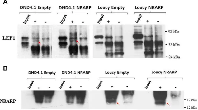
Figure S7.2 Confirmation of LEF1 pull-down ... 36
xiv
Abbreviation Index
ºC: Degrees Celsius
7AAD: 7-Aminoactinomycin D
AEBSF: 4-(2-Aminoethyl) Enzenesulfonyl Fluoride Hydrochloride
AKN: Ankyrin repeat
AKT (or PKB): Protein kinase B ALL: Acute Lymphoblastic Leukemia AML: Acute Myeloid Leukemia APC: Adenomatosis Polyposis Coli Axin: Axis inhibition protein Axin2: Axis inhibition protein 2
BCL11B: B-cell lymphoma/leukemia 11B CBF-1 (or RBP-Jκ): Recombining binding protein suppressor of hairless
CCND1: Cyclin D1 CCND3: Cyclin D3
CDK: Cyclin dependent kinase CK1α: Casein Kinase 1α
CLP: Common Lymphoid Progenitor CNS: Central Nervous System
CREB: Cyclic AMP-responsive element-binding protein
CSL-family: CBF-1/ Suppressor of hairless/ Longevity-assurance gene-1 family
Dll: Delta-like ligand DMSO: Dimethyl Sulfoxide DN: Double negative
DNA: Deoxyribonucleic Acid dNTP: deoxyribonucleotide DP: Double positive
Dsh: Dishevelled DTT: Dithiothreitol DTX1: Deltex1
EDTA: Ethylenediamine tetraacetic acid ETP: early T lineage progenitor
FBS: Fetal Bovine Serum
FBW7: F-box and WD repeat domain-containing 7
GFP: Green Fluorescent Protein GSI: Gamma (γ)-secretase inhibitor GSK3: Glycogen Synthase Kinase 3 HD: Heterodimerization Domain HEPES: 4-(2-hydroxyethyl) -1-Piperazineethanesulfonic Acid HES: Hairy enhancer of split HMG: High mobility group HOXA genes: Homebox A genes HSC: Hematopoietic Stem cells IL7R: Interleukin 7 receptor IP: Immunoprecipitation Jag: Jagged
JAK: Janus Kinase KD: Knock-down
LAG-1: Longevity-assurance gene-1 LEF1: Lymphoid Enhancer Binding Factor 1
LIC: Leukemia initiating cells LYL1: Lymphoblastic Leukemia associated hematopoietic regulator 1 mA: milliampere
mAb: monoclonal antibody MAML1: Mastermind-like-1
MHC: Major histocompatibility complex min: minutes
mL: milliliter
MPP: multipotent progenitors
xv MYC: Myelocytomatosis Viral Oncogene
Homolog
NECD: Notch Extracellular Domain NF-
κ
B: Nuclear Factor kappa B ng: nanogramNICD: Notch Intracellular Domain NK: Natural Killer
nm: nanometer
NP: nonyl phenoxypolyethoxylethanol
NRARP: NOTCH Regulated Ankyrin Repeat protein
OE: Overexpression
PBS: Phosphate Buffer Saline PCR: Polymerase Chain Reaction
PEST motif: Proline, glutamic acid, serine, threonine rich motif
PI3K/AKT: Phosphoinositide-3-kinase– protein kinase B/Protein kinase B
PTCRα: pre-T-cell antigen receptor alpha PTEN: Phosphatase and tensin homolog PLA: Proximity Ligation Assay
RAM: RBPJκ-associated molecule RNA: Ribonucleic Acid
rpm: rotations per minute RT: room temperature
RT-qPCR: Real Time Quantitative Polymerase Chain Reaction
SDS-PAGE: Sodium Dodecyl Sulfate-Polyacrylamide Gel Electrophoresis sec: seconds
shRNA: short-hairpin RNA SP: Single positive
STAT: Signal transducer and activator of transcription
Su(H): Suppressor of Hairless
TAD: Transcriptional Activation Domain T-ALL: T-cell Acute Lymphoblastic Leukemia
TBS: Tris Buffered Saline TCF: T-cell factor
TCR: T-cell receptor
TCRα: T-cell antigen receptor alpha TM: Transmembrane Domain V: volt WB: Western Blot WT: Wild Type μg: microgram μL: microliter μM: micromolar
1
1. Introduction
T-cell Development
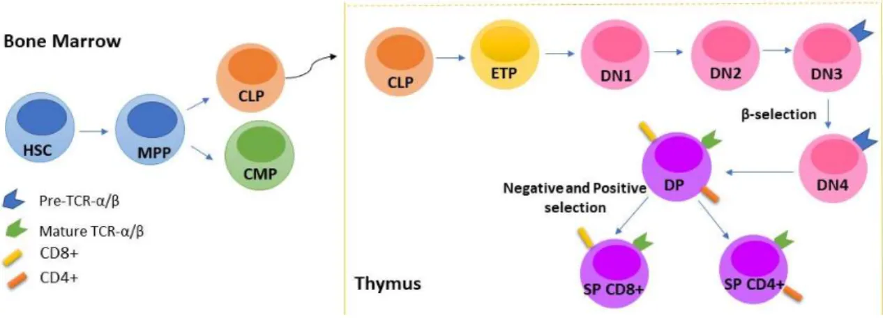
All blood cells are derived from a common pool of pluripotent cells called hematopoietic stem cells (HSC). Bone marrow hematopoietic stem cells give rise to multipotent progenitors (MPPs) that differentiate either into common myeloid progenitors (CMPs) or common lymphoid progenitors (CLPs). CLPs are precursors of lymphoid cells, which includes T, B and natural killer (NK) cells, and CMPs originate myeloid cells, such as granulocytes, monocytes, erythrocytes and megakaryocytes (1,2).
CLPs that migrate from the bone marrow, via blood, to the thymus undergo differentiation and maturation processes into functional T-cells (Figure 1.1) (2,3). CLPs enter the thymus at the cortico–medullary junction and by interacting with the thymic epithelium enter into the early T lineage progenitor (ETP) stage of T-cell fate specification, losing their potential to originate B or NK cells. This loss of potential results from the activation of the Notch signaling pathway that leads to the upregulation of the transcription factor T-cell factor 1 (TCF1), which in turn induces the expression of several lineage genes including GATA3, BCL11b and components of the T-cell receptor (TCR) (4).
ETPs originate double negative (DN) thymocytes, characterized by the absence of CD4 and CD8 T-cell markers expression. These T-cell precursors can be further subdivided into four sequential stages of differentiation (DN1, DN2, DN3 and DN4) (5). At the DN2 stage occurs an
expansion of thymocytes that requires Wnt signaling activation (6) that is followed, at the transition from the DN2 to DN3 stage, by the αβ versus γδ T cell fate specification step (3,7). This step involves Notch signaling activity (8). The cells that undergo αβ TCR differentiation start by expressing a preTCR-α, encoded by a non-rearranged locus, that later on pairs with a TCR β-chain. At this stage, cells undergo β-selection that selects cells that have a properly rearranged TCR-β chain locus. β-selection is followed by a new thymocyte proliferation phase, also promoted by Wnt signaling (5,6). In the meantime, thymocytes begin to co-express the receptor proteins CD8 and CD4 and eventually, form a large population of CD4 and CD8 double-positive (DP) TCR-expressing immature cells (3,5). Wnt signaling is sought to play a role in DN to DP transition, since inhibition of the interaction between β-catenin and TCF1/LEF1, the main drivers of Wnt signaling, results in a block at DN to DP transition (6).
In DP stage, TCRα chain is rearranged in order to produce a mature TCR αβ that is functionally tested for the recognition of self-MHC molecules (positive selection) and the absence of reactivity against self-antigens (negative selection). During this stage, Wnt signaling is involved in DP thymocytes survival and also in the regulation of positive and negative selections (9). Afterward, T-cells with a functional TCR αβ receptor evolve to mature single positive (SP) T-cells (CD4+ or CD8+), concluding its maturation process (4,6).
2
Figure 1.1. Schematic representation of T-cell development. Hematopoietic stem cells (HSC) go through a set of differentiation stages in the bone marrow and thymus to give rise to mature T-cells.
T-cell acute lymphoblastic leukemia (T-ALL)
Acute Lymphoblastic Leukemia (ALL) comprises a group of hematological malignancies characterized by the abnormal proliferation of lymphoid progenitors in the bone marrow, blood, and other organs. It accounts for 80% of childhood leukemias and 20% of adult leukemias (10).
When malignant transformation occurs in T-cell progenitors it gives rise to a specific type of ALL, named T-cell acute lymphoblastic leukemia (T-ALL). T-ALL is an immature lymphoid malignancy characterized by bone marrow and thymus infiltration of transformed hematopoietic cells expressing immature T-cell markers (11), accounting for 10–15% of pediatric and 20–25% of adult cases of ALL (12). Clinically, patients with T-ALL typically present hyperleukocytosis and hematopoietic failure, with neutropenia, anaemia and thrombocytopenia. In addition, T-ALL patients frequently show mediastinal thymic masses and meningeal infiltration of the central nervous system (CNS) (12,13).
Currently, cure rates reach 80% in pediatric and 60% in adult cases as a result of intensive chemotherapy protocols. However, 25-30% of pediatric and 60% of adult T-ALL patients relapse due to primary or acquired therapy resistance. These patients have extremely poor prognosis, underscoring the need for more specific and efficient therapies (13–16).
T-ALL is the result of a multistep transformation process in which the accumulation of genetic alterations in T-cell-specific transcription factors, proto-oncogenes and/or tumor suppressor genes as well as in these genes transcription regulatory regions, are responsible for the disruption of normal thymocyte development. These transcription factors include: T-cell acute lymphocytic leukemia (TAL1 and TAL2), lymphoblastic leukemia associated hematopoiesis regulator 1 (LYL1), LIM domain only (LMO1 and LMO2), the T-cell leukemia homeobox 1 (TLX1 and TLX3), NK2 homeobox (NKX2-1 and NKX2-2) and homeobox A genes (HOXA). Regarding proto-oncogenes, the Avian Myelocytomatosis Viral Oncogene Homolog (MYC) and MYB must be considered (13,16). In addition, mutations and deletions in tumor suppressor genes such as Wilms Tumor 1 (WT1), B-cell CLL/Lymphoma 11B (BCL11B), GATA binding protein 3 (GATA3) or the cyclin-dependent kinase inhibitor 2A (CDKN2A) are also recurrent events in T-ALL(16–18).
3 Moreover, the deregulation of signaling pathways involved in T-cell development and cell proliferation such as IL-7R/JAK/STAT, PI3K/ AKT, Notch and Wnt also contribute to T-ALL development (13,16,18).
Notch signaling
Notch signaling is a highly evolutionary conserved pathway responsible for cell fate specification by regulating cell proliferation, differentiation, apoptosis and survival (8,13,19). This pathway plays a critical role in the development and maintenance of embryonic and adult tissues (17). In the hematopoietic system, Notch signaling is essential for the generation of hematopoietic stem cells and is involved in the regulation of myeloid and lymphoid lineage differentiation, having a crucial role in T-cell development (20).
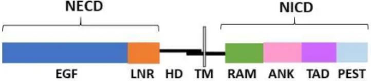
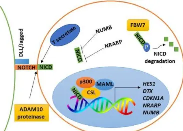
In mammals, there are four NOTCH receptors (NOTCH 1- 4), however T-cells only express NOTCH 1, 2 and 3 (21). NOTCH receptors consist of a large extracellular domain (NECD), a heterodimerization domain (HD), a transmembrane domain (TM) and an intracellular domain (NICD). NECD is composed by epidermal growth factor (EGF)–like repeats, containing the ligand-binding sites of the receptor, followed by Lin12-Notch repeats (LNR). The NICD part, which is involved in relaying signal to the nucleus, contains a RBPJk-associated molecule (RAM) region, followed by an ankyrin domain (ANK), a transactivating domain (TAD) and a C-terminal PEST motif (Figure 1.2) (22,23). Regarding the ligands, there are three Delta-like ligands (Dll1, Dll3, and Dll4) and two ligands of the Jagged family (Jag1 and Jag2) (24). In what regards T-cell development, Notch signaling is triggered upon the interaction of a NOTCH receptor on the surface of T-cells with a NOTCH ligand expressed on the surface of thymic stroma cells (Figure 1.3) (21).
In the canonical pathway, when a NOTCH receptor interacts with a NOTCH ligand, it occurs the proteolytic cleavage of the receptor, first by a disintegrin and metalloproteinase domain-containing protein 10 (ADAM10) and then by the γ-secretase complex (Figure 1.3) (17). Following this latter cut, NICD is released from the membrane and translocated to the nucleus, where it interacts with the CBF-1/Suppressor of Hairless (Su(H))/ Longevity-assurance gene-1(LAG-1) (CSL)DNA-binding protein (Figure 1.3). NICD displaces the corepressors bound to CBF region, such as CtBP1 (C-terminal-binding protein 1) and SMRT (retinoid or
thyroid-hormone receptors), and recruits coactivators, such as mastermind-like protein 1 (MAML1) and
p300 (an histone acetyltransferase), inducing the transcription of Notch target genes such as the HES (hairy enhancer of split) family of transcriptional repressors, the Notch-related ankyrin repeat protein (NRARP), NUMB, c-MYC, Deltex (DTX) and others (Figure 1.3) (2,25). NICD is short-lived, it gets phosphorylated on its PEST domain, ubiquitinated by FBW7 (F-box and WD repeat domain containing 7) and subsequently targeted for degradation by the proteasome pathway (2,13,26).
Figure 1.2. Simplified representation of NOTCH 1, 2 and 3 receptors structure. These receptors are expressed in the surface of T-cells, and the proteolytic cleavage of its NICD part triggers Notch signaling activation.
4
Figure 1.3. Schematic representation of Notch signaling canonical pathway. Ligand-receptor interaction promotes the proteolytic cleavage of NOTCH receptor and the translocation of NICD to the nucleus, where it displaces corepressors and recruits coactivators leading to the transcription of Notch target genes. NICD is rapidly degraded by the proteasome, ending Notch signaling.
Considering the crucial role of Notch signaling in developmental processes, it is not surprising that deregulation of this pathway is found in various diseases (27). Besides T-cell leukemia, it is also deregulated in breast (28), prostate (29), colorectal and lung (30) cancers, and also in central nervous system malignancies (17,31,32).
Notch signaling in T-ALL
NOTCH1, a key regulator of T-cell fate specification and thymocyte development, is found mutated in the majority of T-ALL cases, making of Notch signaling deregulation an hallmark of T-ALL pathogenesis (33,34).
Aberrant constitutively active NOTCH1 was first identified in rare T-ALLs harboring the t(7;9)(q34;q34.3) chromosomal translocation, which leads to the expression of a truncated and constitutively active form of the receptor. However, in more than 60% of T-ALLs, NOTCH1 is activated by gain-of-function mutations (17).
NOTCH1 is activated by disruption of the domains responsible for controlling the initiation and termination of signaling. The majority of mutations in NOTCH1 receptor occur mainly in HD and PEST domains, leading respectively, to ligand-independent activation of the receptor or impaired NICD degradation, inducing a constitutive and aberrant activation of Notch signaling. In addition, 8–30% of T-ALLs harbor mutations in the FBXW7 gene, impairing NICD degradation (19,35).
Furthermore, paracrine mechanisms that result in the upregulation of Notch signaling and rare NOTCH3 receptor mutations can also contribute to T-ALL as well as the aberrant expression of NOTCH ligands (18,36,37).
NOTCH1 promotes leukemia cell growth through direct transcriptional upregulation of anabolic pathways, such as ribosome biosynthesis, protein translation and nucleotide and amino acid metabolism (18). The growth-promoting effects of NOTCH1 are enhanced by the upregulation of
c-
MYC oncogene that is involved in the regulation of cell cycle, cell metabolism and DNA replication (19). In T-ALL, c-MYC is highly expressed and required for T-ALL cell growth, proliferation, and leukemia-initiating cell activity (38). HES1, a transcriptional repressor that functions downstream of NOTCH1, plays also a part in leukemogenesis (13). The5 upregulation of HES1 has been implicated in the activation of PI3K/AKT pathway and consequent transcriptional downregulation of PTEN, a tumor suppressor gene (39,40).
NOTCH1 can further interact and activate other signaling pathways involved in T-ALL, such as the PI3K/AKT/mTOR axis. In particular, NOTCH1 regulates the expression of proteins upstream of PI3K, including the IL-7 receptor (IL-7R), the pre-T-cell receptor alpha (PTCRA) and the insulin-like growth factor 1 receptor (IGF1R) (41–43). In addition, NOTCH1 stimulates the activation of NF-κB pathway, also known to contribute to T-ALL development (44).
Notch signaling activation further promotes the expression of proteins involved in cell cycle progression such as Cyclin D3 (CCND3) and cyclin-dependent kinases 4 and 6 (CDK4 and CDK6) as well as the S-phase kinase-associated protein 2 (SKP2) responsible for the degradation of the cell cycle inhibitors p27 and p21 (19,42,45).
Targeting NOTCH as a therapeutic approach in T-ALL
The high prevalence of NOTCH1 mutations in T-ALL patients makes of this pathway a potential therapeutic target in T-cell leukemia (19). For this reason, researchers tried to develop therapeutic drugs targeting the Notch pathway. Some of those drugs are currently in pre-clinical and clinical development, including γ-secretase inhibitors (GSI) and monoclonal antibodies against NOTCH1 receptor (14).
GSIs are small molecules that inhibit the activity of the γ-secretase complex, responsible for the final proteolytic cleavage of NICD. If the function of this protein complex is blocked, Notch signaling is not activated, leading to a downregulation of Notch target genes expression and resulting in a G0/G1 cell cycle arrest and decreased cell proliferation (46). However, early phase clinical trials showed that treatment responses are usually incomplete or transient and that these inhibitors cause gastrointestinal toxicity due to simultaneous inhibition of NOTCH1 and NOTCH2 receptors (14,46,47). In addition, studies in T-ALL cell lines showed that chronic GSI exposure results in the development of resistant cells that no longer require NOTCH1 activity to proliferate (47). Overall, GSIs have limited therapeutic activity and severe side effects (14).
Monoclonal antibodies (mAb) against specific NOTCH receptors or receptor domains harboring mutations are an alternative route to overcome GSI toxicity. Pre-clinical studies using these molecules resulted in anti-cancer effects by limiting leukemia cells growth and increasing apoptosis (14). Nonetheless, the clinical therapeutic potential of these molecules needs to be better understood, in particular because of the toxicity associated to them.
In sum, although various therapeutic strategies have been developed to target Notch signaling, none show the anti-leukemia effects necessary and many are associated with severe toxicity. For this reason, there is still the need to identify other therapeutic strategies to target this signaling pathway.
Wnt signaling
Wnt signaling pathway is an evolutionary conserved pathway that plays an important role in embryonic development, adult homeostasis and normal hematopoiesis, particularly in T-cell development (18,48).
The Wnt signaling is divided into canonical and non-canonical pathways. The first involves the regulation of β-catenin-dependent gene expression programs and controls cell renewal,
6 proliferation and differentiation (49). The second operates independently of β-catenin and is responsible for the regulation of cell polarity and cell movement (49).
WNT proteins, the drivers of this pathway, are encoded in humans by 19 genes that activate preferentially either canonical or non-canonical Wnt cascades. For example, WNT1, WNT3A and WNT8 are commonly encountered in canonical signaling and WNT5A and WNT11 are involved in non-canonical signaling (48,50). Here, we will describe only the canonical Wnt pathway.
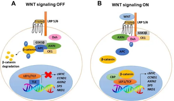
In the canonical Wnt pathway, in the absence of WNT ligands, cytoplasmic levels of β-catenin (CTNNB1) are kept very low due to its constitutively phosphorylation and degradation through the action of a protein destruction complex (49). This destruction complex is composed of two negative regulatory kinases, the glycogen synthase kinase 3 (GSK3β) and the casein kinase 1 (CK1), and at least two anchor proteins, the axis inhibition protein 1 or 2 (AXIN1 or AXIN2) and the adenomatous polyposis coli (APC) protein. APC and AXIN sequester β-catenin in the cytoplasm, allowing its phosphorylation by GSK3β and CK1. Phosphorylated β-catenin is recognized and ubiquitinated by a E3 ubiquitin ligase complex and targeted for destruction by the proteasome (Figure 1.4A) (51). Upon binding of a WNT ligand to the Frizzled receptor (Fz) and LDL receptor related protein (LRP5/6), Fz receptor interacts with Dishevelled (Dsh), a cytoplasmic protein that functions upstream of β-catenin and that is found phosphorylated during Wnt signaling (52). LRP is phosphorylated by GSK3β and CK1, allowing AXIN recruitment to the membrane and inactivation of the destruction complex. Consequently, dephosphorylated β-catenin accumulates in the cytoplasm and migrates to the nucleus where it binds to members of the T-cell factor (TCF)/Lymphoid enhancer binding factor (LEF) transcription factor family and activates transcription (Figure 1.4B) (44–46). TCFs repress transcription in the absence of signaling, by recruiting transducin-like enhancer of split (TLE)/Groucho (Gro) corepressors (53) (Figure 1.4A). The corepressors are displaced by β-catenin which recruits coactivators such as the histone acetyltransferases CREB-binding protein (CBP) and p300 leading to the transcription of Wnt target genes such as cMYC and Cyclin D1 (CCND1) which promote cell proliferation and cell cycle progression, and also SP5, Naked cuticle 1 (NKD1) and AXIN2 which are negative regulators of Wnt signaling (Figure 1.4B) (53–55).
B
A
Figure 1.4. Schematic representation of Canonical Wnt signaling pathway. In the absence of WNT ligands, β-catenin is degraded, inactivating Wnt pathway (A). In the presence of WNT ligands, β-β-catenin is stabilized and migrates to the nucleus where, upon interaction with TCF/LEF1 proteins, activates transcription of Wnt target genes (B).
7 Wnt signaling deregulation has been implicated in various diseases, including neurodegenerative diseases and cancer such as melanoma, colon and breast cancer, as well as T-cell leukemia (56).
Wnt signaling in T-ALL
Wnt signaling often becomes de-regulated in malignancies of the hematological system, including T-ALL, which it is not surprising considering its important role in T-cell development (48,57).
For example, Guo et al. reported that enforced expression of a constitutively active form of β-catenin from the DN3 stage onwards, promotes the accumulation of DP thymocytes predisposed to malignant transformation, ultimately leading to the development of thymic lymphomas. This malignant transformation depends on c-MYC activity, whose ablation blocks lymphomagenesis. Interestingly, β-catenin function was independent of the presence of NOTCH activating mutations (58). In line with these findings, OH and colleagues found that β-catenin mRNA and protein levels are significantly upregulated in more than 85% of childhood T-ALL patients with low NOTCH1 mutation rates. Moreover, the authors also found that β-catenin knock-down promote T-cell apoptosis (50).
In addition, Kaveri et al. showed that upregulation of Wnt signaling promotes the development and maintenance of T-cell leukemia in a mouse model in which high levels of β-catenin are present throughout T-cell development. In this model, abnormal chromosomal rearrangements lead to c-MYC overexpression and PTEN deletion, providing a selective advantage for tumor cell growth (59). Of note, in this report, leukemia development also occurred independently of Notch signaling.
Still in the context of the oncogenic role of Wnt signaling, Giambra et al. verified that Wnt activation is restricted to a minor subpopulations of leukemia cells with potential to promote leukemia development- Leukemia initiating cells (LIC)- and that β-catenin inactivation leads to a decrease of LIC frequency (60).
Besides β-catenin, TCF/LEF1 proteins are also main drivers of Wnt signaling and have been shown to play a role in T-ALL pathogenesis, as well.
TCF1 was identified as an important tumor suppressor gene in T-ALL (61). Tiemessen et al. reported that Tcf1−/− mice spontaneously develop thymic lymphomas/leukemias due to an increase in LEF1 levels, showing that TCF1 can function as a repressor of LEF1 (61,62). Increasing LEF1 protein levels lead to an upregulation of Wnt signaling, predisposing thymocytes to leukemic transformation (62). Supportive of LEF1 oncogenic role, Guo X et al. found that LEF1 is highly expressed in 25% of adult ALL patients and is associated with poor prognosis. Moreover, LEF1 mutations lead to an upregulation of Wnt target genes, such as c-MYC and Cyclin D1 (63).
In what regards LEF1 function as tumor suppressor gene in T-ALL, Gutierrez and colleagues associated LEF1 inactivation with T-cell leukemogenesis development. Further, they found that 11% of the T-ALL samples analyzed carried LEF1 deletions and 7% presented LEF1 inactivating mutations (64).
Importantly, Wnt signaling can also crosstalk with Notch signaling in T-ALL pathogenesis (65). In this regard, Spaulding et al. reported that NOTCH1 regulates directly LEF1 expression in T-cell lymphomas. Therefore, aberrant activation of the Notch pathway accelerates
8 lymphomagenesis by promoting and maintaining increased levels of LEF1, what is essential for survival and expansion of Notch-dependent T-cell lymphomas. Furthermore, the authors found that ectopic LEF1 expression prolonged lymphomas survival after Notch signaling inhibition by GSI treatment (66).
Supporting LEF1 function as an oncogene, although in a different context, Hovanes et al. showed that LEF1 is ectopically activated in colon cancer, due to the upregulation of a promoter for the LEF1 full-length isoform (that binds to β-catenin), at expense of the promoter that drives expression of a dominant negative LEF1 short isoform (which does not possess a β-catenin binding motif). The upregulation of theLEF1 full-length isoform favors a positive feedback loop for Wnt signaling (67). Of note, in T-ALL cells, the expression of both full-length and short LEF1 isoforms has been described (68,69).
NRARP as a regulator of Notch and Wnt signaling
NRARP (Notch-regulated ankyrin-repeat protein) is a downstream target of Notch signaling and encodes a small protein that contains two ankyrin-repeat motifs in its carboxy-terminal domain (70).
NRARP was first identified in Xenopus and later on in mouse as having a role in embryogenesis and in the development of the central nervous system by regulating Notch signaling (70,71). In particular, NRARP was shown to function as a negative regulator of Notch signaling (71) by leading to the decrease of NICD levels via the proteasome (72). In xenopus, NRARP was shown to interact physically and directly with NICD, forming a ternary complex with the CSL protein XSu(H)
(72,73)
.NRARP is expressed in all thymocytes subsets (DN, DP and SP) with highest levels in the DP subset, coincident with the necessary decrease in Notch signaling at this stage of T-cell development (73). Importantly, when overexpressed in murine hematopoietic stem/progenitor cells, NRARP blocks T-cell lineage commitment and impairs the progression through the DN stages of differentiation (73,74). Of note, NRARP overexpression in a mouse thymoma cell line was also shown to inhibit NOTCH function by downregulating CSL mediated transcriptional activation (73).
Interestingly, and although in a zebrafish model, NRARP was described as a positive regulator of the Wnt signaling pathway by preventing LEF1 ubiquitination and degradation (75). The authors further reported that the effects of NRARP as negative regulator of Notch signaling in zebrafish neurogenesis is not dependent of LEF1 levels, showing that NRARP regulates Notch and Wnt signaling independently (Figure 1.5) (75). Corroborating the dual role of NRARP, Chu Bing Feng and colleagues found that NRARP induce angiogenesis and control new vessels stability in mouse and zebrafish, by repressing Notch signaling and promoting Wnt signaling (76).
9
Figure 1.5. Model of NRARP dual role. NRARP modulates Notch signaling by promoting NICD degradation and modulates Wnt signaling by stabilizing LEF1. Adapted from Ishitani T. et al (75).
NRARP in T-ALL
Although not much is known about NRARP, its upregulation has been described in some types of cancer including breast cancer, thyroid carcinoma and lung cancer (77–80).
In fact, while addressing the role of the microRNA181 family in T-ALL, Fragoso and colleagues found that the microRNA mir-181ab1 functions as an oncogene in NOTCH1-induced T-ALL in part by repressing the activity of NRARP (81). This, together with the role of NRARP during T-cell development, suggested that NRARP may play a role in T-ALL pathogenesis. Indeed, recent work from the host laboratory showed that NRARP protein levels are increased in T-ALL cell lines and primary cells in comparison with normal thymocytes. Nonetheless, although increased, NRARP levels are not sufficient to block NOTCH oncogenic signals which led the researchers at the host laboratory to evaluate the functional effects of NRARP overexpression in T-ALL cells. Consistent with its role as a negative regulator of Notch signaling, the researchers observed that NRARP overexpression blocks the Notch pathway by decreasing NICD levels. However, the functional outcome at the proliferation level was not the same in all T-ALL cells. Unexpectedly, NRARP overexpression was found to impair the proliferation of T-ALL cells with NOTCH1 mutations (high basal levels of NICD) while promoting the proliferation of T-ALL cells with lower basal levels of NICD (NOTCH1 WT cells). Moreover, in NOTCH1 WT cells was observed an increase in c-MYC expression, upon NRARP overexpression, what could explain the proliferation increase.
These findings led to the hypothesis that NRARP may modulate cell proliferation and leukemia development in NOTCH1 WT cells through another signaling pathway. The researchers set out to investigate the Wnt signaling pathway having in consideration the role of NRARP in the positive regulation of this pathway and Wnt’s role in T-cell development and T-ALL pathogenesis. In fact, preliminary data showed that NRARP overexpression induces Wnt signaling activation in NOTCH1 WT T-ALL cells, as determined by the accumulation of β-catenin in these cells.
10
2. Objectives
Despite of the significant advances in T-ALL patients’ prognosis, there is still a major need for more effective and less toxic therapies. The long-term side-effects of current therapies and poor prognosis of patients that relapse, underlines the need to a better understanding of the mechanisms and signaling pathways involved in T-ALL pathogenesis, in order to develop new and more efficient therapeutic strategies.
Thus, considering the dual role of NRARP in T-ALL and its opposite functional outcome depending of the signaling pathway regulated (Notch signaling or Wnt signaling), this project aims at dissecting the mechanisms underlying Wnt signaling regulation by NRARP. In line with the overall goal of this project, the main topics of the work were:
1. Functional characterization of Wnt signaling pathway in T-ALL cells: - Evaluate the functional effects of Wnt signaling inhibition in T-ALL cells with and without NOTCH1 activating mutations and upon NRARP overexpression.
2. Mechanistic characterization of Wnt signaling regulation by NRARP in T-ALL: - Functional evaluation of the effects of LEF1 KD in T-ALL cells
- Evaluate if LEF1 protein is directly regulated by NRARP - Investigate if NRARP interacts with a particular LEF1 isoform - Evaluate the impact of NRARP in LEF1/β-catenin interaction.
3. Materials and Methods
Cell culture
Experiments were performed using DND4.1 and Loucy cell lines, which are T-cell acute lymphoblastic leukemia cell lines with different NOTCH1 mutational status. Cells were cultured using RPMI-1640 medium with L-glutamine (Gibco), supplemented with 10% of fetal bovine serum (FBS) (Biowest), 1% of penicillin/streptomycin (Gibco) and 1% of 4-(2-hydroxyethyl)-1-piperazineethanesulfonic acid (HEPES) (Gibco). Cells were kept in culture at a concentration of 0,5 x 10⁶ /ml, at 37ºC and 5% of CO
2.
To establish NRARP overexpressing T-ALL cells, the T-ALL cell lines mentioned above were transduced with lentiviruses with a GFP tagged construct with or without the NRARP gene. These cell lines will be referred to from now on as DND4.1 Empty or NRARP and Loucy Empty or NRARP.
DND4.1 and Loucy cell lines, with and without NRARP overexpression, were subsequently modified to knock-down LEF1. For this purpose, we used shRNAs against LEF1 or a Scramble sequence (shSCR). Culture conditions were described above.
Gene expression analysis
RNA extraction and quantification
Cells were collected (approximately 5 x 10⁶), centrifuged at 3500 rpm for 5 minutes and
resuspended in 1 ml of TriZol® (Ambion™, ThermoFisher Scientific). Lysates were stored at -80º C or follow to RNA extraction.
11 Frozen samples were thawed and incubated at room temperature (RT) for 5 minutes. Then, 200 μl of chloroform were added to each sample followed by 15 seconds of vortexing. Samples were incubated during 3 minutes at RT and centrifuged at 13 000 rpm during 15 minutes at 4ºC, allowing the formation of two phases. The upper colorless phase was recovered into new tubes and to each sample it was added 500 μl of isopropanol. The samples were mixed and incubated on ice for 15 minutes. After a new centrifugation (13 000 rpm, 15 minutes, at 4º C), the supernatant was discarded and the RNA pellet was washed with 1 ml of 75% ethanol. Samples were centrifuged during 10 minutes at 13 000 rpm and the pellet left to dry at RT, for approximately 5 minutes. Then, RNA pellets were resuspended in 50 μl of RNase free water and incubated during 10 minutes in a pre-heated bath (55-65 ºC). RNA was stored at -80ºC.
RNA quantification was performed in a UV-Vis spectrophotometer Thermo Scientific™ NanoDrop 2000, by measuring the absorbance at 230, 260 and 280 nm. RNA was considered with good quality when the absorbance ratios of A260/280 and A260/230 were between 1,8-2,1 and superior to 1,8, respectively.
Complementary DNA (cDNA) synthesis
cDNA synthesis was performed using the Invitrogen kit “Supercripts™ III - First-strand synthesis super mix for qPCR” and following the manufacturer protocol (Invitrogen). cDNA synthesis was performed using 500 ng of RNA.
qPCR (Quantitative PCR)
Gene expression quantification was done by real time PCR using the SYBR Green kit: Power SYBR Green PCR Master Mix (Applied Biosystems, Thermo Fisher). The specific primers sequences were as follow:
hMYC Forward: 5’ GGC TCC TGG CAA AAG GTC A 3’ Reverse: 5’ CTG CGT AGT TGT GCT GAT GT 3’
hLEF1 Forward: 5’ TGC CAA ATA TGA ATA ACG ACC CA 3’ Reverse: 5’ GAG AAA AGT GCT CGT CAC TGT 3’
hAxin2 Forward: 5’ CTC CCC ACC TTG AAT GAA GA 3’ Reverse: 5’ GTT TCC GTG GAC CTC ACA CT 3’
h18S Forward: 5’ GGA GAG GGA GCC TGA GAA ACG 3’ Reverse: 5’ CGC GGC TGC TGG CAC CAG ACT T 3’
PCR reactions were performed in the ViiA™ 7 Real-Time PCR System (Applied Biosystems). Gene expression was normalized to the endogenous gene 18S and mRNA relative expression determined using the 2-ΔΔCt method.
Protein analysis
Protein extraction and quantification
Cells were collected (approximately 5 x 10⁶) and centrifuged at 3500 rpm for 5 minutes at 4ºC.
Then, the cell pellet was resuspended in 50-100 μl of lysis buffer (50mM Tris-Base pH 8.0, 150 mM NaCl, 5mM EDTA, 1 mM NaOV, 10 mM NaF, 10mM Sodium Pyrophosphatase and 1%
12 NP-40), supplemented with the protease inhibitor cocktail Complete Mini (Roche) and the protease inhibitor AEBSF (1mM). Lysates were centrifuged at 13 000 rpm during 20 minutes at 4ºC and the supernatant recovered. Protein extracts were stored at -20ºC. Protein quantification was performed using the Bradford reagent (Bio-Rad) diluted in high purity water in a 1:5 ratio and obtained using the GeneQuant pro (Amersham Biosciences) spectrophotometer at a 595 nm wavelength (Protein595 Program).
Western Blot
Protein extracts (50 µg of protein) were diluted in laemmli buffer and lysis buffer and boiled for 5 minutes in a dry bath to promote protein denaturation. Samples were spin down and loaded on a SDS-PAGE gel with different acrylamide concentrations (10, 12 and 16% depending on the molecular weight of the proteins to separate). Samples ran at 60 Volts (V) for 30 minutes and then at 120V during different periods of time depending on the molecular weight of the proteins of interest. Next, proteins were transferred to a nitrocellulose membrane at constant amperage (400 mA) for 90 minutes at 4ºC. Proteins successful transfer was confirmed using the Ponceau S solution (Sigma). Membranes were blocked with 3% (w/v) milk diluted in Tris-Buffered Saline with 0.1% Tween 20 (TBS-T) and incubated with primary antibodies, diluted in TBS-T, overnight at 4ºC under agitation. The primary antibodies used include: LEF1 (Cell signaling) 1:1000; β-catenin 1:1000 and NRARP 1:1000 (Santa Cruz biotechnology). Next day, membranes were washed 3 times for 15 minutes with TBS-T and incubated at RT during 1 hour with secondary antibodies: anti-Rabbit IgG and anti-Mouse IgG (Promega) diluted 1:5000 in 3% milk TBS-T. After incubation, membranes were washed 3 times for 15 minutes with TBS-T and treated with Pierce ECL Plus Western Blotting Substract (Thermo Fisher Scientific). Membranes were exposed to a X-ray film (FujiFilms) and the blot was detected by chemiluminescence using the Curix60 (AGFA HealthCare) apparatus.
Membrane stripping
Western blot membranes were incubated with stripping buffer (15 mM Tris Base, 100 mM 2-β-mercaptoethanol (Sigma), 2% SDS, pH=6.7) supplemented with 2-2-β-mercaptoethanol, in a dilution of 1:1000. Membranes were incubated during 30 minutes at 56º C with agitation. Afterwards, membranes were washed with H2O at least 10 times and washed with TBS-T during
15 minutes with agitation
.
Co-immunoprecipitation
Cells were harvested and centrifuged at 3500 rpm for 5 minutes, at 4º C. Then, cells were washed twice with ice-cold PBS 1X and centrifuged between washes, using the same conditions. The pellet was resuspended in PBS 1X and 1/10 of the resuspension was used as input, which represents the total cellular lysate (protein extraction was performed as described in “Protein extraction and quantification”). The remaining cell suspension was treated with IP lysis buffer (20 mM HEPES; 10 mM KCl; 0,1% NP40; 6 mM MgCl2 and 20% glycerol), incubated on ice
during 15 minutes and centrifuged at 13000 rpm for 20 minutes. The supernatant was recovered, incubated with 50 μl of Protein A/G plus agarose beads (Santa Cruz Biotechnology) and kept rotating during 30 minutes at 4º C, to pre-clear the samples. Then, pre-clearing beads were discarded (centrifugation at 3500 rpm during 1 minute) and the sample was divided into the desired number of conditions (immunoprecipitation condition and negative condition). Primary antibodies against the proteins intended to be pulled down (LEF1, Cell Signaling; NRARP and β-catenin, Santa Cruz Biotechnology), were added (3 μg) plus 500 μl of IP buffer (20 mM HEPES;
13 10 mM KCl; 0,2% Tween20; 1,5 mM MgCl2; 10% glycerol and 0,5 mM DTT). Samples were
kept rotating overnight. Next day, 50 μl of Protein A/G plus agarose beads were added to each sample and left rotating for at least 2 hours. Afterwards, beads were pelleted down by centrifugation (1 minute, 3500 rpm) and the supernatant discarded. The complex protein-antibody-beads was washed with wash buffer 1 (20 mM HEPES; 50 mM KCl; 0,2% Tween20; 1,5 mM MgCl2; 10% glycerol and 0,5 mM DTT) and wash buffer 2 (20 mM HEPES; 100 mM
KCl; 0,2% Tween20; 1,5 mM MgCl2; 10% glycerol and 0,5 mM DTT) and samples kept rotating
for 10-15 minutes between washes. In the final wash, the supernatant was discarded and the complex resuspended in 2x laemmli buffer. Co-immunoprecipitated proteins were analyzed by western blot, following the protocol described above.
Duolink Proximity Ligation assay
In situ proximity ligation assay (PLA) is a protein detection technique used to detect the interaction between two proteins with high specificity and sensitivity. This method is very reliable because it requires the binding in close proximity (30-40 nm apart) of two antibodies conjugated with oligonucleotides to different epitopes of two proteins in a complex (82,83). These assays were performed using fixed samples and analyzed by fluorescence microscopy. The principle of PLA is described in detail in Figure S7.1.
Cells were immobilized in 1 cm2 coverslips (Normax) coated with Poly-D-lysine
(Sigma-Aldrich). Then cells were fixed and permeabilized with methanol cool down at -20ºC, during 10 minutes on ice. Fixed cells were then subjected to the PLA assay using the Duolink red kit (Sigma). First of all, samples were blocked for 30 minutes using the Duolink blocking solution, followed by the incubation with the primary antibodies diluted 1:200 in TBS-T, during 1 hour at RT, in a humidity chamber. The primary antibodies used include anti-LEF1 Rabbit mAb (Sigma), anti-β-catenin mouse mAb and anti-NRARP goat mAb (Santa Cruz biotechnology). Next, the coverslips were incubated with PLA specific probes (consisting of secondary antibodies conjugated with oligonucleotides) during 1 hour, at 37º C, in a humidity chamber. Then, connector oligonucleotides (plus ligase enzyme) were added to the coverslip to hybridize with PLA probes. If probes are in close proximity, a circular DNA molecule will be generated. This incubation step occurs in a humidity chamber at 37ºC, for 30 minutes. To induce amplification (rolling-circle amplification) of the DNA product previously formed, the amplification solution (consisting of nucleotides and fluorescently labelled oligonucleotides - Alexa Fluor 594), together with the polymerase enzyme were added to the coverslips, for 100 minutes at 37ºC. The fluorescently labelled oligonucleotides hybridize to the amplified DNA product and the signal is visible as a distinct red fluorescent dot. The washing steps were performed according to the manufacturer’s instructions. The coverslips were mounted onto slides with Duolink mounting media with DAPI (4′,6- diamidino-2-phenylindole) and samples were analyzed in a Confocal Laser Point-scanning Microscope (Zeiss LSM 710), using the integrated software ZEN 2012. Cell counting and image configuration were performed using Fiji/ImageJ software.
Functional assays
To assess the importance of Wnt signaling in the proliferation and viability of T-ALL cells with and without NRARP overexpression (DND4.1 and Loucy) we performed functional assays using a canonical Wnt signaling pathway inhibitor, the PRI-724 (Selleckchem). This inhibitor
14 binds to CBP, blocking the interaction of CBP with β-catenin, impairing this way the transcription of Wnt target genes (84). Cells were treated with two different concentrations of PRI-724 (1 μM and 2 μM). In control conditions, cells were treated with Dimethyl sulfoxide (DMSO), the vehicle used to dilute the PRI-724 inhibitor.
Functional assays to assess the proliferation levels of these cell lines (DND4.1 and Loucy) when LEF1 is knock-down and NRARP is overexpressed were also performed.
Proliferation assays
To perform the proliferation assays, cells were plated in a 24-well plate (2,0 x 10⁵ cells per
well) at a concentration of 0,5 x 10⁶ /mL and cultured for different periods of time (24, 48, 72 and
96 hours). At each time point, the total number of cells was determined by flow cytometry (BD LSRFortessa) using counting beads (Beckmon Coulter). Beads were previously diluted in PBS 1X and counted using a hemocytometer. Flow cytometry data analysis was performed using FlowJo™ and cell number determined using the following equation:
Equation 3.1
𝑁𝑢𝑚𝑏𝑒𝑟 𝑜𝑓 𝑐𝑒𝑙𝑙𝑠 =
×
×where the 𝑁𝑢𝑚𝑏𝑒𝑟 𝑜𝑓 𝐺𝐹𝑃 𝑐𝑒𝑙𝑙𝑠 corresponds to the number of GFP positive events and the 𝑁𝑢𝑚𝑏𝑒𝑟 𝑜𝑓 𝑐𝑜𝑢𝑛𝑡𝑒𝑑 𝑏𝑒𝑎𝑑𝑠 represents the number of beads events acquired in the cytometer. Finally, the 𝑁𝑢𝑚𝑏𝑒𝑟 𝑜𝑓 𝑏𝑒𝑎𝑑𝑠/𝑚𝐿 and the volume of beads added (𝑉 ) corresponds to the number of beads added to each sample.
Viability assays
To determine leukemia cells viability at different time points (24h, 48h, 72h and 96h) we used the same experimental conditions used for the proliferation assays. Upon harvesting, the cells were stained with AnnexinV and with 7AAD to assess apoptosis and cell death, respectively. In detail, cells were washed twice with ice-cold PBS 1X and resuspended in 100 μl of annexinV binding buffer 1X (Promocell). Cells were then incubated with 1 μl of AnnexinV/APC (eBiosciences) and with 2 μl of 7AAD (Pharmigen) dyes during 15 minutes in the dark, at RT. Afterwards, 200 μl of annexinV binding buffer 1X were added to each sample. Cell viability was determined by flow cytometry (BD LSRFortessa) and analyzed using FlowJo™.
15
4. Results
Functional characterization of Wnt signaling pathway in T-ALL cells
Evaluate the functional effects of Wnt signaling inhibition in T-ALL cells
Preliminary work performed in the host laboratory shows that NRARP plays a dual role in T-ALL, either inhibiting or promoting T-ALL cells proliferation, depending on NOTCH1 mutational status/NICD levels. In cells with NOTCH1 mutations and consequently higher levels of NICD, NRARP overexpression inhibits leukemia cells growth through Notch signaling inhibition. On the other hand, in NOTCH1 WT T-ALL cells (lower levels of NICD) NRARP overexpression promotes leukemia cell proliferation. The upregulation of β-catenin and cMYC protein levels in these cells suggests that NRARP, in this cellular context, may promote proliferation through activation of the Wnt signaling pathway.
Thus, to functionally characterize the role of Wnt signaling in T-ALL cells growth induced by NRARP overexpression we evaluated leukemia cell proliferation and viability in the presence of a Wnt signaling inhibitor.
The inhibitor used to block Wnt signaling, PRI-724 (or ICG-001) has been reported as a potent inhibitor of cell proliferation and cancer growth, both in vitro and in vivo (85–87). As described in materials and methods, this drug binds to CBP blocking its interaction with β-catenin and inhibiting transcription of Wnt downstream target genes (84,86).
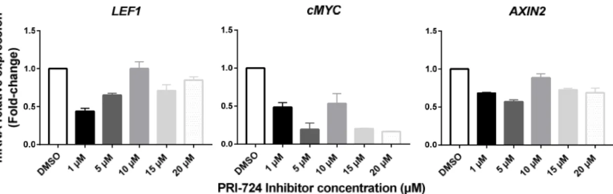
We started by performing a preliminary assay in order to choose the concentration(s) of the inhibitor that block(s) the transcription of Wnt signaling target genes without causing cytotoxicity. For that purpose, we exposed T-ALL cells to 1, 5, 10, 15 and 20 μM of PRI-724 and, 24h after treatment, the expression of the Wnt target genes cMYC, LEF1 and AXIN2 was quantified by real time PCR. Overall, the different inhibitor concentrations induced a decrease in the mRNA levels of all target genes analyzed (Figure 4.1). Nonetheless, since these effects were stronger and consistent for all target genes using concentrations equal or lower than 5 μM, we chose 1 μM and 2 μM to proceed with the functional assays.
Figure 4.1. Relative expression of Wnt signaling transcriptional targets upon treatment with different concentrations of PRI-724.The housekeeping gene used was the 18S ribosomal RNA gene and expression quantification was performed using ΔΔCt method. Samples were normalized to the control of the experiment (DMSO condition).
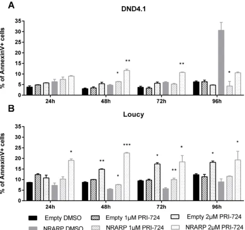
Proliferation and viability assays were performed using NRARP overexpressing cells with or without NOTCH1 mutations (DND4.1 and Loucy cell lines respectively). Cells were treated with 1 μM and 2 μM of the inhibitor or with DMSO in the control condition. Cell number and viability were analyzed by flow cytometry 24,48, 72 and 96 hours after treatment.
16 Consistent with our hypothesis that Wnt signaling plays a role in the growth of NOTCH1 WT cells upon NRARP overexpression, our results showed that Loucy NRARP cells are more sensitive to the inhibitor than DND4.1 NRARP cells (Figure 4.2A). Although both concentrations significantly affect the proliferation of both DND4.1 and Loucy NRARP cells starting as early as 24h after treatment, we verified that DND4.1 NRARP cells start recovering between 48 and 72 hours after treatment. Of note, at 96 hours, DND4.1 NRARP cells treated with the lower dose of the inhibitor proliferate significantly more than cells not treated (Figure 4.2A). Importantly, contrary to DND4.1 NRARP cells, Wnt signaling inhibition impaired Loucy NRARP cells proliferation throughout the experiment.
To evaluate the impact of Wnt signaling inhibition in parental T-ALL cells we have further treated T-ALL cells without NRARP modulation. The proliferation of both DND4.1 and Loucy cells was not affected in a statistically significant manner by the lower concentration of PRI-724 (Figure 4.2B). Regarding the higher dose of the Wnt inhibitor, although it initially inhibits the proliferation of both cell lines, DND4.1 cells start recovering after 72h of treatment (Figure 4.2B). Moreover, this recovery capacity was not observed in Loucy Empty cells.
Overall, in what concerns NOTCH1 WT cells, we observed that Loucy NRARP cells are more sensitive to Wnt signaling inhibition than Loucy Empty cells. While the proliferation of NRARP overexpressing cells significantly decreased upon treatment with both concentrations of the inhibitor, Empty cells were only affected by the higher concentration of PRI-724 (Figure 4.2A and 4.2B), which suggests that NRARP may strength/sustain Wnt signaling activity. In NOTCH1 mutant cells, we verified that although the proliferation of both Empty and NRARP overexpressing cells is initially affected, these cells seem to recover overtime (Figure 4.2A and 4.2B). Of note, this capacity to recover is more evident in DND4.1 NRARP cells whose proliferation recovery starts earlier and is faster than in DND4.1 Empty cells.
Figure 4.2. Effect of Wnt signaling inhibition in the proliferation of T-ALL cells overexpressing (A) or not (B) NRARP. The figure depicts the mean ± SEM of the fold-change in cell proliferation induced by the Wnt signaling inhibitor PRI-724 (ratio between cell count in treatment conditions and cell count of control condition, DMSO). The results presented are a combination of three independent assays (each conducted in triplicate). Statistical analysis was done using t-tests. P-values ≤ 0,05 were considered statistically significant and represented as *p<0.05; **p<0.01; ***p<0.001.
17 The analysis of NRARP overexpressing cells viability upon Wnt signaling inhibition showed that the percentage of apoptotic cells (AnnexinV+ cells) is higher in Loucy than in DND4.1 cells, for both inhibitor concentrations used (Figure 4.3A and 4.3B). In the Empty condition, Loucy cells showed a statistically significant increase in the percentage of apoptotic cells when treated with 2 µM of the inhibitor, while DND4.1 cells presented no alterations in viability upon treatment (Figure 4.3A and 4.3B).
Looking only at Loucy cells (NOTCH1 WT), we observed that the viability of NRARP overexpressing cells is more affected by Wnt inhibition than Empty cells (Figure 4.3B), which suggests that, as observed for proliferation, NRARP through Wnt signaling activation may regulate Loucy cells viability.
Regarding NOTCH1 mutant cells, DND4.1 NRARP are more sensitive to the inhibitor than DND4.1 Empty cells (Figure 4.3A). However, it is important to consider that in this cell line, NRARP overexpression by itself induces apoptosis, what may make these cells more susceptible to the inhibitor. Having this in consideration, it is also important to note that at 96 hours after treatment, while the viability of the cells not treated decreased considerable, the same was not verified in cells treated with the inhibitor (Figure 4.3A), suggesting that Wnt signaling has a negative impact in the viability of these cells.
Figure 4.3. Effect of Wnt signaling inhibition in the viability of T-ALL cells with and without NRARP overexpression. Representation of mean ± SEM of the percentage of Annexin V positive cells in each experimental conditional, regarding NOTCH1 mutant cell lines, DND4.1 (A) and NOTCH1 WT cell lines, Loucy (B). The results presented are a combination of three independent assays (each conducted in triplicate). Statistical analysis using t-tests were performed to analyze the differences between control and treatment conditions. p-values ≤ 0,05 were considered statistically significant and represented as *p<0.05; **p<0.01; ***p<0.001.
Overall, these results suggest that NRARP regulates both proliferation and viability of Loucy cells through Wnt signaling pathway.