Intestinal Epithelial Cells
Bruno Bezerra Lima,a
Bárbara Faria Fonseca,b
Nathália da Graça Amado,b
Débora Moreira Lima,a
Ronaldo Albuquerque Ribeiro,a José Garcia Abreu,b
Gerly Anne de Castro Britoa,c
Departamento de Fisiologia & Farmacologia, Faculdade de Medicina, Universidade Federal do Ceará (UFC), Fortaleza, Brazila; Instituto de Ciências Biomédicas, Universidade Federal do Rio de Janeiro (UFRJ), Rio de Janeiro, Brazilb; Departamento de Morfologia, Faculdade de Medicina, Universidade Federal do Ceará (UFC), Fortaleza, Brazilc
Clostridium difficiletoxins A and B (TcdA and TcdB) are homologous glycosyltransferases that inhibit a group of small GTPases within host cells, but several mechanisms underlying their pathogenic activity remain unclear. In this study, we evaluated the effects of TcdA on the Wnt/-catenin pathway, the major driving force behind the proliferation of epithelial cells in colonic crypts. IEC-6 and RKO cells stimulated with Wnt3a-conditioned medium were incubated with 10, 50, and 100 ng/ml of TcdA for 24 h, resulting in a dose-dependent inhibition of the Wnt signaling, as demonstrated by a T-cell fac-tor (TCF) reporter assay. This was further confirmed by immunofluorescence staining for nuclear localization of-catenin and Western blotting for-catenin and c-Myc (encoded by a Wnt target gene). Moreover, our Western blot analysis showed a decrease in the-catenin protein levels, which was reversed by z-VAD-fmk, a pan-caspase inhibitor. Nonethe-less, TcdA was still able to inhibit the Wnt/-catenin pathway even in the presence of z-VAD-fmk, lithium chloride (a GSK3inhibitor), or constitutively active-catenin, as determined by a TCF reporter assay. Furthermore, preincubation of RKO cells with TcdA for 12 h also attenuated Wnt3a-mediated activation of Wnt signaling, suggesting that inactivation of Rho GTPases plays a significant role in that inhibition. Taken together, these findings suggest that attenuation of the Wnt signaling by TcdA is important for TcdA antiproliferative effects.
T
oxins A and B (TcdA and TcdB) are the main virulence factors of the Gram-positive bacillusClostridium difficile(1). Both TcdA and TcdB bind (2) and enter the cells through receptor-mediated endocytosis (3), translocate, and then cleave their cata-lytic domain into the cytosol, which permanently inactivates the Rho GTPases (Rho, Rac1, and Cdc42) by glycosylation at Thr-35/ Thr-37 (4). Inhibition of those critical signaling molecules even-tually leads to actin cytoskeleton disruption, intestinal epithelial cell damage, and apoptosis by caspase activation (5–9).TcdA and TcdB exert important antiproliferative effects in vitroas well (10–12). In part, this is due to actin reorganization and consequent inhibition of contractile ring formation in cyto-kinesis triggered by Rho glycosylation, leading to binucleation and cell cycle arrest at the G2-M level (11,13–16). TcdA and TcdB also block the G1-S transition (10,12). Concomitant inhibition of Rho, Rac, and Cdc42 by TcdA leads to the blockage of cyclin D1 expres-sion, preventing cell cycle progression through the G1phase (12). TcdA and TcdB alter the expression of genes involved in cell cycle regulation, including several cyclins and cyclin-dependent kinases (CDKs), as determined by microarray analysis. Importantly, G1 arrest occurs even at low toxin doses, while only higher toxin concentrations (100 ng/ml of TcdA or 10 ng/ml of TcdB) stop cell division at the G2-M phase (10). Those results demonstrated that theC. difficiletoxins promote cell cycle arrest at the G1phase by blocking the expression of cyclin D1 and its binding to CDK4 and CDK6, which is a rate-limiting event during progression through G1phase. They did not, however, fully explain the upstream com-ponents causing this. The transcriptional control of the cyclin D1 gene is extremely complex and is influenced by several mecha-nisms, including the Ras-Raf-MEK-extracellular signal-regulated kinase (ERK), the phosphatidylinositol 3 kinase (PI3K)/Akt/
mTOR, and the Wnt/-catenin pathways (17,18). Here, we focus on the effect of TcdA on the Wnt/-catenin pathway.
The Wnt/-catenin pathway is a conserved molecular system that plays a major role in development and homeostatic tissue self-renewal. As such, it is the primary driving force behind the proliferation of human intestine cells (19). In the absence of a Wnt signal,-catenin is targeted for proteasomal degradation through sequential phosphorylations occurring at its N terminus (20). A degradation complex consists of the tumor suppressors axin and adenomatous polyposis coli (APC) as well as the constitutively active kinases glycogen synthase kinase 3(GSK3) and casein kinase I (20–22). This complex regulates the-catenin phosphor-ylation status in a cell (22–24). Otherwise, when Wnt ligands bind their Frizzled and low-density lipoprotein receptor-related pro-tein (LRP) receptors, the destruction complex is inactivated in a manner that is not fully understood (25). As a result,-catenin is no longer phosphorylated, accumulates in the cell, and is trans-ported to the nucleus (26), resulting in its binding to transcription factors of the T-cell factor/lymphocyte enhancer factor (TCF/ LEF) family. TCF/LEF–-catenin constitutes an active
transcrip-Received10 June 2013 Returned for modification11 July 2013
Accepted24 March 2014
Published ahead of print7 April 2014
Editor:B. A. McCormick
Address correspondence to Gerly Anne de Castro Brito, [email protected]. Supplemental material for this article may be found athttp://dx.doi.org/10.1128 /IAI.00567-13.
Copyright © 2014, American Society for Microbiology. All Rights Reserved.
tional complex that activates target genes (e.g., the genes for cyclin D1, c-MYC, and survivin). In the absence of a Wnt signal, tran-scriptional repressors, such as Groucho, bind TCF/LEF and block gene expression (19,23).
TcdA and TcdB likely interfere with Wnt/-catenin signaling. A recent study claimed that Wnt-induced-catenin stabilization is not enough for its nuclear accumulation. Wnt activation of the Rac1 GTPase, which is inhibited by both TcdA and TcdB, is also required (26). Moreover, TcdA and TcdB induce the activation of the caspases’ cascade (6,9,15), an alternate mechanism by which -catenin could be degraded (27). The toxins release NF-B (28, 29), an inactivator of-catenin, thereby inhibiting the transcrip-tion of the Wnt target genes (30,31). Taken together, these data suggest that the inhibition of Wnt/-catenin signaling by TcdA and/or TcdB not only is highly possible but also may contribute to the antiproliferative effects of the toxins.
We therefore investigated whether TcdA interferes with the Wnt/-catenin pathwayin vitroin order to elucidate the mecha-nisms of proapoptotic and antiproliferative effects induced by this toxin.
MATERIALS AND METHODS
Toxins, chemicals, and reagents.All cell culture reagents were purchased from Invitrogen (Carlsbad, CA). Purified TcdA and lithium chloride (LiCl) were purchased from Sigma (St. Louis, MO). Carbobenzoxy-valyl-alanyl-aspartyl-[O-methyl]-fluoromethylketone (z-VAD-fmk) was ob-tained from Promega Corporation (Madison, WI). All solvents and re-agents used in this study were of analytical grade.
Expression plasmids.We used expression plasmids encoding-catenin (CSII⫹-catenin), mutated -catenin S33A (CSII⫹-catenin S33A), which is resistant to GSK3phosphorylation, and Lef1-DeltaN VP16 (cDNA3-Lef1-⌬N VP16), which strongly activates the Topflash reporter independently of Tcf/Lef activation by-catenin. These plasmids were previously described (32–34) and kindly provided by Xi He (Boston Chil-dren’s Hospital/Harvard Medical School, Boston, MA). The transfection was performed in RKO cells using Lipofectamine (Invitrogen) according to the manufacturer’s instructions.
Cell culture.We acquired IEC-6 cells from the American Type Cul-ture Collection. RKO (human colorectal cancer) cells stably cotransfected with pRL-TK (Renilla) and TOPflash were previously described (35) and kindly provided by Xi He (Children’s Hospital/Harvard Medical School). They were both maintained in Dulbecco’s modified Eagle’s medium (DMEM) supplemented with 10% fetal bovine serum (FBS), 120g of penicillin/ml, and 200g of streptomycin/ml in 5% CO2. All cell lines
were used between passages 10 and 20.
CM preparation.To produce Wnt3a-conditioned medium (CM), L cells stably transfected with the Wnt3a gene were cultured in DMEM with 10% FBS for 4 days. The medium was then harvested and sterilized using a 0.22-mm filter. Thereafter, fresh medium was added and the cells were cultured for an additional 3 days. When the medium was collected again, it was combined with the previous medium at a 1:1 ratio. As a control, CM was similarly generated from the untransfected parental cell line.
Transfection and TCF reporter assay.Gene reporter plasmids were transfected into IEC-6 cells using Lipofectamine (Invitrogen) according to the manufacturer’s instructions. After transfection, we added CM with or without TcdA to the cells for approximately 24 h and then determined the luciferase activity. Changes in-catenin/TCF-activated gene expres-sion were measured using the TOPflash and the FOPflash (mutant TOPflash) plasmids obtained from Millipore (Billerica, MA). TOPflash is a TCF reporter plasmid containing two sets of three copies of wild-type TCF binding sites driven by the thymidine kinase minimal promoter and upstream of a luciferase reporter gene. FOPflash contains mutated TCF binding sites driven by the same thymidine kinase promoter and also
upstream of the same luciferase open reading frame as TOPflash. FOPflash is used as a negative control for TOPflash activity (35,36).
IEC-6 cells were cotransfected with the pRL-TK vector (Promega Cor-poration, Madison, WI), which contains theRenillagene under the con-trol of the constitutive active herpes simplex virus thymidine kinase pro-moter and which was used to normalize between different experimental sets (35,36). Luciferase andRenillawere quantified using a dual-luciferase reporter system (Promega), followed by measurement of the lumines-cence signal with a Turner microplate luminometer (Promega).
Immunofluorescence staining and confocal microscopy. Immuno-staining was performed as described previously (37,38). Briefly, IEC-6 cells were fixed in 4% paraformaldehyde in phosphate-buffered saline (PBS) of pH 7.6, washed with PBS, and permeabilized with 0.1% Triton X-100 in PBS for 5 min. Samples were then blocked for 1 h with PBS containing 5% bovine serum albumin. A rabbit anti--catenin (1:200) primary antibody was incubated for 1 h at room temperature. Specific secondary antibodies conjugated with Alexa Fluor 488 (1:10,000) were incubated for 1 h at room temperature. After PBS washes, slides were mounted with VECTASHIELD mounting medium with 4= ,6-diamidino-2-phenylindole (DAPI; Vector Laboratories, Burlingame, CA) and ob-served in a Nikon TE 2000 inverted microscope (Melville, NY). Images were captured using a CoolSNAP-Pro (Media Cybernetics, Bethesda, MD) digital camera. We quantified the total number of cells in each ran-dom field by manually counting all the DAPI-stained nuclei. Thereafter, we calculated the percentage of cells in each random field stained for
-catenin in the nucleus, in the cytoplasm, or in both. Moreover, we determined the percentage of apoptotic cells by manually counting the pyknotic nuclei of DAPI-stained cells and dividing this number by the total amount of DAPI-stained nuclei in each random field.
Western blot analysis.Lysate samples from IEC-6 treated cells were harvested in a sample buffer (0.02 mmol/liter of dithiothreitol, 1.38 mmol/liter of sodium dodecyl sulfate, 125 mmol/liter of Tris-HCl of pH 6.8, and 20% glycerol) as described previously (39). Protein quantification was performed using the Bradford assay, and 30g of the total lysate was separated by 4 to 12% gradient dodecyl sulfate-polyacrylamide (SDS-PAGE) gel (Invitrogen) electrophoresis. Afterwards, cell lysate samples were electroblotted and transferred to a polyvinylidene fluoride mem-brane (HybondTM-P; Amersham Biosciences, São Paulo, SP, Brazil). Thereafter, membranes were preblocked with 5% nonfat dry milk in Tris-buffered saline with 0.001% Tween 20 for 1 h and incubated overnight with anti--catenin (Sigma), anti-c-myc (Sigma), anti-cleaved-caspase-3 (Santa Cruz Biotechnology), or anti-␣-tubulin (Sigma) primary antibod-ies previously diluted in the same preblocking solution. After being washed, the membranes were probed with horseradish peroxidase-conju-gated anti-mouse IgG or anti-rabbit IgG (Sigma) secondary antibodies. The reaction was visualized using the ECL system (Santa Cruz Biotech-nology). Western blot signals were scanned, and the images were analyzed and quantified using the software ImageJ 1.45 (http://imagej.nih.gov/ij/), a public domain Java imaging processing program.
Statistical analysis.Data are represented as means⫾standard errors of the means (SEM) and were analyzed by one-way analysis of variance (ANOVA) with Bonferroni’spost hoctest (GraphPad Software, San Diego, CA). At least three replicates were performed for each experiment. Signif-icance was accepted at aPvalue of⬍0.05.
RESULTS
TcdA attenuates Wnt signaling in cultured intestinal epithelial cell lines.Expression of several Wnt genes, including the Wnt1 and Wnt3a genes, stabilizes-catenin in mammalian cells (23). We established reproducible cell culture models in which we could investigate the effects of TcdA on the Wnt/-catenin path-way. Incubation with TcdA resulted in a dose-dependent decrease of the-catenin transcriptional regulatory response (as demon-strated by the TOPflash/FOPflash ratio) induced by Wnt3a CM (Fig. 1A). To confirm that this response was not restricted to the
IEC-6 cell lineage, we also stimulated RKO cells, a human colon adenocarcinoma cell line, stably cotransfected with TOPflash and Renillaplasmids. A similar concentration-dependent attenuation of the-catenin/TCF-regulated transcription was also observed in the RKO cells (Fig. 1B). To obtain further support for these re-sults, we reduced the amount of time RKO cells were incubated with TcdA (50 ng/ml) to 6 or 12 h and maintained a 24-h group for comparison (Fig. 1C). Nevertheless, we saw a statistically signifi-cant inhibition of the-catenin transcriptional regulatory re-sponse at 6 h (P⫽0.0110) and 12 h (P⫽0.0023), which was comparable to that in the 24-h group (P⫽0.0062).
In addition, we pretreated RKO cells with Wnt3a for 12 h and then incubated them with TcdA for 6, 12, or 24 h (Fig. 1D). We performed this protocol to assess whether preactivation of the Wnt/-catenin pathway by Wnt3a CM would interfere with its further inhibition by TcdA. Even so, TcdA was able to inhibit Wnt signaling in the groups incubated for 12 and 24 h. However, there was no statistically significant difference for the group incubated for 6 h.
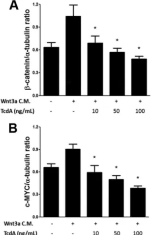
TcdA affects -catenin protein levels and prevents the
c-MYC expression induced by Wnt3a.We next examined the
effect of TcdA on Wnt3a-induced stabilization of-catenin in IEC-6 cells. This stabilization is key in the activation of the down-stream components of the Wnt pathway (23). Thus, we performed Western blotting of total protein extracts from IEC-6 cells to
as-sess the cellular levels of-catenin in response to Wnt3a CM with different concentrations of TcdA.-Catenin protein levels de-creased in response to increasing doses of TcdA (Fig. 2A). Further-more, we evaluated the levels of the protein c-MYC in the same samples. c-myc is a direct target gene of the-catenin/Tcf-Lef complex, and it encodes a transcription factor that has major roles in cell cycle progression and apoptosis. Therefore, c-myc upregu-lation occurs when-catenin is hyperactivated (40). As expected, our pretreatment of IEC-6 cells with Wnt3a CM for 24 h signifi-cantly raised c-MYC protein levels compared to those obtained with the control, untransfected L cell CM. Still, in the groups exposed to TcdA along with Wnt3a CM, the levels of c-MYC were decreased in a concentration-dependent manner (Fig. 2B). Alto-gether, these data not only support the previous results obtained from the TOPflash luciferase reporter assay but also give rise to the hypothesis that TcdA might inhibit Wnt signaling by promoting -catenin degradation.
dramatically reduced, as determined by indirect immunofluores-cence (Fig. 3A). A subset of the cells showed a more significant reduction in nuclear-catenin while still retaining the cytoplas-mic staining of-catenin (Fig. 3B). Importantly, the inhibition of
-catenin nuclear translocation by TcdA is consistent with the TOPflash luciferase reporter assay data.
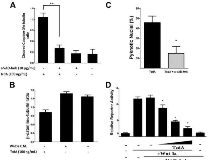
Pan-caspase inhibitor z-VAD-fmk prevents-catenin deg-radation but fails to reverse-catenin/TCF transcription inac-tivation by TcdA.-Catenin is vulnerable to caspase-dependent degradation (31), so we hypothesized that the loss of-catenin protein following treatment with TcdA is a consequence of caspase activation. To assess this, we used z-VAD-fmk, which has successfully reduced activation of the caspases’ cascade by TcdA in previous studies (6, 9, 15). As expected, pretreatment with z-VAD-fmk prevented the cleavage of caspase-3 by TcdA treat-ment of IEC-6 cells (Fig. 4A). Analysis of at least three indepen-dent experiments in IEC-6 cells pretreated with z-VAD-fmk showed a statistically significant protective effect of caspase inhi-bition on downregulation of-catenin protein by TcdA (Fig. 4B). In addition, the pretreatment inhibited morphological signs of apoptosis, including nuclear pyknosis (Fig. 4C). Thereafter, we evaluated if the prevention of-catenin degradation by z-VAD-fmk would reverse the inhibition of the Wnt pathway induced by TcdA (Fig. 4D). However, even in the presence of z-VAD-fmk, TcdA still demonstrated a concentration-dependent inhibition of the Wnt/-catenin pathway in IEC-6 cells in the TCF reporter assay. This suggests that the inhibition of the Wnt/-catenin path-way by TcdA is not secondary to caspase-dependent-catenin degradation.
TcdA attenuates Wnt signaling independently of-catenin destruction complex activity.The intracellular concentration of -catenin is primarily controlled by APC-GSK3-dependent phosphorylation, a key event in targeting the-catenin protein for ubiquitination and proteasomal degradation. Therefore, to inves-tigate the mechanism by which TcdA inhibits Wnt signaling, we assessed the role of the-catenin APC-GSK3destruction com-plex.
Initially, we used LiCl, which mimics members of the Wnt family of signaling proteins by inhibiting the activity of GSK3, causing intracellular accumulation of-catenin (41). RKO cells were incubated with LiCl and TcdA either with or without Wnt3a CM. We assessed the -catenin transcriptional regulatory re-sponse using a TCF reporter assay (Fig. 5). Incubation with TcdA still attenuated the LiCl activation of the Wnt pathway, even in the presence of Wnt3a CM. This suggests that TcdA blocks Wnt sig-FIG 2TcdA downregulates-catenin and c-MYC protein levels in IEC-6 cells.
Total protein extracts were prepared from IEC-6 cells treated with TcdA (10, 50, or 100 ng/ml) in the presence of Wnt3a or L CM and were analyzed by Western blotting with anti-c-MYC and anti--catenin antibodies. The blots were reprobed with anti-␣-tubulin antibody as a loading control. Toxin A was able to cause a dose-dependent downregulation in the levels of-catenin (A) and c-MYC (B) proteins. Values are expressed as means⫾SEM (n⫽3). *,P⬍0.05.
FIG 3TcdA prevents-catenin nuclear translocation induced by Wnt3a in IEC-6 cells, as shown by immunofluorescence confocal microscopy of IEC-6 cells treated with Wnt3a CM with or without TcdA (50 ng/ml). (A)-Catenin signal is in red. Nuclei are in blue (DAPI). (B) Quantification of nuclear or cytoplasmic-catenin immunostaining. The number of DAPI-stained nuclei in randomly chosen microscope fields represents the total cells and was used to calculate the percentage of cells with nuclear (N), cytoplasmic (C), or no (U) staining. The results are shown as the average of three experiments. Values are expressed as means⫾SEM (n⫽3). *,P⬍
0.05.
naling by a mechanism independent of the APC-GSK3 destruc-tion complex. Nonetheless, LiCl has intracellular targets other than GSK3that could potentially have interfered with this result. Hence, we tried a different approach to stimulate the Wnt/ -catenin pathway by transfecting RKO cells with plasmids encod-ing constitutively active-catenin, either wild type (pcatenin) or resistant to GSK3phosphorylation (pcatenin-S33A). We also used a plasmid encoding LEF-DeltaN-VP16-pcDNA3, which binds to DNA and directly activates-catenin-mediated gene ex-pression without the need for-catenin. Thereafter, we assessed -catenin transcriptional regulatory activity by the TCF reporter assay (Fig. 6). TcdA was able to attenuate Wnt signaling even in the presence of pcatenin-S33A, confirming that this inhibition occurs independently of the APC-GSK3destruction complex (Fig. 6). Moreover, TcdA was unable to inhibit the TCF response transcription induced by Lef-DeltaN-VP16.
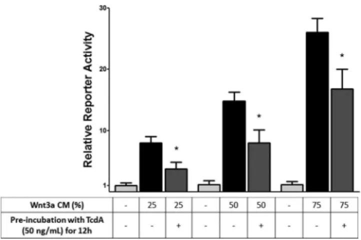
Preincubation with TcdA impairs Wnt signaling activation by Wnt3a.In order to minimize the effects of TcdA on cell viabil-ity, we preincubated RKO cells with TcdA and then washed them twice. This preincubation prevented the-catenin/TCF-mediated transcription induced by different concentrations of Wnt3a CM (Fig. 7). This result strongly suggests that the inhibition of the Wnt FIG 5GSK3inhibitor LiCl is unable to prevent TcdA-induced-catenin/
TCF signaling downregulation, as shown by a TCF reporter assay in RKO cells stably cotransfected with TOPflash andRenilla, incubated with TcdA (50 ng/ dl) for 24 h, and treated with LiCl (20 mM) with or without Wnt3a CM. Values are expressed as means⫾SEM (n⫽3). *,P⬍0.05 versus values for a Wnt3a-treated group that was given z-VAD-fmk.
signaling by TcdA depends on the inhibition of the Rho GTPases (Rho, Rac1, and Cdc42).
DISCUSSION
In this study, we showed that TcdA inhibits Wnt/-catenin signal-ing in a dose-dependent manner in cultured intestinal epithelial cells. This result was consistently demonstrated by three different techniques: TCF reporter assay, immunofluorescence staining for -catenin, and Western blotting for c-MYC (encoded by a target gene).
The activated Wnt pathway is crucial for the maintenance of the rapid turnover of the intestinal epithelium (19). The inhibi-tion of this pathway by TcdA is therefore consistent with previous reports that it exerts antiproliferative effectsin vitro, inducing cell cycle arrest and downregulation of cyclin D1 (encoded by a target gene for Tcf/Lef) (10,12). Unfortunately, the significance of these antiproliferative effects induced by TcdA and TcdB for the patho-genesis ofC. difficile-associated diarrhea (CDAD) is currently not fully understood. However, the inhibition of the intestinal epithe-lial renewal by TcdB may be linked to the development of diarrhea (11). Since the layer of cells covering the human intestine is com-pletely renewed every 3 to 4 days, one may speculate that the impairment of this cellular renewal would compromise the intes-tinal epithelial barrier, contributing to the appearance of diarrhea in the patients afflicted by this infection (10,11,42). In a similar fashion, this mechanism is well established in cancer chemother-apy, as diarrhea represents a frequent side effect of many antican-cer drugs (which also suppress proliferation) (43).
The modulation of the Wnt/-catenin pathway is not re-stricted to TcdA, since it occurs with toxins from other bacteria, thus significantly contributing to their pathogenesis. For example, Salmonella-epithelial cell interactions promote activation of the proinflammatory NF-B signaling pathway, which is associated with degradation of-catenin and upregulation of proinflamma-tory cytokines (interleukin 6 [IL-6], IL-8, and tumor necrosis fac-tor alpha [TNF-␣]) in infected mice (30). Importantly, this
inter-action between the NF-B and Wnt pathways may be strikingly relevant forC. difficileas well, since inflammation plays a central role in the pathogenesis of CDAD. Additionally,Bacillus anthracis edema toxin inhibits the Wnt/-catenin pathway by protein ki-nase A induction. This reduces the levels of phosphorylated GSK3, allowing it to phosphorylate, and thereby inactivate, -catenin and preventing it from binding to the Tcf/Lef transcrip-tion factor (44).
Our results indicate that TcdA decreased the levels of-catenin protein by a mechanism involving the activation of caspases be-cause z-VAD-fmk, a pan-caspase inhibitor, successfully prevented TcdA-induced-catenin degradation. Notably, in previous stud-ies z-VAD-fmk demonstrated some degree of proteasome inhibi-tion, which could also be implicated in its effect of reversing -catenin protein downregulation (45). Nevertheless, diminished intracellular -catenin protein levels did not appear to be the main mechanism by which TcdA attenuates the Wnt signaling, since it occurred even in the presence-catenin upregulation ei-ther by z-VAD-fmk-mediated caspase inactivation or by transfec-tion of constitutively active-catenin plasmids. Altogether, these data indicate that inhibition of the Wnt/-catenin pathway by TcdA is only partially a consequence of the-catenin downregu-lation induced by caspases during apoptosis. Caspases may cleave -catenin to dismantle cell contacts during apoptosis, while there might be other mechanisms induced by TcdA that prevent -catenin translocation to the nucleus and thereby inhibit tran-scription of Tcf/Lef regulated genes.
In addition, the-catenin destruction complex did not appear to have a significant role in the TcdA-induced Wnt signaling in-hibition. LiCl (a GSK3inhibitor) and transfection of mutated GSK3-resistant-catenin (S33A) plasmid were both unable to abrogate the inhibitory effects of TcdA on RKO cells. In contrast, expression of the Lef-DeltaN-VP16 fusion protein strongly en-hanced-catenin/TCF transcription, and this was not reverted by TcdA. This protein lacks the-catenin binding domain and con-stitutively activates transcription from Tcf/Lef-binding elements (32). This finding suggests that the inhibition of RhoGTPases by TcdA may also play a major role in this inhibition, besides its FIG 7RhoGTPases inhibition by preincubation with TcdA attenuates
-catenin/TCF-mediated transcription in RKO cells, as determined by a TCF reporter assay in RKO cells stably cotransfected with TOPflash andRenilla
preincubated with TcdA (50 ng/dl) for 12 h. TcdA was then removed, and cells were treated with Wnt3a CM for 24 h. Values are expressed as means⫾SEM (n⫽3). *,P⬍0.05.
FIG 6TcdA inhibits Wnt/-catenin signaling induced by transfection of con-stitutively active-catenin constructs, as determined by a TCF reporter assay in RKO cells stably cotransfected with TOPflash andRenillaand incubated with TcdA (50 ng/dl) for 24 h under various conditions. Plasmids included pcatenin (wild type-catenin), pcatenin (S33A) (mutated-catenin at S33A, resistant to GSK3phosphorylation), and LEF-DeltaN-VP16 (consti-tutively active Lef1 that directly activates-catenin-mediated gene expres-sion). Values are expressed as means⫾SEM (n⫽3). *,P⬍0.05.
effects on cell viability, which confirms that the effects of TcdA are upstream of Tcf/Lef transcription.
One might speculate that there are other possible mechanisms contributing to the inhibition of the Wnt/-catenin pathway by TcdA. Inhibition of Rac1 expression, by either small interfering RNA (siRNA) or dominant negative Rac1, in ST2 cells impairs nuclear translocation of-catenin induced by Wnt3a CM; thus, no activation of Tcf/Lef target genes could occur (26). Therefore, since Rac1 is critical for-catenin translocation to the nucleus, its permanent glycosylation and inactivation by TcdA could be a hy-pothesis to explain our results.
In conclusion, our study demonstrated for the first time that TcdA inhibits Wnt/-catenin signaling in cultured intestinal epi-thelial cells, affects-catenin protein levels, and prevents c-MYC expression induced by Wnt3a independently of GSK3 inhibi-tion. Although z-VAD-fmk prevents -catenin degradation, it fails to reverse TcdA-induced Wnt/-catenin pathway inhibition. The role of TcdA inhibitory effects on Wnt/-catenin signaling in the pathogenesis of theC. difficile-induced disease may provide novel insight into improving therapies.
ACKNOWLEDGMENTS
We thank Vivaldo M. Neto, a professor at UFRJ), and Fabio Zuim, a lab technician at UFRJ), for their assistance.
This work was supported by grant 573928/2008-8 from the Brazilian Agency for Scientific and Technological Development (INCT-MCT/ CAPES/CNPq).
We declare no conflict of interest.
REFERENCES
1.Kuehne SA, Cartman ST, Heap JT, Kelly ML, Cockayne A, Minton NP. 2010. The role of toxin A and toxin B inClostridium difficileinfection. Nature467:711–713.http://dx.doi.org/10.1038/nature09397.
2.Frisch C, Gerhard R, Aktories K, Hofmann F, Just I.2003. The complete receptor-binding domain ofClostridium difficiletoxin A is required for endocytosis. Biochem. Biophys. Res. Commun.300:706 –711.http://dx .doi.org/10.1016/S0006-291X(02)02919-4.
3.Papatheodorou P, Zamboglou C, Genisyuerek S, Guttenberg G, Akto-ries K. 2010. Clostridial glucosylating toxins enter cells via clathrin-mediated endocytosis. PLoS One 5:e10673. http://dx.doi.org/10.1371 /journal.pone.0010673.
4.Egerer M, Giesemann T, Jank T, Satchell KJ, Aktories K.2007. Auto-catalytic cleavage ofClostridium difficiletoxins A and B depends on cys-teine protease activity. J. Biol. Chem.282:25314 –25321.http://dx.doi.org /10.1074/jbc.M703062200.
5.Nusrat A, von Eichel-Streiber C, Turner JR, Verkade P, Madara JL, Parkos CA.2001.Clostridium difficile toxins disrupt epithelial barrier function by altering membrane microdomain localization of tight junc-tion proteins. Infect. Immun.69:1329 –1336.http://dx.doi.org/10.1128 /IAI.69.3.1329-1336.2001.
6.Brito GA, Fujji J, Carneiro-Filho BA, Lima AA, Obrig T, Guerrant RL. 2002. Mechanism ofClostridium difficiletoxin A-induced apoptosis in T84 cells. J. Infect. Dis.186:1438 –1447.http://dx.doi.org/10.1086/344729. 7.Fiorentini C, Matarrese P, Straface E, Falzano L, Fabbri A, Donelli G,
Cossarizza A, Boquet P, Malorni W.1998. Toxin-induced activation of Rho GTP-binding protein increases Bcl-2 expression and influences mi-tochondrial homeostasis. Exp. Cell Res.242:341–350.http://dx.doi.org/10 .1006/excr.1998.4057.
8.Kim H, Kokkotou E, Na X, Rhee SH, Moyer MP, Pothoulakis C, Lamont JT.2005.Clostridium difficiletoxin A-induced colonocyte apop-tosis involves p53-dependent p21(WAF1/CIP1) induction via p38 mito-gen-activated protein kinase. Gastroenterology129:1875–1888.http://dx .doi.org/10.1053/j.gastro.2005.09.011.
9.Carneiro BA, Fujii J, Brito GA, Alcantara C, Oria RB, Lima AA, Obrig T, Guerrant RL.2006. Caspase and Bid involvement inClostridium diffi-ciletoxin A-induced apoptosis and modulation of toxin A effects by
glu-tamine and alanyl-gluglu-tamine in vivo and in vitro. Infect. Immun.74:81– 87.http://dx.doi.org/10.1128/IAI.74.1.81-87.2006.
10. D’Auria KM, Donato GM, Gray MC, Kolling GL, Warren CA, Cave LM, Solga MD, Lannigan JA, Papin JA, Hewlett EL. 2012. Systems analysis of the transcriptional response of human ileocecal epithelial cells toClostridium difficiletoxins and effects on cell cycle control. BMC Syst. Biol.6:2.http://dx.doi.org/10.1186/1752-0509-6-2.
11. Lica M, Schulz F, Schelle I, May M, Just I, Genth H.2011. Difference in the biological effects ofClostridium difficiletoxin B in proliferating and non-proliferating cells. Naunyn Schmiedebergs Arch. Pharmacol.383: 275–283.http://dx.doi.org/10.1007/s00210-010-0595-5.
12. Welsh CF, Roovers K, Villanueva J, Liu Y, Schwartz MA, Assoian RK. 2001. Timing of cyclin D1 expression within G1 phase is controlled by Rho. Nat. Cell Biol.3:950 –957.http://dx.doi.org/10.1038/ncb1101-950. 13. Halabi-Cabezon I, Huelsenbeck J, May M, Ladwein M, Rottner K, Just
I, Genth H.2008. Prevention of the cytopathic effect induced by Clostrid-ium difficiletoxin B by active Rac1. FEBS Lett.582:3751–3756.http://dx .doi.org/10.1016/j.febslet.2008.10.003.
14. Fiorentini C, Fabbri A, Falzano L, Fattorossi A, Matarrese P, Rivabene R, Donelli G. 1998.Clostridium difficiletoxin B induces apoptosis in intestinal cultured cells. Infect. Immun.66:2660 –2665.
15. Nottrott S, Schoentaube J, Genth H, Just I, Gerhard R.2007. Clostrid-ium difficiletoxin A-induced apoptosis is p53-independent but depends on glucosylation of Rho GTPases. Apoptosis12:1443–1453.http://dx.doi .org/10.1007/s10495-007-0074-8.
16. Ando Y, Yasuda S, Oceguera-Yanez F, Narumiya S.2007. Inactivation of Rho GTPases withClostridium difficiletoxin B impairs centrosomal acti-vation of Aurora-A in G2/M transition of HeLa cells. Mol. Biol. Cell18: 3752–3763.http://dx.doi.org/10.1091/mbc.E07-03-0281.
17. Klein EA, Assoian RK.2008. Transcriptional regulation of the cyclin D1 gene at a glance. J. Cell Sci.121:3853–3857.http://dx.doi.org/10.1242/jcs .039131.
18. Musgrove EA, Caldon CE, Barraclough J, Stone A, Sutherland RL.2011. Cyclin D as a therapeutic target in cancer. Nat. Rev. Cancer11:558 –572.
http://dx.doi.org/10.1038/nrc3090.
19. van der Flier LG, Clevers H.2009. Stem cells, self-renewal, and differen-tiation in the intestinal epithelium. Annu. Rev. Physiol.71:241–260.http: //dx.doi.org/10.1146/annurev.physiol.010908.163145.
20. Kimelman D, Xu W.2006. beta-catenin destruction complex: insights and questions from a structural perspective. Oncogene25:7482–7491.
http://dx.doi.org/10.1038/sj.onc.1210055.
21. Yang J, Zhang W, Evans PM, Chen X, He X, Liu C.2006. Adenomatous polyposis coli (APC) differentially regulates beta-catenin phosphorylation and ubiquitination in colon cancer cells. J. Biol. Chem.281:17751–17757.
http://dx.doi.org/10.1074/jbc.M600831200.
22. Wu G, Huang H, Garcia Abreu J, He X. 2009. Inhibition of GSK3 phosphorylation of beta-catenin via phosphorylated PPPSPXS motifs of Wnt coreceptor LRP6. PLoS One 4:e4926. http://dx.doi.org/10.1371 /journal.pone.0004926.
23. Huang H, He X.2008. Wnt/beta-catenin signaling: new (and old) players and new insights. Curr. Opin. Cell Biol.20:119 –125.http://dx.doi.org/10 .1016/j.ceb.2008.01.009.
24. Zhang B, Abreu JG, Zhou K, Chen Y, Hu Y, Zhou T, He X, Ma JX.2010. Blocking the Wnt pathway, a unifying mechanism for an angiogenic in-hibitor in the serine proteinase inin-hibitor family. Proc. Natl. Acad. Sci. U. S. A.107:6900 – 6905.http://dx.doi.org/10.1073/pnas.0906764107. 25. He X, Semenov M, Tamai K, Zeng X. 2004. LDL receptor-related
proteins 5 and 6 in Wnt/beta-catenin signaling: arrows point the way. Development131:1663–1677.http://dx.doi.org/10.1242/dev.01117. 26. Wu X, Tu X, Joeng KS, Hilton MJ, Williams DA, Long F.2008. Rac1
activation controls nuclear localization of beta-catenin during canonical Wnt signaling. Cell133:340 –353.http://dx.doi.org/10.1016/j.cell.2008.01 .052.
27. Steinhusen U, Badock V, Bauer A, Behrens J, Wittman-Liebold B, Dorken B, Bommert K. 2000. Apoptosis-induced cleavage of beta-catenin by caspase-3 results in proteolytic fragments with reduced trans-activation potential. J. Biol. Chem.275:16345–16353.http://dx.doi.org/10 .1074/jbc.M001458200.
Lima BB, Carvalho AF, Guerrant RL, Ribeiro RA, Brito GA. 2011. Adenosine deaminase inhibition preventsClostridium difficiletoxin A-in-duced enteritis in mice. Infect. Immun.79:653– 662.http://dx.doi.org/10 .1128/IAI.01159-10.
30. Sun J, Hobert ME, Duan Y, Rao AS, He TC, Chang EB, Madara JL. 2005. Crosstalk between NF-kappaB and beta-catenin pathways in bacte-rial-colonized intestinal epithelial cells. Am. J. Physiol. Gastrointest. Liver Physiol.289:G129 –G137.http://dx.doi.org/10.1152/ajpgi.00515.2004. 31. Cho M, Gwak J, Park S, Won J, Kim DE, Yea SS, Cha IJ, Kim TK, Shin
JG, Oh S.2005. Diclofenac attenuates Wnt/beta-catenin signaling in co-lon cancer cells by activation of NF-kappaB. FEBS Lett.579:4213– 4218.
http://dx.doi.org/10.1016/j.febslet.2005.06.049.
32. Aoki M, Hecht A, Kruse U, Kemler R, Vogt PK.1999. Nuclear endpoint of Wnt signaling: neoplastic transformation induced by transactivating lymphoid-enhancing factor 1. Proc. Natl. Acad. Sci. U. S. A.96:139 –144.
http://dx.doi.org/10.1073/pnas.96.1.139.
33. Yost C, Torres M, Miller JR, Huang E, Kimelman D, Moon RT.1996. The axis-inducing activity, stability, and subcellular distribution of beta-catenin is regulated in Xenopus embryos by glycogen synthase kinase 3. Genes Dev.10:1443–1454.http://dx.doi.org/10.1101/gad.10.12.1443. 34. Liu X, Liu T, Slusarski DC, Yang-Snyder J, Malbon CC, Moon RT,
Wang H.1999. Activation of a frizzled-2/beta-adrenergic receptor chi-mera promotes Wnt signaling and differentiation of mouse F9 teratocar-cinoma cells via Galphao and Galphat. Proc. Natl. Acad. Sci. U. S. A. 96:14383–14388.http://dx.doi.org/10.1073/pnas.96.25.14383.
35. Major MB, Camp ND, Berndt JD, Yi X, Goldenberg SJ, Hubbert C, Biechele TL, Gingras AC, Zheng N, Maccoss MJ, Angers S, Moon RT. 2007. Wilms tumor suppressor WTX negatively regulates WNT/beta-catenin signaling. Science 316:1043–1046. http://dx.doi.org/10.1126 /science/1141515.
36. Kumar A, Zloza A, Moon RT, Watts J, Tenorio AR, Al-Harthi L.2008. Active beta-catenin signaling is an inhibitory pathway for human
immu-nodeficiency virus replication in peripheral blood mononuclear cells. J. Virol.82:2813–2820.http://dx.doi.org/10.1128/JVI.02498-07.
37. Garcia-Abreu J, Cavalcante LA, Moura Neto V.1995. Differential pat-terns of laminin expression in lateral and medial midbrain glia. Neurore-port6:761–764.http://dx.doi.org/10.1097/00001756-199503270-00014. 38. Garcia-Abreu J, Moura Neto V, Carvalho SL, Cavalcante LA. 1995.
Regionally specific properties of midbrain glia: I. Interactions with mid-brain neurons. J. Neurosci. Res.40:471– 477.
39. Amado NG, Cerqueira DM, Menezes FS, da Silva JF, Neto VM, Abreu JG.2009. Isoquercitrin isolated fromHyptis fasciculatareduces glioblas-toma cell proliferation and changes beta-catenin cellular localization. An-ticancer Drugs20:543–552.http://dx.doi.org/10.1097/CAD.0b013e32832 d1149.
40. Niehrs C, Acebron SP. 2012. Mitotic and mitogenic Wnt signalling. EMBO J.31:2705–2713.http://dx.doi.org/10.1038/emboj.2012.124. 41. Hedgepeth CM, Conrad LJ, Zhang J, Huang HC, Lee VM, Klein PS.
1997. Activation of the Wnt signaling pathway: a molecular mechanism for lithium action. Dev. Biol.185:82–91.http://dx.doi.org/10.1006/dbio .1997.8552.
42. Kim M, Ashida H, Ogawa M, Yoshikawa Y, Mimuro H, Sasakawa C. 2010. Bacterial interactions with the host epithelium. Cell Host Microbe 8:20 –35.http://dx.doi.org/10.1016/j.chom.2010.06.006.
43. Genth H, Dreger SC, Huelsenbeck J, Just I.2008.Clostridium difficile
toxins: more than mere inhibitors of Rho proteins. Int. J. Biochem. Cell Biol.40:592–597.http://dx.doi.org/10.1016/j.biocel.2007.12.014. 44. Larabee JL, DeGiusti K, Regens JL, Ballard JD.2008.Bacillus anthracis
edema toxin activates nuclear glycogen synthase kinase 3beta. Infect. Im-mun.76:4895– 4904.http://dx.doi.org/10.1128/IAI.00889-08.
45. Canu N, Barbato C, Ciotti MT, Serafino A, Dus L, Calissano P.2000. Proteasome involvement and accumulation of ubiquitinated proteins in cerebellar granule neurons undergoing apoptosis. J. Neurosci.20:589 – 599.