Effects of genetic
manipulation of the
monoaminergic system on
chronic pain and on the
affective disorders
associated with pain
José Carlos Oliveira Ferreira
Mestrado em Bioquímica
Departamento de Química e Bioquímica 2017
Orientador
Doutora Fani Lourença Moreira Neto, Professora auxiliar na Faculdade de Medicina da Universidade do Porto, Investigadora no Grupo Pain Research do Instituto de Investigação e Inovação em Saúde, Universidade do Porto
Co-Orientador
Doutora Maria Isabel Torres Martins, Professora auxiliar na Faculdade de Medicina da Universidade do Porto. Investigadora no Grupo Pain Research do Instituto de Investigação e Inovação em Saúde, Universidade do Porto
Todas as correções determinadas pelo júri, e só essas, foram efetuadas. O Presidente do Júri,
I
Agradecimentos
Terminada esta “maratona” intensiva ao longo da qual se desenvolveu este projeto, repleto de altos e baixos e cujas adversidades foram várias desde o início, gostaria de deixar aqui registado o nome de algumas pessoas que, de alguma forma, foram essenciais durante o meu crescimento científico e, acima de tudo, durante o meu crescimento enquanto pessoa.
À Profª Fani Neto e Profª Isabel Martins, pela orientação, pela disponibilidade e por todo o conhecimento transmitido. Obrigado profª Fani pelo fim de semana intensivo de correções a uma tese que foi ganhando forma e obrigado acima de tudo pela boa disposição quase sempre presente. Obrigado Profª Isabel por arranjar (sempre que possível) tempo para ver tudo ao mais pequeno pormenor e, também, pela boa disposição. Agradeço às duas por todos os conselhos transmitidos e por terem contribuído desta forma para a minha formação.
Ao Professor Pedro Alexandrino Fernandes, Diretor do Mestrado em Bioquímica, pelos conselhos e pelo parecer favorável ao adiamento de entrega desta dissertação que foi, sem dúvida, importante para concluir trabalhos em curso.
À Raquel Silva, por me ter acompanhado desde o primeiro dia em que iniciei este percurso, por me ter sempre tratado como igual e por me ter transmitido tudo o que sabia da melhor maneira possível.
À Raquel Alonso e à Helena Cavaleiro pela amizade e cumplicidade que criamos praticamente desde o início. Foram muitas horas de risos que partilhamos juntos.
Aos meus colegas da Unidade de Biologia Experimental da FMUP, Zé Tiago, Rita Costa, Raquel Oliveira, Marta, Sílvia, Rafael, Inês, Rita Oliveira e Diana pelo companheirismo e pelos bons momentos que passamos juntos.
Aos meus amigos, João Trigo, Guilherme, João Magalhães, André, Carolina, Maria, Joyce, Ana Almeida, Marta, Ana Freitas e Joana por se terem tornado uma segunda família.
Às pessoas que mais importam, os meus pais e a minha irmã, deixo um agradecimento especial. Obrigado por estarem sempre ao meu lado.
II
Abstract
Neuropathic pain is a pathologic and chronic condition that results from damage or dysfunction of the peripheral or central nervous system causing spontaneous pain and a pathologically amplified response to both noxious and non-noxious stimuli. This condition is very afflictive, affecting 7-8% of adult individuals worldwide. The noradrenergic system is of high importance in the neuronal interactions responsible for pain modulation and is also involved in the regulation of emotional/affective behaviours in response to pain.
The Locus Coeruleus (LC) is the main regulator of noradrenergic functions in the central nervous system and is the main source of noradrenergic innervation to the spinal dorsal horn. Besides projecting to the spinal dorsal horn, the LC also projects to many supraspinal brain regions, in particular to pre-frontal cortex areas, and plays a pivotal role in diverse behaviors such as learning, strategic behavior, motivation, and arousal. Furthermore, LC-derived noradrenaline is also causally linked to aversive states like stress, anxiety and depression.
The main goal of this project was to study the effects of selectively knocking-down at the LC the production of Dbh, the rate-limiting enzyme of noradrenaline synthesis, on the induction and development of neuropathic pain and of associated affective disorders, such as anxiety and depression behaviors. This was done by using mutant mice and the Cre-Lox system which allowed to spatially and temporally manipulate the noradrenergic system. For this purpose, mice with a Dbh flox+/+ genotype were used to establish a
protocol for Dbh gene ablation by stereotaxic injection of a lentivirus expressing the Cre recombinase enzyme (LV-Cre), into the LC. The mice underwent then a spared nerve injury (SNI) surgery to induce a well-established model of neuropathic pain (SNI model). The tests to evaluate anxiety- and depression-like behaviors showed similar results in neuropathic pain (SNI) and sham animals. Thus, mice showed a similar exploratory behavior in the open field, as well as a comparable behavior in the Elevated Plus Maze, but with tendency to spend more time in the closed arms than in open arms. On the Marble Burying test, the SNI group buried a few higher number of marbles, while in the forced swimming test, the only one that evaluates depressive-like behaviors, no significant differences were observed. The nociceptive tests confirmed the hypersensitivity typical of a neuropathic pain condition. After stereotaxic injection of LV-Cre, a heterogeneous Dbh ablation was detected in different rostro-caudal areas of the LC. At the behavioral level the data showed different effects on the nociceptive and anxiety and depressive-like behaviors according to the rostro-caudal level of the LC targeted by LV-Cre injection. This suggests that different rostro-caudal regions of the LC
III might be differentially engaged in the processing of the nociceptive and affective components of pain.
IV
Resumo
A dor neuropática é uma condição patológica e crónica que resulta de dano ou disfunção do sistema nervoso central ou periférico, causando dor espontânea e uma resposta patologicamente amplificada a estímulos nóxicos e não nóxicos. Esta condição é muito aflitiva, afetando 7 a 8% das pessoas adultas em todo o mundo. O sistema noradrenérgico é de grande importância nas interações neuronais responsáveis pela modulação da dor e também está envolvido na regulação dos comportamentos emocionais / afetivos em resposta à dor.
O Locus Coeruleus (LC) é o principal regulador das funções noradrenérgicas no sistema nervoso central e é a principal fonte de inervação noradrenérgica para o corno dorsal da medula espinhal. Além de projetar para o corno dorsal da medula espinhal, o LC projeta também para muitas regiões encefálicas supraespinhais, em particular para as áreas do córtex pré-frontal, e desempenha um papel fundamental em comportamentos diversos, como aprendizagem, comportamento estratégico, motivação e atenção focalizada. Além disso, a noradrenalina derivada do LC também está causalmente associada a estados aversivos, tais como ansiedade e depressão.
O principal objetivo deste projeto foi estudar os efeitos de deletar seletivamente no LC a produção de Dbh, a enzima limitante da síntese da noradrenalina, na indução e desenvolvimento de dor neuropática e de distúrbios afetivos associados, tais como comportamentos de ansiedade e depressão. Isto foi feito através da utilização de ratinhos mutantes e do sistema Cre-Lox que permitiram a manipulação espacial e temporal do sistema noradrenérgico. Para este fim, os ratinhos com o genótipo Dbh flox+/+ foram utilizados para estabelecer um protocolo para ablação do gene Dbh por
injeção estereotáxica de, um lentivírus que expressa a enzima Cre recombinase (LV-Cre), no LC. Os ratinhos foram sujeitos depois a uma cirurgia de lesão do nervo ciático para induzir um modelo bem estabelecido de dor neuropática (modelo SNI). Os testes comportamentais para ansiedade e depressão apresentaram resultados semelhantes em animais com dor neuropática (SNI) e sham. Desta forma foi observado um comportamento exploratório em campo aberto semelhante, bem como um comportamento comparável no labirinto Elevated Plus, mas com tendência para permanecerem mais tempo nos braços fechados do que nos braços abertos. No teste dos berlindes, o grupo SNI enterrou um número maior de berlindes, enquanto no teste de natação forçada, o único que avalia o comportamento depressivo, não foram observadas diferenças significativas. Os testes nociceptivos confirmaram a hipersensibilidade típica de uma condição de dor neuropática. Após a injeção estereotáxica das partículas lentivíricas de LV-Cre no LC, uma ablação heterogénea de
V Dbh foi detetada em diferentes áreas rostro-caudais do LC. Ao nível comportamental, os dados mostraram diferentes efeitos nos comportamentos nociceptivos e de ansiedade e depressão, de acordo com o nível rostro-caudal do LC alvejado pela injeção de LV-Cre. Isto sugere que diferentes regiões rostro-caudais do LC poderão estar envolvidas de forma diferencial no processamento dos componentes nociceptivos e afetivos da dor.
VI
Abbreviatures
AAV Adeno-associated virus
ACC Anterior Cingulate Cortex
AMY Amygdala
AP Anteroposterior
AR Adrenergic receptors
ASICs Acid-sensing ion channels
AV Adeno Virus
BA Basolateral Amygdala
BLA Basolateral complex
CCI Chronic Constriction Injury
CeA Central Nucleus of Amygdala
CKO Conditional Knockout
CNS Central Nervous System
COMT Catecol O-Metiltransferase
DAB Diaminobenzidine
DBH Dopamine-β-hydroxylase
DLF Dorsolateral Funiculus
DRG Dorsal Root Ganglia
DRt Dorsal Reticular Nucleus
DV Dorsoventral
EGFP Enhanced Green Fluorescent Protein
EPM Elevated Plus Maze
ERK1/2 Extracellular-signal regulated kinases ½
FST Forced Swimming Test
GFP Green Fluorescent Protein
HFV Human Foamy Virus
HSV Herpes simplex Virus
HSV-1 Herpes Simplex Virus-1
IASP Association for the Study of Pain
IC Insular Cortex
LA Lateral Nucleus of Amygdala
LC Locus Coeruleus
LM Lateromedial
MAO Monoamine Oxidase
MB Marble Burying
VII
NAc Nucleus Accumbens
NGS Normal Goat Serum
NRM Nucleus Raphe Magnus
OF Open Field
PAG Periaqueductal Gray Region
PBS Phosphate-buffered Saline
PBS-T Phosphate-buffered Saline-Triton
PFC Prefrontal Cortex
PSL Partial Sciatic Ligation
rACC Rostral Anterior Cingular Cortex
RVM Rostral Ventromedial Medulla
S1 Primary somatosensory cortex
S2 Secondary somatosensory cortex
SNI Spared Nerve Injury
SNL Spinal Nerve Ligation
TCAs Tricyclic Antidepressants
TG Trigeminal Ganglia
TH Tyrosine Hydroxylase
TRP channels Transient Receptor as Potential-generating channels
VIII
Table of contents
Agradecimentos... I Abstract ... II Resumo ... IV Abbreviatures ... VI I. Introduction ... 1 1. Pain ... 1 1.1. Types of pain ... 2 1.2. Pain transmission ... 3 1.3. Pain modulation ... 51.3.1. The endogenous pain control system ... 5
1.3.2. The noradrenergic System ... 7
1.3.2.1. Locus Coeruleus ... 7
1.3.2.2. Descending noradrenergic modulation... 9
2. Affective component of Pain ... 10
3. Neuropathic Pain ... 12
3.1. Central and peripheral neuropathic pain ... 12
3.2. Neuropathic pain treatment ... 14
4. Experimental Neuropathic Pain ... 14
4.1. Animal models of neuropathic pain ... 15
4.2. Spared nerve injury ... 16
5. Genetic manipulation of the nociceptive system ... 17
II. Aims ... 20
III. Materials and Methods ... 21
1. Generation of mutant mice ... 21
1.1. Generation of chimaeric mice and germ line transmission of the Dbh tm1a allele ... 21
1.2. Generation of the Dbh flox/flox allele ... 21
1.3. PCR genotyping ... 22
1.4. Generation of spatial and temporal conditional KO mice for Dbh (Dbh-CKO) ... 22
2. Experimental design ... 23
3. Lentiviral vectors ... 24
4. Surgical Procedures ... 25
4.1. Stereotaxic Surgeries ... 25
4.1.1. Optimization of the stereotaxic injections protocol ... 25
4.2. Spared nerve injury ... 26
5. Behavioral Assessment ... 27
5.1. Nociceptive Tests ... 27
IX
5.1.2. Acetone Test ... 28
5.2. Anxiety and Depression Behavior Tests ... 28
5.2.1. Open Field ... 28
5.2.2. Elevated Plus Maze ... 29
5.2.3. Marble Burying ... 29
5.2.4. Forced Swimming Test ... 30
6. Tissue preparation and Immunohistochemistry ... 31
6.1. Immunoreaction against DBH ... 31
7. Statistical Analysis ... 32
IV. Results ... 33
1. Evaluation of Anxiety and Depression in Neuropathic Pain Animals ... 33
1.1. Anxiety- and depression-like behavior tests ... 33
1.2. Nociceptive behavior tests ... 35
2. Effects of DBH ablation at the LC of conditional mice ... 36
2.1. Optimization of the experimental conditions ... 36
2.1.1. Optimization of stereotaxic coordinates for LV-Cre injection ... 36
2.1.2. Determination of the virus titer ... 37
2.1.3. Verification of Dbh gene ablation by LV-Cre ... 38
2.2. Characterization of Dbh ablation at the injections sites of the conditional mutant mice ... 39
2.2.1. Nociceptive behavior ... 42
2.2.1.1. Effect of the stereotaxic injection before SNI induction ... 42
2.2.1.2. Time course effects of Dbh ablation at the LC ... 43
2.2.2. Anxiety- and depression-like behaviors ... 47
V. Discussion ... 49
VI. Conclusions and future perspectives ... 55
VII. Appendix ... 57
X
List of Figures
Figure 1- Pain circuits. Schematic overview of the main circuits mediating physiological pain.. .. 4
Figure 2- Schematic representation of pain modularity circuitry.. ... 6
Figure 3- Schematic representation of a noradrenergic synapse.. ... 8
Figure 4- Brain regions and circuits implicated in the comorbidity between pain and depression.. ... 11
Figure 5-The peripheral and central changes induced by nerve injury or peripheral neuropathy. ... 13
Figure 6- Animal models of Neuropathic Pain ... 15
Figure 7- Illustrative image of the von Frey nociceptive test. ... 28
Figure 8 – Illustrative image of anxiety and depression Tests. ... 30
Figure 9- Results for anxiety- and depressive-like behavior tests in neuropathic pain animals . 34 Figure 10-Results of nociceptive tests in neuropathic pain animals ... 35
Figure 11- Optimization of stereotaxic injections into the LC. ... 36
Figure 12- Stereotaxic injections of LV-Cre at two different concentrations.. ... 37
Figure 13- Immunoreaction against DBH revealed with DAB (brown) in the LC 10 days post stereotaxic injection of LV-Cre.. ... 38
Figure 14- Diagrams depicting the LV-Cre injections sites.. ... 40
Figure 15- Immunofluorescence against DBH.. ... 41
Figure 16- Frequency of paw withdrawal in response to Von Frey filaments of the animals before and after LV-Cre injection. ... 42
Figure 17- Frequency of paw withdrawal to von Frey monofilaments... 44
Figure 18- Acetone test. ... 46
Figure 19-Results for the anxiety and depressive-like behavior tests in SNI animals injected with LV-Cre and SNI animals without LV-Cre injection (control). ... 48
List of Tables
Table 1-List of different animal models of neuropathic pain ... 16Table 2 -Viral vector systems ... 18
List of Schemes
Scheme 1 - Experimental Group 1. ... 23Scheme 2 - Experimental Group 2. ... 23
Scheme 3 - Experimental Group 3. ... 24
1
I.
Introduction
Pain is a serious condition and often debilitating for people who live with it, not only for the ones that are suffering with that, but also for their families and society in general1.
However, pain is a common occurrence in the entire life, a survival mechanism that functions as an early warning sign of ongoing or impending tissue damage1, 2. Besides
the association of painwith injury and disease, pain is a subjective symptom and it is that subjectivity the main challenge for current and further investigations.
1. Pain
Until the 1960s, pain was considered an inevitable sensory response to tissue damage without minimal considerations for the affective dimension of the pain experience and none for the effects of genetic differences, past experience, anxiety or expectation3. The
newest advances have been made in our understanding of the mechanisms that underlie pain and in the treatment of people who complain of pain3, 4.
The Melzack-Wall gate control theory, proposed in 1965, demonstrated the mechanisms in the central nervous system that control the perception of the noxious stimulus, and thus integrated afferent, upstream processes, with downstream modulation from the brain 3-5. The gate control theory proposed that the transmission of nerve impulses from
afferent fibers to spinal cord transmission cells is modulated by a gating mechanism in the spinal dorsal horn6. This theory considers the motivational dimension of pain in
addition to its more obvious sensory dimension7. Considering this, the painful sensation
was defined as being comprised by three different components, a sensory-discriminative component that refers to the capacity to analyze the nature, location, intensity, and duration of the nociceptive stimulation; an affective-motivational component that involves the unpleasant character of all the painful perception; and a cognitive-evaluative component related to the past experiences8.
The most complete definition of pain has been provided by the International Association for the Study of Pain (IASP), which established pain as “an unpleasant sensory and emotional experience associated with actual or potential tissue damage, or described in terms of such damage”9.
2
1.1. Types of pain
The existence of many types of pain can be understood by the identification of four broad categories: nociception, perception of pain, suffering, and pain behaviors3. Pain may be
divided into several types according to its duration, etiology, localization or others, contributing to its complexity. Regarding the duration, pain is classified as transient, acute or chronic. Transient pain is elicited by the activation of nociceptive transducers in the skin or other tissues of the body in the absence of any tissue damage. Transient pain has evolved to protect man from physical damage by the environment or by over stress of the body tissues. This is present in every day of life and is rarely a reason to seek health care3. Acute pain is defined as a short duration, phasic and intense physiological
event, which normally has a rapid resolution after the phenomenon that initially originated pain is healed. The majority of cases of acute pain is caused by substantial injury of body tissue with activation of nociceptive transducers at the local of the tissue damage. This injury alters the characteristics of special sensory fibers, called nociceptors, their central connections, and the autonomic nervous system in the region. Chronic pain, in contrast, is a long-lasting persistent pathological pain that is present even when there is no apparent biological cause, or the original cause is no longer present. Indeed, the original injury may be capable of damaging the nervous system in such way so that it is unable to restore itself to a normal state. Other chronic pain syndromes may occur spontaneously without any sign of injury. In all of them, the intensity of pain is out of proportion and leads to health care3. Chronic Pain can be classified into 3 categories:
nociceptive pain, which arises from non-neural tissue damage; neuropathic pain in which there is nervous tissue damage; or a mix of the two categories. Nociceptive pain is the physiological sensation caused by the activation of neuronal pathways by stimuli with sufficient intensity to potentially cause tissue damage. Stimuli detection is named nociception and is a critical protective process that helps to prevent injury by generating a reflex withdrawal from stimuli associated to an unpleasant sensation, namely pain10.
Neuropathic pain is difficult to treat being associated to spontaneous pain, exaggerated responses to nociceptive stimuli (hyperalgesia) and nociceptive responses to stimuli which are normally non-nociceptive (allodynia)11.
3
1.2. Pain transmission
Pain transmission is initiated by the activation of specialized sensory fibers widely distributed throughout the body, called nociceptors. These are high-threshold sensory receptors of the peripheral nervous system capable of transducing mechanical, chemical and thermal noxious stimuli from different organism locations into action potentials. The cell bodies of these neurons are located at trigeminal ganglia (TG) in the cephalic region and at dorsal root ganglia (DRG) in extracephalic regions12, 13. Nociceptors’ cell bodies
possess a unique axon that bifurcates into two branches. A central branch projects to the dorsal horn of the spinal cord while the peripheral branch terminates in one of the many peripheral organs, and constitutes the sensitive fiber. There are three types of fibers, distinguished according to their diameter, myelination degree and conduction velocity (Aβ, Aδ and C)13, 14.These include the two major classes of nociceptors. The
medium diameter myelinated (Aδ) afferents mediate acute, well-localized “first” or fast pain. These fibers differ considerably from the larger diameter and rapidly conducting Aβ fibers that respond to innocuous mechanical stimulation applied to skin, muscle and joints and thus not contribute to pain. The second class of nociceptors includes the small diameter unmyelinated C fibers that convey poorly localized, “second” or slow pain15, 16.
The physiochemical properties of noxious stimuli are converted to electrical activity by ion channels such as transient receptor potential-generating channels (TRP channels) and purinergic channels, and this electrical activity is amplified by sodium channels to elicit action potentials17.
The nociceptive signal is transmitted at central synapses through release of a variety of neurotransmitters that have the potential to excite second-order nociceptive projection neurons in the spinal dorsal horn or hindbrain (Figure 1). These neurons project to supra-spinal sites, which further project to cortical and subcortical regions via third-order neurons, enabling the encoding and perception of the multidimensional pain experience. The transmission of nociceptive signals at the spinal dorsal horn can be modulated by local inhibitory GABAergic and opioidergic interneurons and also by descending serotonergic and noradrenergic projections influencing the response to and perception of pain18.
4
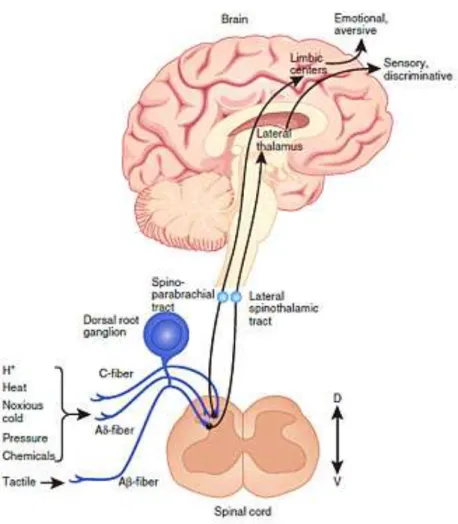
Figure 1- Pain circuits. Schematic overview of the main circuits mediating physiological pain. Nociceptive signals
are transmitted from the periphery by nociceptive sensory neurons (first-order primary afferent neurons) the peripheral terminals of which are clustered with ion channels, including transient receptor potential channel subtypes (TRPA, TRPM and TRPV), sodium channel isoforms (Nav), potassium channel subtypes (KCNK) and acid-sensing ion channels (ASICs). The transduction of external noxious stimuli is initiated by membrane depolarization due to the activation of these ion channels. Action potentials are conducted along the axons of nociceptive Aβ- and C-fibers, through the cell body in the dorsal root ganglion to the axonal terminals, which form the presynaptic element of central synapses of the sensory pathway in the spinal dorsal horn or hindbrain. The central terminals of Aβ- and C-fibers synapse with interneurons and second-order nociceptive projection neurons, primarily within the superficial laminae of the spinal dorsal horn. The axons of second-order nociceptive projection neurons decussate at the spinal cord level, joining the ascending fibers of the anterolateral system, and project to brainstem and thalamic nuclei, transferring information about the intensity and duration of peripheral noxious stimuli. No single brain region is essential for pain, but rather pain results from the activation of a distributed group of structures. Third-order neurons from the thalamus project to several cortical and subcortical regions that encode sensory-discriminative, emotional and cognitive aspects of pain. Several brainstem sites are also known to contribute to the descending modulation of pain; (Adapted from Kunner et al17).
5
1.3. Pain modulation
1.3.1. The endogenous pain control system
Pain transmission at spinal cord can be modulated through a complex process known as endogenous pain modulation. Most of the ascending axons of spinal second order neurons project to the brainstem reticular formation, which includes areas in charge of pain modulation, such as dorsal reticula nucleus (DRt), the rostral ventromedial medulla (RVM), the noradrenergic locus coeruleus( LC) and the periaqueductal gray region (PAG)12. All of these supraspinal sites play an important role in pain modulation but, the
most well characterized pain modulatory areas are the mesencephalic periaqueductal grey (PAG) and the rostral ventromedial medulla (RVM) (Figure 2).
Pain modulation exists in the form of a descending pain modulatory circuit with inputs that arise from multiple areas, including the hypothalamus, the amygdala and the rostral anterior cingular cortex (rACC), feeding to the midbrain periaqueductal gray region (PAG), and with outputs from the PAG to the medulla oblongata12, 19. This modulatory
process works to inhibit or facilitate the spinal nociceptive processing, which ultimately controls the experience of pain. The PAG, a key region for descending inhibition receives projections from regions associated with the processing of emotions and projects to the rostral ventromedial medulla (RVM), which includes the serotonin-rich nucleus raphe magnus (NRM) as well as the nucleus reticularis gigantocellularis pars alpha and the nucleus paragigantocellularis lateralis20. Then, neurons in the RVM project along the
dorsolateral funiculus (DLF) to the dorsal horn21.
The RVM mediates a bidirectional control of nociception by its resident ON- and OFF-cells. OFF-cells are thought to exert descending inhibition of nociception, while ON-cells seem to facilitate nociceptive mechanisms at the spinal dorsal horn12, 19, 21. These cells
are not serotonergic but they can modulate serotonergic neurons of the NRM and the nucleus gigantocellularis. Once serotonin is released in the spinal dorsal horn, it plays inhibitory or facilitatory actions depending on the receptor subtype activated22, 23. Another
important contributor of the modulatory control is the noradrenergic system, which is described more thoroughly in the next section.
6
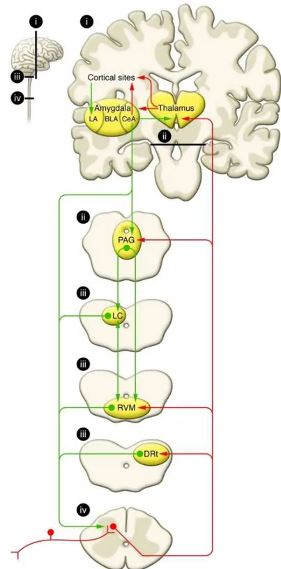
Figure 2- Schematic representation of pain modularity circuitry. Nociceptive inputs enter the spinal dorsal horn
through primary afferent fibers that synapse onto transmission neurons. From dorsal horn, ascending projections, labeling in red, target the thalamus, and collateral projections also target mesencephalic nuclei, including the DRt, the RVM, and the midbrain PAG. Descending pain modulation is mediated through projections, labeling in green, to the PAG, which also receives inputs from other sites, including the hypothalamus (data not shown), and communicates with the RVM as well as other medullary nuclei that send descending projections to the spinal dorsal horn. The noradrenergic Locus Coeruleus receives inputs from the PAG, communicates with the RVM, and sends descending noradrenergic inhibitory projections to the spinal cord. Areas labeled “i–iv” in the small diagram correspond with labeled details of the larger diagram. BA – Basolateral amygdala; CeA – Central nucleus of amygdala; LA – Lateral nucleus of amygdala; (Adapted from Ossipov et al12).
7
1.3.2. The noradrenergic System
The noradrenergic system comprises a vital circuit based on the action of noradrenaline (NA) on different and strategically located adrenergic receptors. NA is involved in the autonomic regulation of various organs24. In the central nervous system (CNS),
noradrenergic neurons are organized in seven clusters, classified from A1 to A7, which
are located in the brainstem25-27.
1.3.2.1. Locus Coeruleus
The Locus Coeruleus (LC or A6), the main structure coordinating the central
noradrenergic system, is located in the upper part of the floor of the fourth ventricle. Rather than being a single brain nucleus, the LC represents a nuclear complex that includes areas located bilaterally within the lateral pontine central grey, as well as scattered catecholamine neurons close to the brachium conjunctivum and within the central tegmental regions of the pontine gray matter28, 29. The main neurotransmitter
synthetized in the LC nucleus is NA, but other substances are also produced in this structure.
Noradrenaline is a neurotransmitter biosynthesized by sequential enzymatic reactions starting with the amino acid tyrosine24, 26. This cascade starts with the conversion of
tyrosine into dihydroxyphenylalanine (L-DOPA) through the rate limiting enzyme action of tyrosine hydroxylase (TH; Fig. 3)24. Then, L-DOPA is converted into dopamine through
the action of the aromatic L-amino acid decarboxylase26 and, only in noradrenergic
neurons, dopamine is posteriorly converted into NA by dopamine-β-hydroxylase (DBH; Fig. 3). After this process, NA is stored into synaptic vesicles and in response to specific electrical input, these vesicles attach to the neuronal membrane through the vesicular monoamine transporter, releasing their content into the synaptic cleft26, 30. The NA
present in the synaptic cleft can bind to post-synaptic adrenergic receptors and activate intracellular signaling cascades and, after transducing their specific signals, NA molecules can undergo reuptake by presynaptic NA transporters or they can be degraded by enzymes in the synaptic cleft and at the nerve terminal26 (Fig. 3).
8
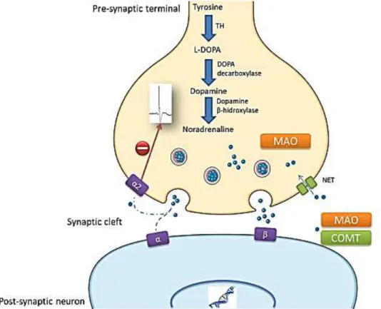
Figure 3- Schematic representation of a noradrenergic synapse. NA is synthesized from the amino acid tyrosine
through a cascade of events. After its synthesis, NA is stored in vesicles at the axon terminal and following specific electrical input, these vesicles attach to the membrane and release the NA content into the synaptic cleft. In the extracellular space, NA exerts its function by binding to α and β adrenergic receptors in the post-synaptic membrane. When the flow of NA into the synaptic cleft is intense, α2-adrenoceptors in the pre-synaptic membrane (auto-receptors) are also activated and they inhibit NA release. At the same time, the extracellular NA content that does not bind to any receptor or that has already exerted its function may be recycled upon reuptake at the pre-synaptic membrane, or it may be degraded: L-DOPA – L-dihydroxyphenylalanine;COMT – Catecol O-Metiltransferase; MAO – monoamine oxidase; TH – tyrosine hydroxylase (adapted from M. Llorca-Torralba et al)26.
Noradrenaline release exerts neuromodulatory effects on synaptic transmission by modifying the membrane potential, synaptic plasticity and the excitability of neurons via G-protein coupled receptors, α1, α2 and β (β1, β2 and β3) adrenoreceptors (ARs)29, 31. The
adrenergic receptors have a different distribution in the different targets of LC projections. The α1 and β receptors are present primarily at postsynaptic sites, whereas α2 receptors
exist both pre- and postsynaptically32. In general, the action of these receptors is
mediated by guanine nucleotide-binding regulatory proteins (G proteins) 26. Although
both categories have been implicated in pain modulation, the α-adrenoreceptors have a key role by mediating the pain regulatory effects of noradrenaline26.
9
1.3.2.2. Descending noradrenergic modulation
The LC (or A6), the A5 and A7 noradrenergic cell groups, are the main sources of
noradrenergic projections to the spinal cord25, 33, 34. These cells groups are connected
with other pain control centers and all of them receive projections from PAG34. LC also
receives projections from the central nucleus of the amygdala (CeA), preoptic area, paraventricular nucleus of hypothalamus and lateral hypothalamus35. These
noradrenergic projections form an important component of descending pain modulation. Electrical or chemical stimulation of noradrenergic nuclei, PAG and RVM produces spinal antinociceptive effects by releasing NA into the spinal cerebrospinal fluid.
At the spinal cord level, NA released from descending pathways may activate several pain inhibitory mechanisms. Direct catecholaminergic innervation of cell bodies of the spinothalamic tracts’ neurons provide a structural basis for post-synaptic noradrenergic inhibition of spinal pain-relay neurons25. Noradrenaline also inhibits transmission of
nociceptive signals in the spinal cord due to action on presynaptic α2-AR36.
Studies with a knockout for the Dbh gene that led to the absence of noradrenaline shows it has a minor effect on pain sensitivity37, supporting the concept that noradrenergic
systems have little influence on baseline pain sensitivity. However, during persistent pain it was shown that noradrenergic systems have a more important role38, 39. Recent studies
suggest that in conditions of nerve injury, the descending noradrenergic system is augmented in an effort to compensate for enhanced nociceptive inputs. Injury is associated with increased synthesis and release of NA along with an enhanced efficacy of spinal α2-AR40.
The noradrenergic system is of high importance in the neuronal interactions responsible for pain modulation and is also involved in the regulation of emotional/affective behaviors. In fact, our group has established that noradrenergic activation by chronic pain increases the descending pain facilitation from the DRt41-43.
10
2. Affective component of Pain
Pain comprises sensory, cognitive and, most importantly, affective components. The affective component includes feelings of annoyance, sadness, anxiety and depression in response to a noxious stimulus44. In chronic pain patients, mood disorders such as
depression and anxiety are frequently observed, with prevalence rates around 30% in neuropathic pain patients45, 46. Although this comorbidity is clinically established, the
underlying mechanisms still remain unclarified.
Clinical imaging studies of acute experimental pain in human subjects provided important information regarding the areas most commonly activated by noxious stimuli, including the primary somatosensory (S1) and secondary somatosensory cortex (S2), anterior cingulate cortex (ACC), insular cortex (IC), prefrontal cortex (PFC), insula, thalamus, amygdala and the mesolimbic reward circuit, which includes the ventral tegmental area (VTA) and nucleus accumbens (NAc). S1 and S2 activations contribute to the sensory-discriminative dimension of pain. The ACC, PFC, IC, NAc, and amygdala, meanwhile, have been implicated in the affective component of pain (Figure 4)47.This interconnected
group of brain regions also forms the basis for understanding the pathophysiology of depression. The NAc receives afferent nociception information through connections with thalamus, parabrachial area, amygdala and ACC. Corticostriatal connections from prefrontal, orbitofrontal and anterior cingulate cortices contribute to an affective, emotional and cognitive control of pain perception and are involved in motivational decision-making. In the NAc, glutamatergic outputs from the amygdala converge on dopaminergic terminals from the VTA and influence motivated behavior in response to stress and anxiety. The dopamine mesolimbic pathway, from the VTA to NAc, also attracted attention in depression research. It is a critical component of ‘brain reward circuits’ which also responds to aversive and nociceptive stimuli48. Additionally, a
descending pathway from the NAc that can modulate spinal nociceptive signals, possibly via the RVM, has been suggested49. The most commonly studied pain modulatory
pathways involve projections from the midbrain PAG to brainstem nuclei, including the rostroventral medulla (RVM) and the locus coeruleus, to the dorsal horn of the spinal cord12, 50. These pathways involve endogenous opioids, noradrenaline and serotonin,
and have both inhibitory and excitatory actions on spinal cord afferent projection neurons51. Interesingly the monoamine serotonergic and noradrenergic systems have
been associated with depression and its treatment. Alba-Delgado et al. explored the role of noradrenergic nucleus locus coeruleus to test the hypothesis that altered LC function
11 might promote depressive and anxiogenic behavior during chronic pain. An increased expression of TH and of NA transporter as well as an α2-AR hypersensitivity has been
observed in a neuropathic animal model, which affect LC firing and NA release in the prefrontal cortex, and is coincident with neuropathic pain-induced anxiodepressive behaviors52. It has also been recently postulated that long-standing chronic pain may
promote higher predisposition for anxiodepressive states53, not only associated with
neuropathy but also with an inflammatory condition. Indeed, Borges et al. demonstrated in monoarthritic rats, that anxiety is associated with this inflammatory condition and is developed within a four-week period after the pain model induction. This finding was accompanied by exacerbated evoked LC responses to noxious stimulation of the inflamed and the healthy paw and increased phosphorylation (activation) of extracellular-signal regulated kinases 1/2 (ERK1/2) in the LC54-56.
Figure 4- Brain regions and circuits implicated in the comorbidity between pain and depression. ACC: anterior
cingulate cortex; AMY: amygdala; IC: insular cortex; NAc: nucleus accumbens; PAG: periaqueductal gray; PFC: prefrontal cortex; RVM: rostral ventromedial medulla; S1: primary somatosensory cortex; S2: secondary somatosensory cortex. (adapted from Doan, L., Manders, T., & Wang, J. (2015)47.
12
3. Neuropathic Pain
The prevalence of chronic pain in Europe is about 25-30%57 and is estimated that about
a fifth of the persons who report chronic pain has neuropathic pain58, 59. Neuropathic pain
is defined by IASP as “pain emerging as a direct consequence of a lesion or a disease of the somatosensory systems”60. This system is involved in the perception of touch,
pressure, pain, temperature, position, movement and vibration. The somatosensory nerves are present in the skin, muscles, joints and fascia and include thermoreceptors, chemoreceptors, pruriceptors and nociceptors that send signals to the spinal cord and eventually to the brain where they will be further processed61.
According to the location of the nerve lesion, neuropathic pain can be divided into two types, central and peripheral neuropathic pain.
3.1. Central and peripheral neuropathic pain
Central neuropathic is caused by a lesion or disease of the spinal cord and/or brain tissue61, 62, including vascular (ischemic or hemorrhagic), infections (abscess,
encephalitis, myelitis), demyelinating (multiple sclerosis), traumatic (brain or spinal cord), or neoplastic disorders. It can also result from syrinx formation in the spinal cord or brainstem62. Cerebrovascular diseases affecting somatosensory pathways (post-stroke
pain) and neurodegenerative diseases are brain disorders that most commonly cause central neuropathic pain63.
For some time, dysfunction of spino-thalamic-cortical pathways, clinically evident as impaired pain (pinprick) and temperature sensation, were considered critical in the development of central neuropathic pain64-66. However, the fact that not all patients with
spinothalamic tract sensory loss suffer central pain suggests a required cofactor to drive its development. Increasing evidences suggest that this cofactor may be the denervation hypersensitivity of surviving spinothalamic tract axons.
By contrast, peripheral neuropathic pain is a condition that develops as a result of damage to the peripheral nervous system. Peripheral changes occur in primary afferent neurons (nociceptive small unmyelinated C-fibers and non-nociceptive myelinated A-fibers) after partial nerve lesion, leading to peripheral sensitization67. Painful generalized
peripheral neuropathies, usually with symmetrical distribution, include those associated with diabetes mellitus, pre-diabetes, and other metabolic dysfunctions, infectious diseases, chemotherapy, immune and inflammatory disorders, inherited neuropathies and channelopathies26.
13 Some axons are damaged and degenerate (upper two axons), whereas others (lower two axons) are still intact and connected with the peripheral end organ. The lesion triggers the expression of sodium channels on damaged C-fibers. Furthermore, products such as nerve growth factor that are associated with Wallerian degeneration are released in the vicinity of spared fibers, triggering channel and receptor expression (sodium channels, TRPV1 receptors, adrenoceptors) on uninjured fibers68.
Indeed, some lesions or diseases of the somatosensory nervous system can lead to altered and disordered transmission of sensory signals into the spinal cord and the brain. Peripheral and central changes induced by nerve injury or peripheral neuropathy are summarized in figure 5.
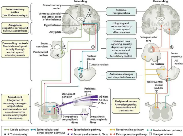
Figure 5-The peripheral and central changes induced by nerve injury or peripheral neuropathy. Damage to all
sensory peripheral fibers alters transduction and transmission due to altered ion channel function. These alterations affect spinal cord activity, leading to an excess of excitation coupled with a loss of inhibition. In the ascending afferent pathways, the sensory componentsof pain are via the spinothalamic pathway to the ventrobasal medial and lateral areas (1), which then project to the somatosensory cortex allowing for the location and intensity of pain to be perceived (2). The spinal cord also has spinoreticular projections and the dorsal column pathway to the cuneate nucleus and nucleus gracilis (3). Other limbic projections relay in the parabrachial nucleus (4) before contacting the hypothalamus and amygdala, where central autonomic function, fear and anxiety are altered (5). Descending efferent pathways from the amygdala and hypothalamus (6) drive the periaqueductal grey, the locus coeruleus, A5 and A7 nuclei and the rostroventral medial medulla. These brainstem areas then project to the spinal cord through descending noradrenaline (inhibition via α2 adrenoceptors), and, in neuropathy, there is a loss of this control and increased serotonin descending excitation via
14
3.2. Neuropathic pain treatment
Several recent studies have shown that NP can adversely affect patients’ overall health-related quality of life, including physical and emotional functioning, as well as the mobility and ability to work, which entails substantial societal costs69. Treatment of neuropathic
pain is challenging as only 50% of the treated patients experience satisfactory pain relief with drug side effects that are considerable. Patients with neuropathic pain continue to experience pain with moderate severity, despite taking prescribed medications70. The
interest group NeuPSIG (Neuropathic Pain Special Interest Group) from IASP recommends as first line treatment, antidepressants, antiepileptics and topical lidocaine. Except in certain clinical circumstances, the use of opioids and tramadol is recommended as second-line treatment, principally for patients who have not responded to the first-line medications70.
Secondary amines of tricyclic antidepressants (TCAs), including nortriptyline and desipramine, and antidepressants with both noradrenaline and serotonin reuptake inhibition (duloxetine, venlafaxine, milnacipran) act by increasing the synaptic serotonin and noradrenaline levels, potentiating the effect of the descending analgesic system 70-72. Antiepileptic drugs, including carbamazepine and calcium channel α
2-δ ligands, such
as gabapentin and pregabalin, function in several ways by modulating voltage-gated sodium and calcium channels, by enhancing the inhibitory effects of GABA and by inhibiting excitatory glutaminergic transmission71. Topical lidocaine is effective for areas
of constant burning pain, especially if they are allodynic.
Tramadol is used as second-line treatment since it is an agonist of the opioid µ-receptor, but it also inhibits the reuptake of serotonin and noradrenaline. Although tramadol has demonstrated efficacy in several neuropathic conditions, it may be less efficacious than strong µ-agonists such as morphine and oxycodone73.
4. Experimental Neuropathic Pain
The evaluation of neuropathic pain in humans is complex if we consider that most of the stimuli required to induce this condition produce irreversible damage. Furthermore, it is almost impossible to recruit a large number of humans for such type of clinical research. Therefore, to understand the mechanisms involved in neuropathic pain and to evaluate the analgesic potential of novel pharmacotherapies for the treatment of neuropathic pain, validated and reproducible animal models of neuropathic pain are necessary74.
15
4.1. Animal models of neuropathic pain
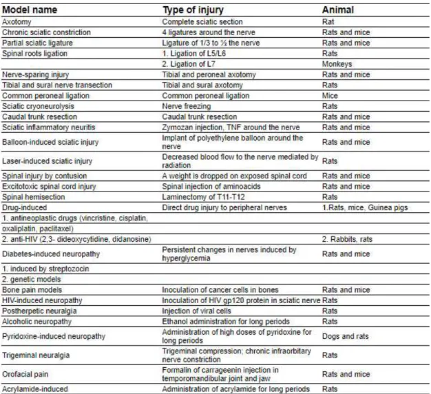
Different models of neuropathic pain have been developed, mostly, in rodents (Table1). Some models aim to replicate peripheral NP mechanisms and others study central mechanisms74. These models are based on most of the known etiologies in humans,
aiming to reproduce peripheral nerve injuries, central injuries, trigeminal neuralgia, diabetic neuropathies, chemo-induced neuropathies, postherpetic neuralgia, and so forth74, 75. Neuropathic pain models fit into three broad classes according to how pain is
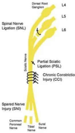
induced: i) traumatic nerve injury, ii) systemic neurotoxicity, or iii) other initiating injuries. Traumatic nerve injuries typically constrict, ligate, crush or transect a nerve, producing a mixture of loss of sensation and gain of pain function. Four different nerve injury models are used76 (Figure 6). In the spinal nerve ligation
(SNL) model, one or more spinal nerves going to the foot are ligated and cut77. In the partial
sciatic ligation (PSL) model, a portion of the sciatic nerve is tightly ligated78. The chronic
constriction injury (CCI) model involves placement of four loose chromic-gut ligatures on the sciatic nerve which triggers an immune response to the sutures leading to nerve swelling and nerve constriction. The fourth nerve injury model is the spared nerve injury (SNI) and in this model, the common peroneal and tibial nerves are cut, while the sural nerve is spared79. In each model, only a portion of the
afferents going to foot are lesioned. All types of injuries induced in these models lead to hyperalgesia, manifested by enhanced responses to mechanical, heat, and/or cooling stimuli. Concomitantly, in these neuropathic pain models, have been shown to induce depression-like behaviors.
Figure 6- Animal models of Neuropathic Pain
16
Table 1-List of different animal models of neuropathic pain (adapted from Jaggi, A.S., et al.2011)74.
4.2. Spared nerve injury
In the SNI model, developed by Decosterd and Woolf79, the tibial and the common
peroneal nerves are sectioned, leaving the sural nerves intact74 (Fig. 6). This procedure
causes a chronic behavioral mechanical and thermal hypersensitivity of the lateral surface of the hindpaw (sural nerve skin area). The areas of the spared sural nerve, and in less extent the saphenous nerve, are both affected, but no changes are observed in the contralateral hindpaw. This change in sensitivity of the intact fibers adjacent to a transected nerve likely results from denervation of the skin normally innervated by the tibial and common peroneal nerves, and by consequent Schwann cells denervation. The difference observed between the saphenous and sural territories is due to the fact that the saphenous nerve is a part of the femoral nerve plexus and enters the spinal cord via a set of dorsal root ganglia different from those of the sciatic nerve79.
17
5. Genetic manipulation of the nociceptive system
The limited therapeutic efficacy and major undesirable side effects frequently associated with current pharmacological treatments of pain explain a strong need of novel therapeutic approaches80-82. Current pharmacological and interventional modalities
available for the treatment of chronic neuropathic pain have variable efficacy and some current therapies include intolerable adverse effects83. The development of gene therapy
has opened the possibility of using nonviral or viral vectors to transduce genes that encode antinociceptive substances to treat chronic pain and study the nociceptive system81, 84. An ideal gene therapy should be efficient in delivering exogenous genes to
target cells that mediate high level and long-term gene expression, and an ideal gene therapy vector should not be able to replicate its own DNA, and should be conductive to long-term gene expression, nonpathogenic and nontoxic85. Viral vectors are generally
created by deleting nonessential genes from the virus while remaining the structural motifs necessary to transfer its genome into the host81, 86. Herpes simplex Virus (HSV),
adeno-associated virus (AAV), adeno virus (AV), lentivirus (LV) and human foamy virus (HFV) can be used as viral vectors for gene therapy of chronic pain. These viral vectors are the most commonly used because of their immunogenicity, natural integration ability and other features. Herpes simplex virus has neurotropic features and naturally maintains lifelong residency in the nucleus of infected neurons, making it suitable for transduction in the nervous system81, 87.Adeno-associated viral vectors are commonly
used to deliver therapeutic genes to target tissues with lower immunogenicity81, 88. They
have a gene carrying capacity of 4.5 kb and can transduce both dividing and nondividing cells89. These viral vectors cannot integrate into the host genome, so there is a low risk
of insertional mutagenicity90. Lentiviral vectors have the advantage of long-term
transgene expression, low immunogenicity, and the ability to accommodate larger transgenes91. LVs belong to a subclass of retroviruses that integrate into the host cell
genome. Due to their natural integration ability, LVs have been extensively utilized for ex vivo gene transfer because of their strong tropism for neuronal stem and progenitor cells92. HFV was the first identified human retrovirus and is nonpathogenic and has
several unique features related to gene transfer, making it a promising vector system for gene therapy93.
18
Table 2 - Viral vector systems1(adapted from Howarth, J. L., Lee, Y. B., & Uney, J. B. (2010)) 94.
The use of viral vectors in neurobiological research has been growing due to their increasing flexibility and the ability to deliver viral particles to brain regions85. In the last
decades, genetic manipulation has been helpful for a better understanding of the molecular mechanisms of the descending pain modulatory system11, 95. It may also
become a versatile tool for chronic pain management, based in three parameters, the vector, the transgene and the promoter. The best combination of these parameters allows to design a gene therapy strategy to study chronic pain11. The vector is the carrier
of the transcriptional cassette and its main function is to deliver its content to specific cell targets. Some of them have the ability to be transported retrogradely, which allows the vector to be uptaken at the nerve terminal and then migrate to the nucleus, often located in remote areas, surgically difficult to access96.
Recent studies targeting the noradrenaline pathway by using gene therapy, specifically noradrenaline synthesis inhibition, allowed a novel approach to pain treatment. Martins et al42 successfully demonstrated a significant decrease in noradrenaline associated with
gene expression in the DRt and also a significant inhibition of mechanical and cold allodynia and mechanical hyperalgesia by using HSV-1 as a vector for delivery of the tyrosine hydroxylase transgene inserted in an antisense orientation into the DRt of the rat brain. This study opened new potential ways to target the noradrenergic modulation during neuropathic pain conditions83.
The understanding of pain-signaling pathways and the specific nerve circuits involved is of major importance for possible therapeutic interventions. Several techniques, classically based on various anterograde and retrograde tracers, electrophysiological recording, viruses and more recently, numerous fluorescent tracers, plant lectins, toxins and fusion proteins, were used for mapping, starting at the periphery and terminating in cortical areas. Although development of approaches allowing the passage of tracer systems across the synapses was an important evolution, the real break-through in this field was the set up of transgenic systems based on inducible tracer expressions. The
1Asterisks indicate the relative ease of production or the relative level of immune response, i.e. a greater ease of
19 most current conditional gene-targeting systems are based on the use of the site-specific recombinase Cre (cyclization recombination) which catalyzes recombination between two 34-bp DNA recognition sites named loxP (locus of crossing [x-ing]-over of bacteriophage P1). The basic strategy for Cre-lox-directed gene Knockout experiments is to flank, or “flox”, an essential exon of the gene of interest with two loxP sites (by homologous recombination in embryonic stem cells), and then to “deliver” Cre that excises the intervening DNA including the exon from the chromosome, thus generating a null allele in all cells where Cre is active. The bacterial Cre recombinase activity may be provided either by crossing mice carrying the floxed target gene with transgenic Cre-expressing mice or by using viral vectors Cre-expressing the Cre transgene97.
20
II.
Aims
The LC is the principal noradrenergic nucleus in the CNS and is the main source of noradrenergic innervation to the spinal dorsal horn. The LC also projects almost to the entire neuraxis and plays a pivotal role in diverse behaviors such as learning and memory, motivation and emotions with LC-derived noradrenaline being linked to aversive states like stress, anxiety and depression. However, the mechanisms and neuronal pathways linking neuropathic pain and depression/anxiety are still unknown and solid and reliable information is still quite sparse. This project aims at providing insight on the effects of the noradrenergic system on nociception and chronic pain-induced anxiety and depression during neuropathic pain conditions. For that purpose, it is imperative to optimize all procedures including the animal model. By using temporal and spatial conditional knockout (CKO) mice for the noradrenaline biosynthetic enzyme Dbh at the LC, and the SNI model of neuropathic pain, the main objectives of this thesis were:
1. To establish a protocol for Dbh gene ablation at the LC of transgenic mice for the successful generation of a temporal and spatial conditional KO animal model for NA production at the LC, to be used in this project;
2. To optimize the lentiviral stereotaxic injection conditions to allow complete Dbh ablation at the LC (viral particles concentration, injection volume, stereotaxic coordinates and latency time for maximum virus expression).
3. To study the effects of Dbh KO at the LC, and therefore of NA impaired synthesis, on the induction and development of neuropathic pain;
4. To study the effects of Dbh KO at the LC, and therefore of NA impaired synthesis, on the depressive and anxiety-like behaviors during neuropathic pain;
21
III.
Materials and Methods
1. Generation of mutant mice
Mutant mice allowing the conditional knockout of the Dbh gene by the Cre-Lox recombination system were developed by members of our Laboratory of Support to Research in Molecular Medicine (LAIMM) following the steps 1.1, 1.2 and 1.3, as follows:
1.1. Generation of chimaeric mice and germ line transmission of
the Dbh
tm1aallele
Ready-targeted Dbh tm1a(EUCOMM)Wtsi ES cells (clone EPD0800_2_E05, Dbh knockout-first
with conditional potential allele, European Conditional Mouse Mutagenesis Program-EUCOMM Consortium) were microinjected into C57BL/6J 8-cell uncompacted morulae using a Leica AM6000 Advanced Micromanipulation system.
Injected embryos were implanted into the uteri of pseudopregnant CD-1 females and the born chimaeric animals were readily identified by the coat colour (Agouti in white CD-1 background). Chimaeric males were mated with C57BL/6J females to obtain germ line transmission of the tm1a allele. Heterozygous mutant progeny was easily discriminated by the Agouti coat color and following molecular genotyping by PCR of genomic DNA isolated from ear punch or tail biopsies at weaning.
1.2. Generation of the Dbh
flox/floxallele
The knockout-first tm1a allele was converted into the floxed Dbh allele (Dbh flox/flox) following recombination in vivo by crossing the heterozygous mutant with a constitutive FLP deleter strain (B6.129S4-Gt(ROSA)26Sortm1(FLP1)Dym/RainJ, The Jackson
Laboratory). Successful recombination of the FRT sites by the FLP recombinase was confirmed by PCR in the double heterozygous progeny. Experimental Dbh flox/flox
homozygous mice, as well as control wild type littermates, were obtained from heterozygote intercrosses.
22
1.3. PCR genotyping
Genomic DNA isolated from ear punch or tail biopsies was PCR amplified using two sets of primers as follows: a) detection of the tma1 (KO-first allele) - Pair 1, Fw GGGGAAACTCAAACACTGCT, Rv ACCACCTCATCAGAAGCAGG, 60 ºC annealing, 35 cycles; b) detection of Dbh floxed allele - pair 2, Fw GGGGAAACTCAAACACTGCT, Rv GGAGGCAGGGGAAAGGTATT, 60 ºC annealing, 35 cycles. Primer pair 1 produces an amplicon of 313 bp encompassing endogenous sequence and part of the tma1 targeting cassette; the WT does not amplify. Pair 2 anneals to genomic sequence only amplifying a fragment of 118 bp within the Dbh locus of wild type animals and a fragment of 298 bp encompassing a LoxP site and remainings of the targeting cassette flanked by the endogenous sequence in floxed animals; the Tm1a allele (KO-first) does not amplify.
1.4. Generation of spatial and temporal conditional KO mice for
Dbh (Dbh-CKO)
Experiments were carried out in the Dbh flox+/-, Dbh flox-/- or the Dbh flox/flox homozygous
C57BL/6 mice (now named as Dbh flox+/+) generated as described above. The Dbh
flox+/+-mice were used for the spatial and temporal generation of conditional KO flox+/+-mice for Dbh (Dbh-CKO). The conditional knockdown of the Dbh gene was induced at a specific time point of the experiments and was circumscribed specifically to the LC area by using the Cre-LoxP recombinant system, and by injecting a lentiviral vector possessing the Cre recombinase gene (LV-Cre: hSyn-eGFP-Cre) specifically into the LC region.
Animals were hosted in Biotério Geral do Centro de Investigação Médica da Faculdade de Medicina da Universidade do Porto under controlled conditions of temperature and humidity, 23 ± 2ºC and 50 ± 10%, respectively, with food and water freely available and darkness/light periods of 12 hours.
The experimental procedures were performed in accordance with the ethical guidelines for the study of experimental pain in conscious animals98, and the European Council
Directive 2010/63/EU, and were approved by the Ethical Committee for Animal Experimentation at our institution (ORBEA) and by Direção Geral de Alimentação e Veterinária (DGAV), Portugal. The mice physical condition was monitored throughout the experiment, with attention to the presence of stress signs, illness or poor physical condition, such as loss or gain of excessive weight, dehydration, aggressive social behavior, low mobility, bleeding and poor wound healing, infection of the sutures and opening of the stitches in the post-surgery period.
23
2. Experimental design
Experimental procedures comprised three different groups of mice weighting between 25 to 30g. Groups 1 and 2 were used as controls and Group 3 served as the experimental group. All mice were submitted to surgical and behavioral assessment procedures as described in detail below. At the end of the experimental period, all the animals were sacrificed for histological and molecular analyses.
Experimental group 1 consisted of Dbh flox+/- or Dbh flox-/- mice that underwent either sham
(N= 7) or spared nerve injury (SNI) surgeries (N= 5), and were then subjected to anxiety and depression-like behavior tests at six weeks after the surgeries. This was followed by nociceptive behavior tests, which were performed a week later, to ensure that all the animals had developed the typical hypersensitivity symptoms associated with a neuropathic pain condition (Scheme 1).
Scheme 1 - Experimental Group 1 used for preliminary evaluation of anxiety- and depression-like behaviors in neuropathic
pain animals, and as a control of the experimental conditions in the for anxiety and depressive-like behavioral tests.
In Experimental group 2 (N=5), mice with the Dbh flox+/+ genotype were subjected to the
SNI surgery to induce a neuropathic pain condition. Nociceptive behavior tests were performed before the SNI surgery (baseline) and then once a week for 28 days (Scheme 2).
Scheme 2 - Experimental Group 2 was a control for neuropathic nociceptive behavior and the evolution of this condition
throughout time.
Experimental group 3 (N=8) was used for the evaluation of the efficacy of conditional Dbh ablation at the LC of the mice, and of the effects on the nociceptive and anxiety-
24 and depression-like behaviors in those mice upon induction of a neuropathic pain condition. For this, animals with the Dbh flox+/+ genotype underwent first a stereotaxic
injection for delivery into the LC of a lentivirus containing the Cre recombinase gene (LV-Cre: hSyn-eGFP-Cre) in order to induce the Dbh gene ablation. Fourteen days later, they were submitted to the SNI surgery to induce the neuropathic pain model. Nociceptive baselines were obtained two days before stereotaxic injection (Baseline 1) and one day before spared nerve injury induction (Baseline 2). Then, the behavioral tests were conducted once a week for 4 weeks. The development of anxiety- and depression-like behaviors was also studied. After the last nociceptive behavioral test, mice were allowed to repose for two weeks. This period was planned to reduce stress and other environmental factors that may interfere with behavioral testing. Then at the 6 weeks’ time point after SNI induction (42 days), mice were submitted to anxiety and depressive-like behavior testing (Scheme 3).
Scheme 3 - Experimental Group 3 was the main experimental group with Dbh gene ablation.
3. Lentiviral vectors
To knockout the DBH floxed gene we used a lentiviral vector purchased from the Gene Vector and Virus Core of the Stanford University (CA, USA). The lentiviral vector named LV-Cre carries an expression cassette that contains the human synapsin promoter (hSYN-p), the Cre recombinase cDNA coupled to the enhanced green fluorescent protein (EGFP) reporter gene and the woodchuck hepatitis virus post-transcriptional regulatory element (WPRE; Scheme 4). The WPRE was used to improve transgene expression. The lentiviral particles were produced by transfection of human embryonic kidney 293T cells with the LV-Cre vector, a packaging plasmid (pCMVΔR8.92), a plasmid encoding the rev protein (pRSV-Rev) and a plasmid encoding the vesicular stomatitis virus G glycoprotein (pMD.G).
25
Scheme 4 - Schematic diagrams of the LV-Cre vector. The lentiviral vector contains the human synapsin promoter
(hSYN-p), the coding region of EFGP coupled to Cre recombinase in the same orientation) and the woodchuck hepatitis virus post-transcriptional regulator.
4. Surgical Procedures
4.1. Stereotaxic Surgeries
At the start of the surgical procedures, mice weighing 25 to 30g were deeply anesthetized with an intraperitoneal injection of a mixture containing ketamine hydrochloride (IMALGENE™ 1000, Merial, Chile; 60 mg/kg) and medetomidine (Medetor, Virbac; 10 mg/kg) and were placed on a rat stereotaxic frame with a mouse adapter (Stoelting Instruments, USA) for bilateral injection of lentiviral vectors (LV_Cre) into the LC by using a Hamilton syringe. Preliminary experiments aimed at optimizing the procedure conditions, namely the accurate stereotaxic coordinates, volume and concentration of injected LV-Cre virus and latency time for maximum lentivirus expression, were firstly performed as described below.
4.1.1. Optimization of the stereotaxic injections protocol
a) Establishment of accurate LC coordinates for injections
After identifying a brain region to be studied, coordinates for stereotaxic injections must be optimized. Firstly, it was necessary to determine initial coordinates (anteroposterior (AP); lateromedial (LM); dorsoventral (DV)) by locating the injection target in the brain atlas Paxinos and Franklin's the Mouse Brain in Stereotaxic Coordinates (4th Edition). The chosen initial coordinates were: anteroposterior (AP) -5.3mm, lateromedial (LM) +/-1.0, dorsoventral (DV) 3.6mm in relation to bregma.
Then a sky-blue dye was injected bilaterally at LC at the determined coordinates followed by animal sacrifice by decapitation under deep anesthesia in order to verify if these theoretical coordinates were on the desired target. The coronal sections surrounding the region of injection were collected and stained with formol-thionin for microscopic observation. For that tissue sections encompassing the LC were obtained in a cryostat at 30µm and mounted on poly-L-lysine coated glass slides, the slices were dried for 1h at 37ºC and immersed in a solution containing 4 volumes of acetone to 1 volume glacial acetic acid for 5 minutes. Washes were made with distilled water then the sections were stained for a period of 20 seconds in a formol-thionin solution (0,1g of thionin in 100mL