Probable acute
disseminated
encephalomyelitis
due to
Haemophilus
influenzae
meningitis
Pedro Beleza*MD, Department of Neurology; Manuel RibeiroMD, Department of Neuroradiology; João PereiraMD;
Carla FerreiraMD; Maria José JordãoMD;
Fátima AlmeidaMD, Department of Neurology, São Marcos Hospital, Braga, Portugal.
Correspondence to first author atServiço de Neurologia, Hospital São Marcos, Largo Carlos Amarante, Apartado 2242, 4701-965 Braga, Portugal.
E-mail: beleza.76@gmail.com
DOI: 10.1111/j.1469-8749.2008.02052.x Published online 18th March 2008
We report the case of a 17-year-old male on long-term steroid therapy for minimal lesion glomerulopathy who, after an upper respiratory infection, presented with Haemophilus influenzaetype b meningitis. Twenty-four hours later he developed depression of consciousness which progressed to coma and left hemiparesis. Brain magnetic resonance imaging (MRI) revealed multiple lesions (hyperintense on T2 and slightly hypointense on T1) involving mainly white matter suggestive of inflammation. MRI features were compatible with acute disseminated encephalomyelitis (ADEM), although a differential diagnosis included cerebritis or vasculitis, secondary to bacterial meningitis. The patient was treated with high-dose steroids which resulted in a gradual improvement followed by complete clinical recovery. We propose a diagnosis of ADEM was the best diagnosis because of the radiological features and response to steroids. The occurrence of ADEM associated with acute meningitis, however rare, represents an important diagnostic challenge for the clinician.
Neurological complications of bacterial meningitis presenting with brain lesions include venous sinus thrombosis, arterial stroke, subdural empyema, vasculitis, abscesses, and acute disseminated encephalomyelitis (ADEM).1We report a case illustrating the difficulty in establishing a differential diagnosis between ADEM and cerebritis or vasculitis on the basis of the current, nonspecific neuroradiological criteria of ADEM. The association of ADEM with meningitis is rare; however, a high level of awareness is needed to reach a timely diagnosis and initiate appropriate treatment.
Case report
A 17yearold male was admitted to the Emergency Depart -ment of the São Marcos Hospital, Braga, Portugal with a dull occipital progressive headache of rapid onset that began 6 hours earlier. This clinical picture occurred in the setting of an upper respiratory infection which had lasted for 1 week, and had been treated with acetaminophen. His medical history was remarkable for minimal lesion glomerulopathy, at that time in remission with prednisolone (10mg administered daily). The patient had been vaccinated against Haemophilus influenzae type b (Hib) at 2, 4, 6, and 18 months, in accordance with the national immunization programme. On examination in the emergency department he was found to have neck rigidity and hyperthermia (38.5˚C). Blood tests showed leukocytosis (17.6×10⁹/L), neutrophilia (0.85), and increased reactive C protein (75.52mg/L). Chest Xray and brain computed tomo -graphy (CT) were normal. Cerebrospinal fluid (CSF) revealed 1120 cells/µL (78% neutrophils, 15% monocytes, 7% lymph -ocytes), 1.44g/L proteins, and 41mmol/L glucose; Hib was disclosed with a latex particle agglutination antigen test, but no bacteria were detected with Gram’s stain.
388 Developmental Medicine & Child Neurology 2008, 50: 388–391
Case report
Case Report 389
We were, therefore, facing acute bacterial meningitis, proba-bly caused by Hib, in an immunocompromised young male. Antibiotic therapy against Hib was initiated with cefotaxime (2g IV 4-hourly for 14d).
At 24 hours clinical worsening occurred with depression of consciousness and bilateral pyramidal syndrome which progressed further the following day to coma and left hemi-paresis. Brain CT at that time showed multiple non-contrast-enhancing hypodensities, located on both hemispheres, involving white matter and right internal capsule.
In the light of acute bacterial meningitis complicated by multiple brain lesions, we considered the following differen-tial diagnoses: cerebritis, multiple arterial or venous strokes in the context of vasculitis, and ADEM. Therefore, methyl-prednisolone (1g IV/d) was initiated and the antibiotic spec-trum was widened by adding to the cefotaxime a high dose of ampicillin (2g IV 4-hourly for 21d) and gentamicin (100mg IV 8-hourly for 12d), with the aim of enhancing the coverage of Gram-negative bacteria. Within 24 hours the patient regained consciousness and exhibited left hemiparesis with no lan-guage impairment. After 48 hours he was apyretic and from then on underwent a gradual clinical improvement in neuro-logical deficits.
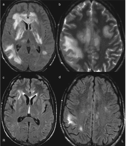
The remaining infection (blood and CSF cultures, poly-merase chain reaction–human immunodeficiency virus), biochemistry (glucose, aspartate aminotransferase, alanine amino transferase, creatinine, urea, 3,3′,5-tri-iodothyronine and thyroxine levels) and immunology (immunoglobulin [Ig] G, IgM, complement [C3, C4, and CH50], antinuclear antibodies, anticardiolipin antibodies, antineutrophil cyto-plasmic antibodies, and double-stranded DNA) studies were negative. A CSF examination repeated on day 4 showed signs of recovery, with 114 cells/µL (60% neutrophils, 30% lym-phocytes), 1.03g/L protein, 59mmol/L glucose, and negative culture tests. Brain MRI performed on day 6 revealed multi-ple T2 hyperintense and T1 slight hypointense lesions with little mass effect and involving the subcortical white matter, basal ganglia, and internal capsule bilaterally (Fig. 1). No dis-ruption of the blood–brain barrier, restriction of diffusion, or abnormality in the major arterial vessels were present. These imaging findings were compatible with areas of inflamma-tion. Afterwards, the patient showed a clinical recovery asso-ciated with improvement of laboratory indices with no further recurrences for a period of more than 1 year. On day 16 he was asymptomatic with a normal neurological exami-nation; CSF showed 14 cells/µL (95% lymphocytes) and hypoglycorrachia (28mmol/L; 34% glycemia) with normal proteinorrachia (0.43g/L). MRI performed on day 18 showed a volume decrease of the lesions (Fig. 1 c,d) and by month 7 no new lesion was found.
Discussion
In the context of acute bacterial meningitis supported by clinical and laboratory data, the detection of Hib in CSF was based on a latex-particle agglutination antigen test because Gram’s staining and a bacterial study were negative. This method has proved to be highly sensitive (95.7%) and specif-ic (100%) for the diagnosis of Hib meningitis,2even in blood-stained CSF specimens.3On the basis of this knowledge, we provided antibiotic therapy directed against Hib, which is supported by other studies.4
Hib meningitis is a rare finding in adults, accounting for
1.8% of meningitis cases.5 In our patient, Hib meningitis might well have resulted from the occurrence of a respiratory infection in an immunosuppressed patient related to pro-longed steroid therapy. Predisposing factors have been found in 74% of adults with Hib meningitis6 and include pneumonia, otitis, diabetes, and alcoholism.7 In addition, prolonged steroid therapy provides a low risk of infections in general and not specifically meningitis or cerebritis.8How -ever, it increases susceptibility to infections due to encapsu-lated bacteria, such as Hib.9
In the reported patient, meningitis had a surprising deterio-rating course, despite continuing therapy: within 48 hours the patient had become comatose with multiple brain lesions on brain CT. The differential diagnosis included cerebritis, vasculi-tis, and ADEM. MRI was consistent with ADEM, because it showed multiple large, asymmetric, mainly white matter T2 and fluid-attenuated inversion recovery (FLAIR) hyperintense lesions.10No MRI criterion has yet been identified that is specif-ic to ADEM,11and T2 and FLAIR hyperintense lesions may also be seen in cerebritis.12Vasculitis was less likely to be involved because MRI did not show restricted diffusion and the angioMRI was normal; it would also be unlikely for vasculitic lesions to resolve completely after only a single course of steroids. The brain lesions were probably of an inflammatory nature because they were T2 hyperintense and T1 hypointense and resolved with little scarring.10Solely on the basis of MRI, it is not possible to distinguish ADEM with allergic inflammatory perivenous encephalitis from cerebritis with inflammatory lesions related to bacterial meningitis. This raises the issue of the specificity of the actual diagnosis of ADEM based on clini-cal–imagiological grounds. The diagnosis of ADEM has evolved significantly, mainly as a result of MRI. One proposed radiolog-ical classification of ADEM includes four patterns of cerebral involvement: (1) ADEM with small lesions (less than 5mm); (2) ADEM with large, confluent lesions; (3) ADEM with additional symmetric bithalamic involvement; and (4) acute haemorrhag-ic encephalomyelitis.13This heterogenity in neuroradiological criteria of ADEM has led to increasing recognition but has also led to a significant blurring of the definition of the disease. Studies reported in the literature consider different definitions of ADEM11and it is possible that not all these syndromes corre-spond to pathological ADEM.
should be located at the grey–white matter junction.12More -over, the lesions lacked mass effect and showed no restricted diffusion. The diagnosis of probable ADEM was corroborated by the dramatic and rapid clinical improvement associated with the laboratory and imagiological recovery seen after the initiation of methylprednisolone, with no further recurrences. Although less frequently than viruses, some bacterial agents have been found to be related to ADEM. These include Borrelia burgdorferi,16Chlamydia pneumoniae,17Legionella
pneumophila,18Mycoplasma pneumoniae,19Rickettsia
rick-ettsi,20 Streptococcus,21 and Pasteurella multocida.14 How -ever, to our knowledge only two other case reports have described ADEM due to bacterial meningitis, and none was associated with Hib.14,19
Here we have shown, for the first time, to our knowledge, that ADEM can occur in association with acute Hib meningitis. In the setting of an acute bacterial meningitis, complicated with depression of consciousness and focal neurological deficits, the hypothesis of ADEM must be considered and should prompt further neurological investigation. Timely diag-nosis and early treatment with high-dose corticoids may improve the prognosis of ADEM.
Accepted for publication 15th January 2008.
Acknowledgements
The authors would like to thank José Armando Leitão for linguistic advice.
References
1. Van de Beek D, de Gans J, Tunkel AR, Wijdicks EF. Community-acquired bacterial meningitis in adults. N Engl J Med2006;
354:44–53.
2. Camargos PA, Almeida MS, Cardoso I, et al. Latex particle agglutination test in the diagnosis of Haemophilus influenzae type B, Streptococcus pneumoniae and Neisseria meningitidis A and C meningitis in infants and children. J Clin Epidemiol 1995;
48:1245–50.
3. Camargos PA, Almeida MS, Filho GL, Batista KW, Carvalho AG, Pereira CL. Blood stained cerebrospinal fluid responsible for false positive reactions of latex particle agglutination tests. J Clin Pathol1994; 47:1116–17.
4. Das BK, Gurubacharya RL, Mohapatra TM, Mishra OP. Bacterial antigen detection test in meningitis. Indian J Pediatr2003;
70:799–801.
5. Tang LM, Chen ST, Wu YR. Haemophilus influenzae meningitis in adults. Diagn Microbiol Infect Dis1998; 32:27–32.
6. Bol P, Spanjaard L, van Alphen L, Zanen HC. Epidemiology of Haemophilus influenzae meningitis in patients more than 6 years of age. J Infect1987; 15:81–94.
7. Spagnuolo PJ, Ellner JJ, Lerner PI, et al. Haemophilus influenzae meningitis: the spectrum of disease in adults. Medicine (Baltimore)
1982; 61:74–85.
390 Developmental Medicine & Child Neurology 2008, 50: 388–391
Figure 1:(a, b)brain magnetic resonance images (MRIs) performed on day 6. Axial fluid-attenuated inversion recovery (FLAIR) and axial T2–TSE show bilateral asymmetric confluent hyperintensity involving subcortical white matter, corpus callosum, and deep grey matter. (c, d) MRIs performed on day 18. FLAIR shows a decrease in lesion load.
a b
c d
Case Report 391 8. Wilckens T, De Rijk R. Glucocorticoids and immune function:
unknown dimensions and new frontiers. Immunol Today1997;
18:418–24.
9. Vinuesa CG, de Lucas C, Cook MC. Clinical implications of the specialised B cell response to polysaccharide encapsulated pathogens. Postgrad Med J2001;77:562–69.
10. Menge T, Hemmer B, Nessler S, et al. Acute disseminated encephalomyelitis: an update. Arch Neurol2005;62: 1673–80. 11. Tardieu M, Mikaeloff Y. What is acute disseminated
encephalomyelitis (ADEM)? Eur J Paediatr Neurol2004;
8:239–42.
12. Falcone S, Post MJ. Encephalitis, cerebritis, and brain abscess: pathophysiology and imaging findings. Neuroimaging Clin N Am2000; 10:333–53.
13. Tenembaum S,Chitnis T, Ness J, Hahn JS; International Pediatric MS Study Group. Acute disseminated encephalomyelitis.
Neurology2007; 68(Suppl. 2):S23–S36.
14. Proulx NL, Freedman MS, Chan JW, Toye B, Code CC. Acute disseminated encephalomyelitis associated with Pasteurella multocida meningitis. Can J Neurol Sci 2003; 30:155–58. 15. Shahar E, Andraus J, Savitzki D, Pilar G, Zelnik N. Outcome of
severe encephalomyelitis in children: effect of high-dose methylprednisolone and immunoglobulins.J Child Neurol
2002; 17:810–14.
16. van Assen S, Bosma F, Staals LM, et al. Acute disseminated encephalomyelitis associated with Borrelia burgdorferi. J Neurol
2004; 251: 626–29.
17. Heick A, Skriver E. Chlamydia pneumoniae-associated ADEM.
Eur J Neurol2000; 7:435–38.
18. Spieker S, Petersen D, Rolfs A, et al. Acute disseminated encephalomyelitis following Pontiac fever. Eur Neurol 1998;
40:169–72.
19. Riedel K, Kempf VA, Bechtold A, Klimmer M. Acute disseminated encephalomyelitis (ADEM) due to Mycoplasma pneumoniae infection in an adolescent. Infection2001; 29:240–42. 20. Wei TY, Baumann RJ. Acute disseminated encephalomyelitis
after Rocky Mountain spotted fever. Pediatr Neurol1999;
21:503–05.
21. Dale RC, Church AJ, Cardoso F, et al. Poststreptococcal acute disseminated encephalomyelitis with basal ganglia involvement and auto-reactive antibasal ganglia antibodies. Ann Neurol 2001;
50: 588–95.
List of abbreviations
ADEM Acute disseminated encephalomyelitis FLAIR Fluid-attenuated inversion recovery Hib Haemophilus influenzae type b