Neurological complications after H1N1
influenza vaccination: magnetic resonance
imaging findings
Complicações neurológicas após vacinação para H1N1: achados de ressonância
magnética.
Ronaldo Lessa1, Maurício Castillo2, Renata Azevedo3, Fernanda Azevedo4, Hildo Azevedo5
ABSTRACT
Objective:To report 4 different neurological complications of H1N1 virus vaccination.Method:Four patients (9, 16, 37 and 69 years of age) had neurological symptoms (intracranial hypertension, ataxia, left peripheral facial palsy of abrupt onset, altered mental status, myelitis) starting 4-15 days after H1N1 vaccination. MRI was obtained during the acute period.Results:One patient with high T2 signal in the cerebellum interpreted as acute cerebellitis; another, with left facial palsy, showed contrast enhancement within both internal auditory canals was present, however it was more important in the right side; one patient showed gyriform hyperintensities on FLAIR with sulcal effacement in the right fronto-parietal region; and the last one showed findings compatible with thoracic myelitis.Conclusion:H1N1 vaccination can result in important neurological complications probably secondary to post-vaccination inflammation. MRI detected abnormalities in all patients.
Keywords:central nervous system, influenza A virus, H1N1, magnetic resonance imaging, encephalitis after vaccination.
RESUMO
Objetivo:Relatar quatro diferentes complicações neurológicas da vacina contra o vírus H1N1.Método:Quatro pacientes (9, 16, 37 e 69 anos) tinham sintomas neurológicos (hipertensão intracraniana, ataxia, paralisia facial esquerda de inicio abrupto, estado mental alterado e mielite) iniciando-se 4 a 15 dias após vacinação contra H1N1. RM foi realizada em quatro pacientes na fase aguda e em um paciente na fase crônica.Resultados:Dos quatro pacientes, um apresentou hipersinal em T2 no cerebelo, interpretado como cerebelite aguda; um, com paralisia facial esquerda, tinha realce dos condutos auditivos internos, maior à direita; um tinha hipersinal em T2 no cortex parieto-occipital direito; um apresentou sinais compatíveis com mielite torácica. Conclusão: A vacinação contra o H1N1 pode resultar em importantes complicações neurológicas, provavelmente secundárias a inflamação pós-vacinal. A RM detectou anormalidades em todos os pacientes.
Palavras-chave:sistema nervoso central, virus influenza H1N1, imagem de ressonância magnética, encefalite pós-vacinal.
Neurological complications of influenza occur in about 4:100,000 of patients per year1. Encephalitis due to H1N1 influenza has been described with the first report dating back to the 1918 influenza pandemia2,3,4,5,6. The most common clinical findings of encephalitis are high fever, seizures, intra-cranial hypertension, altered consciousness, and coma7,8. Although some encephalitis patients fully recover without any neurological sequelae, about 30% die8. In influenza-related encephalitis neuroimaging studies may be normal,
but T2 and T2 FLAIR hyperintense lesions in the white matter and thalami, cerebral edema, hemorrhages, and a pattern similar to that of acute necrotizing encephalitis have been described9,10.
Vaccination is an important prophylaxis against H1N1 influenza decreasing the infection rate and preventing severe complications11. Although rare, CNS complications of vac-cination against H1N1 influenza may occur. In this article we describe the clinical and neuroimaging findings in 4
1Setor de Radiologia, Universidade de Pernambuco, Recife PE, Brazil;
2University of North Carolina, Chapel Hill, North Carolina, United States;
3Setor de Neurologia, Hospital Santa Joana, Recife PE, Brazil;
4Setor de Neurologia, Hospital Correia Picanço, Recife PE, Brazil;
5Universidade de Pernambuco, Recife PE, Brazil.
Correspondence:Ronaldo Lessa; Rua de Casa Forte, 65/1901 Casa Forte; 52061-460 Recife PE, Brasil; E-mail: [email protected] Conflict of interest:There is no conflict of interest to declare.
Received 24 November 2013; Received in final form 02 April 2014; Accepted 22 April 2014.
DOI:10.1590/0004-282X20140064
ARTICLE
patients who developed variety of imaging manifestations that are different from those of other reports describing complications after H1N1 influenza vaccination.
METHOD
Patients and MR Studies
We reviewed imaging findings in 4 patients (age range: 9-69 years) who were vaccinated against H1N1 influenza. Neurologic symptoms started, on average, 9.8 days (range: 4-15 days) after vaccination and unlike other case series clin-ical presentations in our patients were protean. All four patients underwent brain MRI within the first week of symp-toms. The patients had a minimum of sagittal and axial non-contrast T1 images, axial T2 and T2 FLAIR images, and axial and coronal post contrast T1 images. One patient, who has been previously reported in the literature by 1 of us, received pre (sagittal T1, T2, and STIR and axial T1) and post contrast (sagittal and axial T1) imaging of the cervical and thoracic spine12.
Case 1
A 16-year-old girl presented with headaches, vomiting, and cerebellar ataxia, which had developed over 24 hours. There was no history of viral infection or other associated disorders except that she had been vaccinated against H1N1 12 days before. Laboratory tests including CSF ana-lysis showed no significant abnormalities.
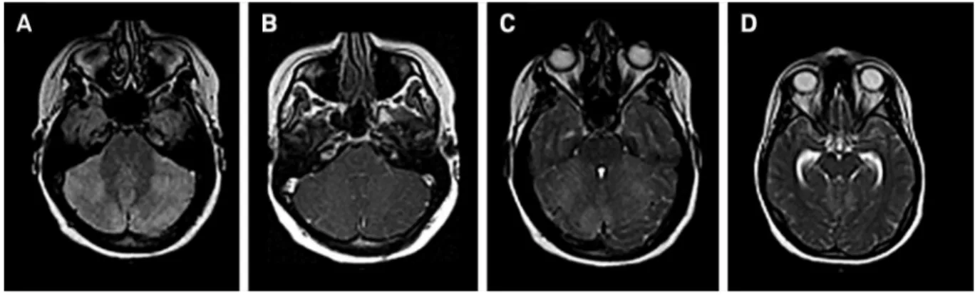
Brain MRI showed cortical foci of hyperintensity on FLAIR in the cerebellar hemispheres with significant mass effect on the 4th ventricle and distal aspect of the cerebral aqueduct with resulting hydrocephalus (Figures 1A and 1C). The supratentorial brain was normal with the exception of ventricular dilatation.
The patient was treated with corticosteroids and follow-up MRI one week later showed decreased hydrocephalus.
After 60 days, clinical examination showed that the patient’s ataxia and headaches had resolved. Follow up MRI showed a significant regression of the abnormalities. No further imaging follow up was deemed necessary.
Case 2
A 9-year-old boy presented with a left peripheral facial palsy of abrupt onset. He had no history of viral illness, head-ache, vomiting, fever or other neurological symptoms but he had been vaccinated against H1N1 9 days prior to the onset of the facial palsy. Laboratory tests including CSF analysis were normal.
Brain MRI showed contrast enhancement in both internal auditory canals and in the labyrinthic and petrous portions of the facial nerves, more marked on the right side (Figures 2A and 2C). No other abnormalities were seen. He was treated with corticosteroids and significant improve-ment in his facial palsy was observed 8 days later. No follow up MRI was obtained.
Figure 1.Axial FLAIR (A) and corresponding post contrast T1 image (B) show areas of high signal in both gray and white matter without enhancement. Note mass effect compressing the 4th ventricle. Axial T2 weighted images show areas of high signal and mass effect on the 4th ventricle and distal aspect of the cerebral aqueduct with resulting hydrocephalus (C-D).
Figure 2.Axial T1 post contrast shows enhancement in the right internal auditory canal (white arrow). Contrast enhance-ment in the opposite side is not shown.
Case 3
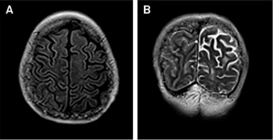
A 69-year-old woman presented with behavioral changes and declining mental status with no focal neurologic symp-toms. The patient had been vaccinated against H1N1 15 days before the onset of neurological symptoms. Brain MRI showed gyriform hyperintensities on FLAIR with sulcal effacement in the right fronto-parietal region (Figures 3A and 3C). No other abnormalities or contrast enhancement were noted. The patient was treated with corticosteroids resulting in improvement of the neurological signs. MRI follow-up 4 weeks later was normal.
Case 4
A 37-year-old female presented with fever and ascending paresthesias which started in her legs and progressed to a T3 level 4 days after H1N1 vaccination. MRI of the spine showed a focal short area of high T2 signal and cord expan-sion at C6-7 (Figure 4) with little if any contrast enhance-ment (not shown). CSF analysis was normal (including herpes virus, CMV, oligoclonal bands and others) and con-servative treatment was prescribed. One month later symp-toms had resolved and follow up MRI showed complete resolution of findings.
DISCUSSION
The H1N1 virus of influenza type A is a combination of several genetics segments of the human, avium, and swine virus that traditionally infect pigs but may spread to humans as thus it is categorized as a“variant”virus13. Only 16 cases of H1N1 infection have been documented in the USA since 200514. But in 2009 the World Health Organization declared a global H1N1 influenza pandemia. Complications following H1N1 infection may occur and the most common are those involving the respiratory tract. Neurological complications may be seen in up 10% of children with H1N1 infection the most common include acute disseminated encephalo-myelitis (ADEM), acute necrotizing encephalitis (ANEC), influenza encephalitis, and Guillain-Barré syndrome have also been described13,15,16,17,18,19.
H1N1 vaccination is an important prophylatic tool to decrease the incidence, intensity, and morbidity of the disease20. Cases of different types of encephalitis and myelitis after vaccination against the H1N1 have been described4,10,11,20,21. The pathogenesis of this encephalitis is not yet fully explained and is believed that immune-mediated alterations resulting in the production of
Figure 3.Axial T2 FLAIR and Coronal T2 images show mild gyriform hyperintensities with sulcal effacement in the right fronto-parietal gray matter and some hyperintensities in the white matter of the opposite hemisphere which were thought to be related to small vessel disease (A–B).
Figure 4.Midsagittal T2 image in the cervical spine shows a focal, well-defined hyperintense region (arrow) at the C6-7 level (used with permission from: Docampo J, Mariluis C, Castillo M, Bruno C, Morales C. Mielitis transversa asociada a vacinacion antigripal. RAR 2011; 75: 15-18).
antibodies and/or the emergence of de novo inflammatory process are responsible20,21,22. Types of encephalitis seen after vH1N1 vaccination include a non-specific pattern, an ADEM pattern, an ANEC pattern, and an influenza-like pattern. Clinical presentations of CNS complications of H1N1 vaccination can be diverse as seen in our patients but the most common symptom are seizures which were not seen in any of our patients. Our first patient presented cerebellar ataxia and intracranial hypertension as a result of diffuse cerebellar edema and hydrocephalus. The second patient was a boy with left facial palsy in whom MRI showed bilateral facial nerve involvement. The third patient presented with behavioral changes and a decline in mental status. All were treated with steroids and showed significant imaging and clinical improvement. The fourth case was a 37 year old woman with cervical medullary involvement in whom symptoms resolved spontaneously 4 weeks after and a follow up MRI was normal. We believe that our cases are worth reporting because the clinical and imaging manifestations were varied and different
to those reported in larger series implying the complications of H1N1 vaccination could be more protean than previously described.
Although our patients were only evaluated with conven-tional MRI sequences, one report describes the use of MR spectroscopy (MRS) in the setting of acute H1N1-related encephalitis23
. In that article, 10 patients were assessed with MRS which showed minimal and non-specific metabolic alterations with only a slight elevation of taurine seen; a find-ing of uncertain significance.
Based on this small series of patients and other reported cases, encephalitis is the most common, albeit rare, complica-tion of H1N1 vaccinacomplica-tion and although as seen in 3 patients reported herein, cerebellitis, neuritis and myelitis may also occur. MRI is the imaging method of choice to assess the finding which tend to be non-specific but are related in time with the vaccine administration. As seen in our cases, most patients will have a favorable prognosis. We concluded that CNS involvement of H1N1 vaccination related complications is more varied than previously reported.
References
1. Newland JG, Laurich VM, Rosenquist AW, et al. Neurological complications in children hospitalized with influenza: characteristics, incidence, and risk factors. J Pediatr 2007;150:306-310.
2. Ekstrand JJ. Neurologic complications of influenza. Semin Pediatr Neurol 2012;19:96-100.
3. Talan J. Two case reports of encephalopathy associated with H1N1. Neurology Today 2010;18:6-7.
4. Athauda D, Andrews TC, Holmes PA, et al. Multiphasic acute disseminated encephalomyelitis (ADEM) following influenza type A (swine specific H1N1). J Neurol 2012;259:775-778.
5. Martin A, Reade EP. Acute necrotizing encephalopathy progressing to brain death in a pediatric patient with novel influenza A (N1N1) infection. CID 2010;50:50-52.
6. Jelliffe S. Nervous and mental disturbances of influenza. N Y Med J 1918;108:725-728.
7. Sugaya N. Influenza-associated encephalopathy in Japan. Semin Pediatr Infect Dis 2002;13:79-84.
8. Togashi T, Matsuzono Y, Narita M, et al. Influenza- associated acute encephalopathy in Japanese children in 1994-2002. Virus Res 2004;103:75-78.
9. Kulkarni R, Kinikar A. Encephalitis in a child with H1N1 infection: first case report from India. J Pediatr Neurosci 2010;5:157-159.
10. Zeng H, Quinet S, Huang W, et al. Pediatri Radiol, 2013;43:1182-1189.
11. Willi B, Fahnenstich H, Weber P. Encephalitis after vaccination against H1N1 influenza virus. 2011;15:276-277.
12. Docampo J, Mariluis C, Castillo M, Bruno C, Morales C. Mielitis transversa asociada a vacinacion antigripal. RAR 2011;75:15-18.
13. Steckelberg JM. http://www.mayoclinic.com/health/influenza-a.
14. http://www.cdc.gov/flu/swineflu/variant-cases-us.htm, accessed 09/23/13.
15. Calitri C, Gabiano C, Garazzino S, et al. Clinical features of hospitalized with 2009 H1N1 influenza virus infection. Eur J Pediatr 2010;169:1511-1515.
16. Hoshino T, Uchiyama Y, Ito E, et al. Simultaneous development of acute disseminated encephalomyelitis and Guillain-Barré syndrome associated with H1N1 09 influenza vaccination. Intern Med 2012;51:1595-1598.
17. Poland GA, Poland CM, Howe CL. Influenza vaccine and Guillain-Barré syndrome: making informed decisions. The Lancet 2013;381:1437-1438.
18. Wang J, Duan S, Zhao J, et al. Acute disseminated encephalomyelitis associated with Influenza A H1N1 infection. Neurol Sci 2011; 32: 907-909.
19. Lee ST, Choe YJ, Moon WJ, et al. An adverse event following 2009 H1N1 influenza vaccination: a case of acute disseminated encepha-lomyelitis. Korean J Pediatr 2011;54:422-424.
20. Maeda K, Idehara R. Acute disseminated encephalomyelitis following 2009 H1N1 influenza vaccination. Intern Med 2012;51:1931-1933.
21. Isai A, Durand J, Meur SL, et al. Autoimmune disorders after immunization with Influenza A/H1N1 vaccines with and without adjuvant: EudraVigilance data and literature review. Vaccine 2012;30:7123-7129.
22. Schatter A. Consequence or coincidence? The occurrence, patho-genesis and significance of autoimmune manifestations after viral vaccines. Vaccine 2005;23:3876-86.
23. Tomiyasu M, Aida N, Watanabe Y, Mori K, et al. Monitoring the brain metabolites of children with acute encephalopathy caused by the H1N1 virus responsible for the 2009 influenza pandemic: a quantitative in vivo HMR spectroscopy study. Mag Res Imaging 2012;30:1527-1533.