Minas Gerais Center for Investigation of Multiple Sclerosis (CIEM), Federal University of Minas Gerais Medical School, Belo Horizonte MG, Brazil and The Brazilian Committee for Treatment and Reseach in Multiple Sclerosis (BCTRIMS): 1Resident in Neurology; 2Neurologist;3Associate Professor of Neurology and Ophthalmology.
Received 8 May 2003, received in final form 5 September 2003. Accepted 4 October 2003.
Dr. Marco A. Lana-Peixoto - Rua Padre Rolim 769/1301 - 30130-090 Belo Horizonte MG - Brazil. E-mail: [email protected]
Acute disseminated encephalomyelitis (ADEM)
is a rare inflammatory demyelinating disease of the central nervous system (CNS) affecting predomi-nantly children and young adults1-3. It is usually
pre-ceded by an infectious illness, such as measles, mumps, rubella, and respiratory infections, inclu-ding influenza A or B, Mycoplasma pneumoniae,
Legionella,ChlamydiaorStreptococci1,2,4. Vaccines
mainly for rabies and smallpox have also been re-ported to precipitate ADEM. In fact, approximate-ly 70% of patients report a precipitating infection or vaccination1,2. ADEM was originally believed to
represent a delayed but direct invasion of the CNS by a virus. Nevertheless, no virus or other infectious agent has been isolated from the CSF or brain in cases of ADEM. Furthermore, its pathology, charac-terized by perivascular inflammation and demyeli-nation, is quite different from that of infectious encephalitis. ADEM is now believed to result from a transient autoimmune response towards myelin-oligodendrocyte antigens possibly via molecular mimicry, or by non-specific activation of autoreac-tive T cell clones1,2,5. Few patients can develop
acu-te hemorrhagic leukoencephalitis (AHLE), a more
ACUTE HEMORRHAGIC LEUKOENCEPHALITIS
MIMICKING HERPES SIMPLEX ENCEPHALITIS
Case r
eport
Henrique Milhomem Mar
tins
1, Antônio Lúcio Teixeira-Jr
2,
Marco Aurélio Lana-Peixoto
3ABSTRACT - Acute hemorrhagic leukoencephalitis (AHLE) is a more severe form of acute disseminated encephalomyelities (ADEM) characterized by a fulminant clinical course and the presence of hemorrhag-ic necrosis of the white matter. We report the case of a 57-year-old woman who developed delirium fol-lowing a respiratory infection. Magnetic resonance imaging of the brain disclosed signal abnormalities in the frontal and temporal lobes, usually found in herpes simplex encephalitis (HSE). Gram stain, India ink and acid-fast bacilli staining were all negative in CSF as was a polymerase chain reaction (PCR) for herpes simplex virus. A diagnosis of AHLE was made and the patient was treated with IV methylprednisolone 1g/day for 5 days. Despite treatment, the patient developed several neurological sequelae compatible with the severity of her illness.
KEY WORDS: acute disseminated encephalomyelitis, acute hemorrhagic leukoencephalitis, herpes simplex virus encephalitis.
Leucoencefalite hemorrágica aguda mimetizando encefalite herpética: relato de caso
RESUMO - Leucoencefalite hemorrágica aguda (AHLE) é forma grave da encefalomielite disseminada agu-da, caracterizada por curso clínico fulminante e necrose hemorrágica da substância branca. Relatamos o caso de uma paciente de 57 anos de idade que desenvolveu estado confusional agudo uma semana após infecção respiratória. Ressonância magnética do encéfalo mostrou alterações de sinal bilateralmente em lobos frontal e temporal sugestivas de encefalite herpética. O estudo microbiológico do líquor foi negati-vo, assim como a reação de cadeia da polimerase (PCR) para o vírus herpes simplex. Diagnosticou-se AHLE e a paciente foi tratada com metilprednisolona 1g/dia durante 5 dias. Apesar do tratamento, a paciente apresentou sequelas neurológicas compatíveis com a gravidade de seu quadro clínico.
140 Arq Neuropsiquiatr 2004;62(1)
severe form of ADEM, which is characterized by hemorrhagic necrosis of the white matter and a fulminant clinical course6,7.
We report a case of AHLE that followed a res-piratory infection and mimicked on clinical and ini-tial imaging grounds herpes simplex encephalitis (HSE).
CASE
A previously healthy 57-year-old black woman was admitted to the hospital for evaluation of an acute inhibited delirium associated with fever. One week pri-or to the onset of these symptoms, she had presented a flu-like episode that resolved spontaneously in few days. Her medical and family history was unremarkable. The initial laboratory work-up disclosed no abnormality. Computerized tomography (CT) of the head was ureveal-ing. Analysis of the cerebrospinal fluid (CSF) showed 1 lymphocyte/ mm3; 2 RBC/ mm3; protein concentration 34
mg/dl and glucose concentration 82 mg/dl. Chest x-rays showed a diffuse interstitial infiltrate. The patient was given ceftriaxone and clarithromycin as treatment of a presumptive atypical pneumonia.
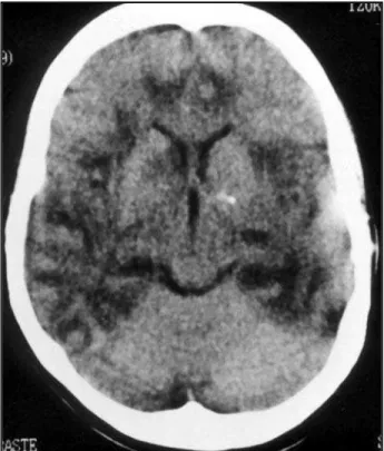
Despite treatment the patient evolved with mental status deterioration and coma but no sign of respirato-ry failure. On the fourth day of disease, she was transfer-red to our hospital and put on mechanical ventilation in the intensive care unit. On examination she could react to pain stimulation bilaterally. Her pupils measured 3/3 mm, the light reflex and oculocephalic reflex were intact. The deep tendon reflexes were brisk and a left Babinski sign could be easily elicited. A new head CT scan was performed demonstrating bilateral poorly defined areas of fronto-temporal hipodensities (Fig 1). Repeated lum-bar puncture disclosed CSF with 58 WBC (85% lympho-cytes); 1 RBC; protein concentration 285 mg/dl and glu-cose concentration 62 mg/dl. Gram stain, India ink and acid-fast bacilli staining were all negative. Blood cell count, coagulogram, blood urea nitrogen and creatinine concentrations were all within normal range values. Se-rology for human immunodeficiency virus, syphilis and hepatitis B were negative.
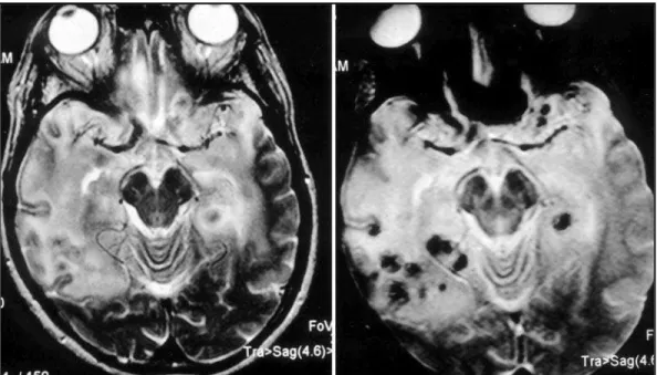
A presumptive diagnosis of HSE was made and the patient was treated with acyclovir (30 mg/Kg/ day). On the second intensive care day, she developed decerebrate posturing bilaterally and was given IV mannitol and put on hyperventilation. As serum and CSF enzyme-lin-ked immunoassay and CSF Polymerase Chain Reaction (PCR) for HSV 1 and 2 were negative, acyclovir was inter-rupted. Magnetic resonance imaging (MRI) of the brain was then performed revealing a T2-hyperintense sign in both frontal and temporal lobes as well as signs of hemorrhages (Fig 2).
The diagnosis of acute hemorrhagic leukoencepha-litis (AHLE) was made and the patient was treated with IV methylprednisolone (1 g/day for five days). She
recov-ered consciouness but exhibited global aphasia and assymetric tetraparesis, more severe in left side. She was discharged two months later following treatment of nosocomial pulmonary and urinary tract infections. On follow-up examination her neurological condition remains unchanged.
DISCUSSION
The diagnosis of ADEM is based on history of
acute onset focal neurological disturbances, fre-quently associated with mental changes following a febrile illness, usually an upper respiratory tract infection. Exclusion of a better explanation for the neurological symptoms is necessary. The main conditions to be considered in the differential dia-gnosis are other inflammatory disorders includ-ing vasculitis, ischemic vascular disease, tumors such as lymphoma and glioma, paraneoplastic disorders, infectious diseases and exposure to toxic agents1,8.
Our patient had no history or clinical evidence of any systemic disease except of an upper respira-tory infection antedating the onset of neurologi-cal signs. It may be significant that atypineurologi-cal pneumo-nia was suspected in our patient as infection by Mycoplasma pneumoniaehas already been descri-bed in association with ADEM and AHLE5,9,10.
Other disorders predominantly affecting the white-matter were ruled out on grounds of an
unremarkable past medical history. The diagnosis of multiple sclerosis, the most common among the demyelinating diseases, requires the presence of relapsing or progressive neurological disturban-ces with spatial dispersion11-13. More agressive
vari-ants of multiple sclerosis, such as Schilder’s disea-se and Balo’s concentric sclerosis, usually take months or years but rarely weeks to develop.
A more difficult differential diagnosis to be ru-led out in the current case was herpes simplex en-cephalitis (HSE). Clinical features, such as fever, de-lirium and the presence of focal neurological signs, cannot differentiate the two diseases. In fact, our patient presented many features commonly seen in HSE. The patient’s age would preferentially point to HSE14as the majority of ADEM cases occur in
chil-dren7. Typical neuroimaging findings in HSE include
abnormalities in the orbitofrontal and temporal lobe areas associated with variable degrees of mass effect, edema and, occasionally, hemorrhage15.
The T2-weighted images show signal hyperinten-sity in the orbitofrontal and temporal lobes15. Our
patient presented similar changes on her head CT scan and MRI studies, although its noteworthy that her first CT scan was normal (Fig 1 and 2). Interes-tingly, CT scans performed in a early stage of the HSE can also be normal. On the other hand, in ADEM CT scans are often normal early in the course of the illness, but over one-half of the patients develop white matter lucencies later on. MRI is more sensitive for diagnosing ADEM and
demon-strates areas of increased signal intensity on T2-weighted images, mainly in the white matter16.
Some of these areas exhibit gadolinium-enhance-ment on T1-weighted images16. However, in some
case reports of AHLE in the literature, MRI demons-trates a more diffuse compromise of the brain, affecting not only the white matter, but also exten-sive cortical areas like our patient17.
The patient’s initial CSF examination failed to demonstrate any abnormality. A repeated lumbar puncture, however, disclosed CSF with marked pleo-cytosis, increased protein concentration and nor-mal glucose concentration. This CSF profile may be seen in a number of conditions such as viral menin-gitis and encephalitis, as well as post-infectious en-cephalomyelitis. As the CSF PCR for herpes simplex virus was negative, the diagnosis of HSE was ru-led out. It is well established that CSF PCR for her-pes simplex virus has a high sensitivity (98%) and specificity (94%) being considered the method of choice for the diagnosis of HSE18,19. However, in
so-me circumstances a negative PCR assay can not ex-clude the diagnosis of HSE as pointed out by some authors20,21. One reason for negative PCR might be
an advanced stage of disease as CSF PCR becomes negative over time, especially in immunocompetent patients20. This seems unlikely in our case as the CSF
for PCR analysis was collected in the fourth day fol-lowing the onset of the neurological symtoms. Anyway, brain biopsy remains as the gold stan-dard method for the definite diagnosis of HSE20,21. Fig 2. Axial T2-weighted MR image demonstrates areas of T2-hyperintense sign in both frontal and
142 Arq Neuropsiquiatr 2004;62(1)
In a Brazilian series of 61 patients with clinical-ly suspected HSE, onclinical-ly 29.5% of them had the diagnosis of HSE confirmed by CSF PCR, while 29.5% received an alternative diagnosis, such as demye-linating diseases and other CNS infections (bacte-rial meningitis and neurocysticercosis)22. In the
re-maining patients, the diagnosis was undetermined. Only two patients had non-herpetic viral encephali-tis. This low frequency of non-herpetic viral ence-phalitis in that series probably reflects a non syste-matic search for CNS viruses22. However, even in a
larger Brazilian series of 383 patients with aseptic meningitis and encephalitis, the use of PCR pro-tocols for the presence of 17 infectious agents, in-cluding enteroviruses, herpes simplex virus, vari-cella zoster virus, Epstein-Barr virus (EBV), cytome-galovirus, measles and mumps virus, yielded a pos-itive result in just 12% of the patients23.
Interestin-gly, as 32.6% of patients in this series were known to have HIV infection, a large number of CSF sam-ples were positive for EBV, a virus whose role in pa-thogenesis of CNS disease still remains uncertain23.
It is therefore not probable that the search for oth-er viruses by PCR analysis of the patient’s CSF would turn out more revealing.
ADEM and AHLE are the only two diseases of the white matter with a monophasic clinical course associated with a rapidly progressive demyelina-tion. As both conditions share many features they are considered part of a spectrum of diseases rather than distinct entities6,7. The more severe and more
acute clinical symptomatology discriminates AHLE from ADEM8,24. In addition, MRI may establish the
diagnosis in the appropriate clinical setting17. Of
note is the fact that until recently AHLE was mis-diagnosed as a viral encephalitis17.
AHLE is a rare demyelinating disease first des-cribed by Hurst25in 1941. Although considered a
form of viral encephalitis for many years it is now considered a severe variant of ADEM. Until 1991, approximately 70 cases had been reported in the literature5,26. Rust7believes that AHLE is still
becom-ing less frequent with the development and disse-mination of safer programs of immunization. Its clinical features are usually very characteristic with previous history of a respiratory tract infection one or a few weeks before the onset of the neu-rological symptoms26. Following this prodromal
illness, neurological symptoms such as mental chan-ges, signs of meningeal irritation and variable fo-cal signs start abruptly in association with fever. Death from brain edema is common in the first
week26. Although there has been no established
guidelines for treatment of AHLE, the use of var-ious combinations of immunossupressive agents, including corticosteroids, immunoglobulin, plas-mapheresis and cyclofosfamide have been proved beneficial for some patients5,27,28. Occasionally
pa-tients may be discharged with no neurological sequelae5,26. Recently two case reports17,24
demons-trated that treatment with high-dose intravenous corticosteroid (e.g. methilprednisolone 1 g/day for 3 days24) was followed by full recovery. The authors
suggest that patients with AHLE must be put on high-dose corticosteroid therapy as early as possi-ble17,24. Unfortunately in spite of the use of this
ther-apy our patient remains with severe neurological deficits.
The present case illustrates that AHLE must be considered in patients with clinical and imaging sus-picion of HSE. Repeated CSF examination and ima-ging studies may provide evidences to establish the differential diagnosis and allow early and effective treatment.
REFERENCES
1. Tselis AC, Lisak RP. Acute disseminated encephalomyelitis and isolat-ed central nervous system demyelinative syndromes. Curr Opin Neurol 1995;8:227-229.
2. Victor M, Ropper AH. Multiple sclerosis and allied demyelinative dis-eases. In Adams and Victor’s Principles of Neurology, 7th edition. New York, McGraw Hill, 2001:954-982.
3. Reis F, Kobayashi E, Maciel EP, et al. Ressonância magnética e carac-terísticas clínicas em adultos com doenças desmielinizantes monofási-cas. Encefalomielite aguda disseminada ou uma variante de esclerose múltipla? Arq Neuropsiquiatr 1999;57:853-859.
4. Dale RC, Church AJ, Cardoso F, et al. Poststreptococcal acute dissemi-nated encephalomyelitis with basal ganglia involvement and auto-reactive antibasal ganglia antibodies. Ann Neurol 2001;50:588-595. 5. Seales D, Greer M. Acute hemorrhagic leukoencephalitis: a successful
recovery. Arch Neurol 1991;48:1086-1088.
6. Russell DS. The nosological unity of acute haemorrhagic leukoence-phalitis and acute disseminated encephalomyelitis. Brain 1955;78:369-376.
7. Rust RS. Multiple sclerosis, acute disseminated encephalomyelitis, and related conditions. Semin Pediatric Neurol 2000;7:66-90.
8. Case Records of the Massachusetts General Hospital. Case 1-1999. N Engl J Med 1999;340:127-135.
9. Donnet A, Dufour H, Gambarelli D, Bruder N, Pellissier JF, Grisoli F. Leucoencéphalite aiguë hémorragique et nécrosante de Weston Hurst. Rev Neurol (Paris) 1996;152:748-751.
10. Kumada S, Kusaka H, Okaniwa M, et al. Encephalomyelitis subse-quent to mycoplasma infection with elevated serum Gal C anti-body. Pediatr Neurol 1997;16:241-244.
11. Noseworthy JH, Lucchinetti C, Rodriguez M, Weinshenker BG. Multiple sclerosis. N Engl J Med 2000:343:938-952.
12. Dale RC, Sousa C, Chong WK, Cox TC, Harding B, Neville BG. Acute disseminated encephalomyelitis, multiphasic disseminated encephalo-myelitis and multiple sclerosis in children. Brain 2000;123:2407-2422. 13. Hartung HP, Grossman RI. ADEM: distinct disease or part of the MS
spectrum? Neurology 2001;56:1257-1260.
14. Whitley RJ, Lakeman FD. Herpes simplex virus infections of the cen-tral nervous system: therapeutic and diagnostic considerations. Clin Infect Dis 1995;20:414-420.
16. Caldemeyer KS, Harris TM, Smith RR, Edwards MK. Gadolinium enhancement in acute disseminated encephalomyelitis. J Comp Assit Tomogr 1991;15:673-675.
17. Klein CJ, Widjicks EFM, Earnest F. Full recovery after acute hemorrhag-ic leukoencephalitis (Hurst’s disease). J Neurol 2000;247:977-979. 18. Lakeman FD, Whitley RJ. Diagnosis of herpes simplex encephalitis:
appli-cation of polymerase chain reaction to cerebrospinal fluid from brain-biopsed patients and correlation with disease. J Infect Dis 1995;171:857-863.
19. Domingues RB, Lakeman FD, Mayo MS, Whitley RJ. Application of com-petitive PCR to cerebrospinal fluid samples from patients with herpes simplex encephalitis. J Clin Microbiol 1998;36:2229-2234.
20. DeBiasi RL, Kleinschmidt-DeMasters BK, Weinberg A, Tyler KL. Use of PCR for the diagnosis of herpesvirus infections of the central nerv-ous system. J Clin Virol 2002;25:S5-S11.
21. Sauerbrei A, Wutzler P. Laboratory diagnosis of the central nervous sys-tem infections caused by herpesviruses. J Clin Virol 2002;25:S45-S51. 22. Domingues RB, Pannuti CS, Fink MCD, Tsanaclis AMC. Diagnósticos
alternativos em pacientes com suspeita de encefalite por herpes
sim-plex e negativos à reação em cadeia por polimerase (PCR). Arq Neuropsiquiatr 2000;58:1073-1080.
23. Chesky M, Scalco R, Failace L, Read S, Jobim LF. Polymerase chain reac-tion for the laboratory diagnosis of aseptic meningitis and encephali-tis. Arq Neuropsiquiatr 2000;58:836-842.
24. Meilof JF, Hijdra A, Vermeulen M. Successful recovery after high-dose intravenous methylprednisolone in acute hemorrhagic leukoencephali-tis. J Neurol 2001;248:898-899.
25. Hurst EW. Acute haemorrhagic leuco-encephalitis: a previously unde-fined entity. Med J Aust 1941;2:1-6.
26. Geerts Y, Dehaene I, Lammens M. Acute hemorrhagic leucoencephali-tis. Acta Neurol Belg 1991;91:201-211.
27. Huang C, Chu N, Chen T, Shaw C. Acute haemorrhagic leucoencephali-tis with a prolonged clinical course. J Neurol Neurosurg Psychiatry 1988;51:870-874.