Antitumor Effect of the Essential Oil from Leaves of
Guatteria pogonopus
(Annonaceae)
byJose´ Eraldo do N. Fontesa),Rosana P. C. Ferrazb),Anny C. S. Brittob),Adriana A. Carvalhoc),
Manoel O. Moraesc),Claudia Pessoac),Emmanoel V. Costaa), andDaniel P. Bezerra*b)
a) Department of Chemistry, Federal University of Sergipe, Sa˜o Cristo´va˜o, Sergipe, Brazil b) Department of Physiology, Federal University of Sergipe, Sa˜o Cristo´va˜o, Sergipe, Brazil
(phone:þ55-79-2105-6644; e-mail: danielpbezerra@gmail.com)
c) Department of Physiology and Pharmacology, School of Medicine, Federal University of Ceara´, Fortaleza, Ceara´, Brazil
Guatteria pogonopus Martius, a plant belonging to the Annonaceae family, is found in the remaining Brazilian Atlantic Forest. In this study, the chemical composition and antitumor effects of the essential oil isolated from leaves ofG. pogonopuswas investigated. The chemical composition of the oil was determined by GC-FID and GC/MS analyses. Thein vitrocytotoxicity was evaluated against three different tumor cell lines (OVCAR-8, NCI-H358M, and PC-3M), and thein vivoantitumor activity was tested in mice bearing sarcoma 180 tumor. A total of 29 compounds was identified and quantified in the oil. The major compounds were g-patchoulene (13.55%), (E)-caryophyllene (11.36%), b-pinene (10.37%), germacrene D (6.72%), bicyclogermacrene (5.97%),a-pinene (5.33%), and germacrene B (4.69%). The essential oil, but neither (E)-caryophyllene norb-pinene, displayedin vitrocytotoxicity against all three tumor cell lines tested. The obtained averageIC50values ranged from 3.8 to 20.8mg/ml.
The lowest and highest values were obtained against the NCI-H358M and the OVCAR-8 cell lines, respectively. Thein vivotumor-growth-inhibition rates in the tumor-bearing mice treated with essential oil (50 and 100 mg/kg/d) were 25.3 and 42.6%, respectively. Hence, the essential oil showed significantin vitroandin vivoantitumor activity.
Introduction.
– The genus
Guatteria
(Annonaceae) comprises
ca.
300 species and is
distributed from southeastern Mexico to southern Brazil [1] [2]. Numerous biological
properties have been reported for the plants belonging to this genus, including
antioxidant [3], antimicrobial [4], antimalarial [5], antileishmanial [6], insecticide [7],
and cytotoxic effects [8]. In particular, cytotoxic activity has been found for
Guatteria
hispida
[8],
G. blepharophylla
[8],
G. boliviana
[9], and
G. friesiana
[10].
G. pogonopus
Martius
is a tree (4 – 10-m-tall) characterized by very large leaves,
which often have a rounded base. It was reported to grow in the Brazilian states of
Bahia, Esprito Santo, and Minas Gerais [11]. In addition, in this work, we were able to
find it in the Brazilian state of Sergipe. Several beneficial biological activities had been
reported for
Guatteria
species [3 – 10], but up to now, no chemical or pharmacological
scientific research was published for the species
G. pogonopus
.
Hence, in this study, the chemical composition of the essential oil from leaves of
G.
pogonopus
was characterized by GC-FID and GC/MS analyses, and the
in vitro
and
in
vivo
antitumor effects of this oil were investigated.
Results and Discussion.
–
Chemical Composition.
Hydrodistillation of
G.
pogono-pus
leaves gave a crude, red essential oil with a yield of 0.28
0.00% (
v
/
w
, based on the
dry weight of the plant material). As shown in
Table 1
, 29 compounds were identified
by GC-FID and GC/MS analyses. The major compounds were
g-patchoulene
(13.55%), (
E
)-caryophyllene (11.36%),
b-pinene (10.37%), germacrene D (6.72%),
bicyclogermacrene (5.97%),
a-pinene (5.33%), and germacrene B (4.69%). The
presence of some of these major compounds, along with spathulenol (3.57%), was also
detected in other essential oils from
Guatteria
species [4] [7] [12] [13], indicating that
G.
pogonopus
is a typical member of the Annonaceae family. However, recent chemical
Table 1. Chemical Composition of the Essential Oil Isolated from Leaves ofGuatteria pogonopus
Compound name and class RIexpa) RIlitb) Content [%]c)
(E)-Hex-3-enol 841 844 1.850.41
(Z)-Hex-2-enol 854 859 0.770.20
a-Pinene 930 932 5.331.10
b-Pinene 975 974 10.371.71
o-Cymene 1023 1022 0.580.10
Sylvestrene 1028 1025 2.520.26
b-Phellandrene 1029 1025 1.030.11
(E)-b-Ocimene 1046 1044 2.900.30
Linalool 1099 1095 0.400.03
d-Elemene 1339 1335 0.550.01
a-Ylangene 1373 1373 0.540.04
a-Copaene 1379 1374 0.580.03
b-Elemene 1391 1389 0.890.03
(E)-Caryophyllene 1421 1417 11.360.50
g-Elemene 1430 1434 3.550.03
cis-Muurola-3,5-diene 1450 1448 0.350.02
Spirolepechinene 1454 1449 1.900.10
a-Humulene 1457 1452 0.660.03
Germacrene D 1483 1484 6.720.15
g-Amorphene 1495 1495 0.730.04
Bicyclogermacrene 1497 1500 5.970.05
g-Patchoulene 1507 1502 13.550.41
d-Cadinene 1519 1522 0.980.03
Germacrene B 1561 1559 4.690.20
Spathulenol 1578 1577 3.570.27
Globulol 1588 1590 0.761.31
Viridiflorol 1591 1592 1.661.44
Rosifoliol 1595 1600 1.070.45
Alloaromadendrene epoxide 1634 1639 0.360.06
Alcohols 2.62
Monoterpenes 23.13
Sesquiterpenes 60.44
Total identified 86.19
a)RIexp: Retention index determined on a Rtx-5MS column rel. to thet
investigations have demonstrated significant variations in the chemical composition of
the essential oils from species belonging to this genus.
Maia et al.
[12] analyzed the chemical composition of four Amazon
Guatteria
species (
G. juruensis
,
G. microcalyx
,
G. poeppigiana
, and
G. blepharophylla
) and
observed variations in their chemical composition. Indeed, the main compounds found
in the leaf oil of
G. juruensis
were spathulenol (77.5%) and
a-pinene (4.5%). The leaf
oil of
G. microcalyx
was dominated by caryophyllene oxide (44.2%),
a-pinene
(11.9%), and
b-pinene (6.3%). The major constituents identified in the leaf oil of
G.
poeppigiana
were spathulenol (53.0%), kushinol (10.9%), and humulene epoxide II
(5.7%), whereas those found in the leaf oil of
G. blepharophylla
were caryophyllene
oxide (51.0%), humulene epoxide II (6.8%), and (
E
)-14-hydroxy-9-epicaryophyllene
(4.1%).
Aciole et al.
[7] also analyzed the chemical composition of three Amazon
Guatteria
species (
G. blepharophylla
,
G. friesiana
, and
G. hispida
) and found significant
differences in their chemical composition. Caryophyllene oxide (70.0%) predominated
in the essential oil from the leaves of
G. blepharophylla
, while
a-,
b-, and
g-eudesmol
(15.1, 52.0, and 24.0%, resp.) were the main compounds in the essential oil from the
leaves of
G. friesiana
. The major constituents identified in the leaf oil of
G. hispida
were
a- and
b-pinene (31.0 and 36.0%, resp.) and (
E
)-caryophyllene (21.0%). These results
are in agreement with those reported by
Costa et al.
[4].
Palazzo et al.
[13] analyzed the chemical composition of three
Guatteria
species
from Costa Rica and also observed differences in their chemical composition. The
essential oil from leaves of
G. costaricensis
was constituted mainly of
a- and
b-pinene
(36.3 and 48.2%, resp.) as well as (
E
)-caryophyllene (5.4%). The leaf oil of
G.
diospyroides
was composed principally of germacrene D (46.4%), (
Z
)-b-ocimene
(17.4%), (
E
)-b-ocimene (12.0%), and (
E
)-caryophyllene (10.3%). Germacrene D
predominated in the leaf oil of
G. oliviformis
(73.3%), but
a- and
b-pinene (3.4 and
4.4%, resp.) and bicyclogermacrene (4.5%) were also detected in considerable
amounts.
These significant variations in the major oil constituents as well as the varying
contents of all oil components of the various
Guatteria
species might be explained by
the different climate conditions of these regions. Nevertheless, the presence at high
concentrations of
a- and
b-pinene, (
E
)-caryophyllene, germacrene D, and
bicycloger-macrene appears to be a common characteristic of the essential oils of
Guatteria
species.
In vitro
Cytotoxicity.
The
in vitro
cytotoxicity of the essential oil isolated from the
leaves of
G. pogonopus
and its components (
E
)-caryophyllene and
b-pinene (
Fig. 1
)
was evaluated against three different human tumor cell lines,
i.e
., OVCAR-8,
NCI-H358M, and PC-3M, using the MTT assay.
Table 2
summarizes the
IC
50values for the
cytotoxic activity. The essential oil, but neither (
E
)-caryophyllene nor
b-pinene,
showed
in vitro
cytotoxicity against all tested tumor cell lines. The obtained average
promising [16 – 19]. Therefore, the
G. pogonopus
essential oil was considered to possess
potent cytotoxic activity. On the other hand, (
E
)-caryophyllene and
b-pinene were
regarded as compounds without cytotoxic potential (
IC
50>
5
m
g/ml). The cytotoxic
activity of (
E
)-caryophyllene and
b-pinene have previously been tested, and they
showed
IC
50values higher than 25
m
g/ml and of
ca
. 24
m
g/ml, respectively [8]. In
another study, (
E
)-caryophyllene showed cytotoxic activity against a renal
adenocar-cinoma (ACHN) and an amelanotic melanoma (C32) cell line with
IC
50values of
ca
.
20
m
g/ml [20]. Probably, the potent cytotoxic activity of the
G. pogonopus
essential oil
tested might be attributed to additive and/or synergic effects of its main and minor
constituents.
The cytotoxic activity of some essential oils from
Guatteria
species have been
previously investigated by us. The antitumor effects of
G. friesiana
essential oil have
been studied using both
in vitro
and
in vivo
models, and these effects seem to be
assigned to its main components
a-,
b-, and
g-eudesmol [10]. The essential oils isolated
from
G. blepharophylla
and
G. hispida
have also shown potent cytotoxic activity. In
contrast to the
G. friesiana
oil, but similar to the
G. pogonopus
oil, the association of the
activity of main and/or minor constituents seems to be responsible for their cytotoxic
activity [8].
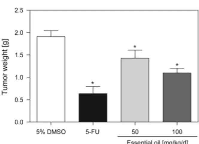
In vivo
Antitumor Activity.
For the study of the
in vivo
antitumor activity of
G.
pogonopus
leaf essential oil, mice were subcutaneously transplanted with sarcoma 180
cells and treated with oil, by the intraperitoneal route, once a day for seven consecutive
days. The effects of the essential oil on mice transplanted with sarcoma 180 tumor are
presented in
Fig. 2
. On the eighth day, the average tumor weight of the control mice was
1.91
0.14 g. In the presence of the essential oil (50 and 100 mg/kg/d), the average
Fig. 1.Chemical structures of (E)-caryophyllene andb-pineneTable 2. In vitroCytotoxic Activity of the Leaf Essential Oil ofGuatteria pogonopus
IC50[mg/ml]a)
OVCAR-8 NCI-H358M PC-3M
Essential oil 20.8 (16.1 – 26.9) 3.8 (2.6 – 5.5) 17.0 (12.3 – 23.4)
b-Pinene >5 >5 >5
(E)-Caryophyllene >5 >5 >5
Doxorubicinb) 1.2 (0.9 – 1.6) 0.9 (0.6 – 1.3) 1.6 (1.1 – 2.4)
a) TheIC
tumor weights were 1.43
0.18 and 1.10
0.11 g, respectively. Hence, the
tumor-growth-inhibition rates were 25.3 and 42.6%. The tumor inhibition was significant for
both doses compared to the control group (
p
<
0.05). At a dose of 25 mg/kg/d, the
positive control 5-fluorouracil (5-FU) reduced the tumor weight by 66.8% within the
same time period.
Some systemic toxicological parameters were also examined in the essential
oil-treated mice. The treatment with essential oil did not significantly affect the body mass,
the macroscopic structure of the organs (liver, kidney, and spleen), and the blood
leukocyte counts (
p
>
0.05, data not shown). However, anal ulcers were observed at the
end of the treatment in mice receiving essential oil at the dose of 100 mg/kg/d. In
contrast, the positive control 5-FU reduced the body weight of mice as well as the
spleen weight, and it induced a decrease in the total leukocytes (
p
<
0.05, data not
shown).
In conclusion, the essential oil isolated from leaves of
G. pogonopus
presented as
major constituents
g-patchoulene, (
E
)
-
caryophyllene,
b-pinene, germacrene D,
bicy-clogermacrene,
a-pinene, and germacrene B and showed significant
in vitro
and
in vivo
antitumor activity.
Experimental Part
Plant Material.TheGuatteria pogonopusleaves were collected in February 2012 in the Itabaiana Mountain National Park, Municipality of Itabaiana, Sergipe, Brazil (coordinates: 10845’16.8’’ S, 37820’32.7’’W). The leaves were obtained from a flowering and fructifying plant. The plant material was identified by Dr.Ana Paula do Nascimento Prata, a plant taxonomist from the Department of Biology, Federal University of Sergipe, Brazil, and a voucher specimen (No. 22793) has been deposited with the Herbarium of the Federal University of Sergipe.
Fig. 2. In vivoantitumor effect of the leaf essential oil ofGuatteria pogonopus. Mice were injected with sarcoma 180 tumor cells (2.0106cells/animal,s.c.) and treated by intraperitoneal administration of essential oil (50 and 100 mg/kg/d) or the positive control 5-fluorouracil (5-FU, 25 mg/kg/d) for seven consecutive days, starting one day after tumor implantation. The negative control group was treated with the vehicle used for the dilution of the tested substances (5% DMSO). Data are presented as mean
SEM of 8 – 12 animals. Significant differences compared to control group (ANOVA followed by
Chemical Compounds and Reagents. 5-Fluorouracil (5-FU, purity >99%), doxorubicin (purity >98%), and 3-(4,5-dimethylthiazol-2-yl)-2,5-diphenyl-2H-tetrazolium bromide (MTT) were purchased fromSigma Chemical Co. (St Louis, MO, USA).RPMI 1640Medium, fetal bovine serum, penicillin, and streptomycin were obtained fromCultilab(Campinas, SP, Brazil), and CO2was purchased fromWhite Martins(Rio de Janeiro, Brazil). The compounds (E)-caryophyllene (purity 86 %) andb-pinene (purity97 %) were obtained fromAldrich Chemical Company, Milwaukee, Wisconsin, USA. All other reagents were of analytical grade.
Cells.The cytotoxicity assay was performed using OVCAR-8 (ovarian adenocarcinoma), NCI-H358M (bronchoalveolar lung carcinoma), and PC-3M (metastatic prostate carcinoma) human tumor cell lines, all obtained from the National Cancer Institute, Bethesda, MD, USA. The cells were grown in
RPMI-1640 medium supplemented with 10% fetal bovine serum, 2 mm glutamine, 100mg/ml streptomycin, and 100 U/ml penicillin and incubated at 378in a 5% CO2atmosphere.
The sarcoma 180 tumor cells, which had been maintained in the peritoneal cavity ofSwissmice, were obtained from the Laboratory of Experimental Oncology, Federal University of Ceara´.
Animals.A total of 40 Swiss mice (males, 25 – 30 g) obtained from the central animal house of the Federal University of Sergipe, Brazil, were used. The animals were housed in cages with free access to food and water and kept under a standard light-dark cycle of 12 h (lights on at 6:00 a.m.). The animals were treated according to the ethical principles for animal experimentation of theSBCAL(Brazilian Association of Laboratory Animal Science), and the experimental protocol (No. 08/2012) was approved by the Animal Studies Committee of the Federal University of Sergipe.
Hydrodistillation of the Volatile Constituents.Portions ofG. pogonopusleaves (3200 g) were dried in a stove with circulating air at 408for 72 h and submitted to hydrodistillation for 4 h using aClevenger -type apparatus (Amitel, Sa˜o Paulo, Brazil) [21]. The essential oil was dried (anh. Na2SO4), and its yield in % (v/w) was calculated on the basis of the dry weight of the plant material. The essential oil was stored in a freezer until analyses. The hydrodistillation was performed in triplicate.
GC-FID and GC/MS Analyses.The GC-FID and GC/MS analyses were performed with aGC-2010 Plusand aGCMS-QP2010 Ultra(Shimadzu Corporation, Kyoto, Japan) apparatus, resp., equipped with anAOC-20i(Shimadzu) autosampler and anRtx-5MS(Restek) fused-silica cap. column (5% diphenyl/ 95% dimethylpolysiloxane; 30 m0.25 mm i.d., film thickness 0.25mm). The oven temp. was
programmed isothermal at 408 for 1.5 min, then rising from 40 to 2308at 48/min, and finally kept isothermal at 2308for 5 min (total analysis time, 54 min); carrier gas, He (99.999%; 1.2 ml/min); split ratio, 1 : 10; injection volume, 0.5ml of the essential oils in AcOEt (5.0 mg/ml).
The MS and FID data were simultaneously acquired employing a detector splitting system with a split-flow ratio of 4 : 1 (MS/FID). Restrictor tubes (capillary columns) of 0.62 m0.15 mm i.d. and 0.74 m0.22 mm i.d. were used to connect the splitter to the MS and the FID detector, resp. The injector and ion-source temp. were 250 and 2008, resp. MS Spectra were taken at 70 eV with a scan interval of 0.3 s over the mass range 40 – 350 Da. The FID temp. was set to 2508, and the gas supplies for the FID were H2, air, and He at flow rates of 30, 300, and 30 ml/min, resp.
The content of each constituent was estimated by FID peak-area normalization (%). The analyses of the essential oil were performed in triplicate.
Identification of the Oil Constituents.The essential oil components were identified by comparison of
i) their retention times (tR) with those of standard compounds analyzed under identical conditions,ii) their retention indices (RIs, determined on aRtx-5MScolumn rel. to thet
Rof a series ofn-alkanes, according toVan Den DoolandKratz[14]) with those published in the literature [14], andiii) their mass spectra with those listed in theNISTand Wiley mass spectral libraries and those published in the literature [15].
In vitroCytotoxicity Assay.The tumor cell growth was determined by the ability of living cells to reduce the yellow dye MTT to a purple formazan product, as described byMosmann[22]. For all experiments, cells were seeded in 96-well plates in 100ml of medium (0.7105cells/ml for adherent cells and 0.3106cells/ml for suspended cells). After 24 h, the essential oil or compounds to be tested (0.78 – 50mg/ml for the oil and 0.078 – 5mg/ml for isolated compounds) were dissolved in pure DMSO and added
control. At the end of incubation, the plates were centrifuged and the medium was replaced by 150ml
fresh medium containing 0.5 mg/ml MTT. After 3 h, the formazan product was dissolved in 150ml DMSO
and the absorbance was measured using a multiplate reader (DTX 880 Multimode Detector,Beckman Coulter Inc., Fullerton, CA, USA). The effects of the oil and test compounds were expressed as percentage of the absorbance of reduced dye at 595 nm of the control.
In vivoAntitumor-Activity Assay.Thein vivoantitumor effect was evaluated using sarcoma 180 ascites tumor cells and following protocols previously described [10] [16] [23] [24]. Ten-day old sarcoma 180 ascites tumor cells (2106cells per 500
ml) were implanted subcutaneously into the left hind groin of
mice. The essential oil was dissolved in 5% DMSO and given to mice intraperitoneally once a day for seven consecutive days. At the beginning of the experiment, the mice were divided into four groups of 8 – 12 animals as follows:Group 1, animals treated by injection of vehicle (5% DMSO;n¼12);Group 2, animals treated by injection of 5-FU (25 mg/kg/d;n¼10);Group 3, animals treated by injection of the essential oil (50 mg/kg/d;n¼10);Group 4, animals treated by injection of the essential oil (100 mg/kg/d;
n¼8). The treatments were started one day after tumor injection. The dosages were determined based on previous studies. On day eight, the animals were sacrificed by cervical dislocation, and the tumors were excised and weighed. The effects of the oil and test compounds were expressed as percent inhibition of tumor growth compared to the control (Group 1).
Systemic-Toxicity Evaluation.Body mass loss, organ weight alterations, and changes in the leukocyte counts were determined at the end of the in vivoantitumor-activity assay as previously described [10] [16] [24]. Peripheral blood samples of the mice were collected from the retro-orbital plexus under light ether anesthesia, and the animals were sacrificed by cervical dislocation. After sacrifice, the liver, kidney, and spleens were removed and weighed. For the hematological analysis, total leukocyte counts were determined by standard manual procedures using light microscopy.
Statistical Analysis. Data are presented as meanSEM (or SD) or as IC50 values with 95% confidence intervals (CI 95%) obtained by nonlinear regression. The differences between experimental groups were compared by ANOVA (analysis of variance) followed by theStudentNewmanKeulstest (p<0.05). All statistical analyses were performed using the GraphPad program (Intuitive Software for Science, San Diego, CA, USA).
This work was financially supported by Capes (Coordenadoria de Apoio a Pesquisa e Ensino Superior), CNPq (Conselho Nacional de Desenvolvimento Cientifico e Tecnolo´gico), FUNCAP (FundaÅa˜o Cearense de Apoio ao Desenvolvimento Cientfico e Tecnolo´gico), and FAPITEC/SE (FundaÅa˜o de Amparo a` Pesquisa e a` InovaÅa˜o Tecnolo´gica do Estado de Sergipe). The authors are also grateful to Prof. Dr.Ana Paula do Nascimento Prata, Department of Biology, Federal University of Sergipe, for the botanical identification.
REFERENCES
[1] K. Barringer,Ann. Missouri Bot. Gard.1984,71, 1186.
[2] R. H. J. Erkens, L. Y. T. Westra, P. J. M. Mass,Blumea2008,53, 467.
[3] E. V. Costa, M. L. Pinheiro, A. Barison, F. R. Campos, M. J. Salvador, B. H. Maia, E. C. Cabral, M. N. Eberlin,J. Nat. Prod.2010,73, 1180.
[4] E. V. Costa, S. D. Teixeira, F. A. Marques, M. C. T. Duarte, C. Delarmelina, M. L. B. Pinheiro, J. R. Trigo, B. H. L. N. Sales Maia,Phytochemistry2008,69, 1895.
[5] B. Weniger, R. Aragon, E. Deharo, J. Bastida, C. Codina, A. Lobstein, R. Anton,Pharmazie2000,
55, 867.
[6] J. E. Correa, C. H. Ros, A. del Rosario Castillo, L. I. Romero, E. Ortega-Barra, P. D. Coley, T. A. Kursar, M. V. Heller, W. H. Gerwick, L. C. Rios,Planta Med.2006,72, 270.
[7] S. D. G. Aciole, C. F. Piccoli, J. E. Duque L, E. V. Costa, M. A. Navarro-Silva, F. A. Marques, B. H. L. N. Sales Maia, M. L. B. Pinheiro, M. T. Rebelo,Rev. Colomb. Entomol.2011,37, 262. [8] S. S. Ribeiro, A. M. de Jesus, C. S. Dos Anjos, T. B. da Silva, A. D. Santos, J. R. de Jesus, M. S.
M. L. Pinheiro, A. P. Prata, A. F. Blank, R. Silva-Mann, V. R. Moraes, E. V. Costa, P. C. Nogueira, D. P. Bezerra,Planta Med.2012,78, 1601.
[9] V. Mahiou, F. Roblot, A. Fournet, R. Hocquemiller,Phytochemistry2000,54, 709.
[10] A. C. Britto, A. C. de Oliveira, R. M. Henriques, G. M. Cardoso, D. S. Bomfim, A. A. Carvalho, M. O. Moraes, C. Pessoa, M. L. Pinheiro, E. V. Costa, D. P. Bezerra,Planta Med. 2012,78, 409. [11] P. J. M. Maas, H. M. Kamer, L. Junikka, R. Mello-Silva, H. Rainer,Rodrigue´sia2001,52, 61. [12] J. G. S. Maia, E. H. A. Andrade, L. M. M. Carreira, J. Oliveira, J. S. Arau´jo,Flavour Fragrance J.
2005,20, 478.
[13] M. C. Palazzo, H. L. Wright, B. R. Agius, B. S. Wright, D. M. Moriarity, W. A. Haber, W. N. Setzer,
Rec. Nat. Prod.2009,3, 153.
[14] H. Van Den Dool, P. D. Kratz,J. Chromatogr., A1963,11, 463.
[15] R. P. Adams, Identification of Essential Oil Components by Gas Chromatography/Mass Spectrometry, 4th edn., Allured Publishing Corporation, Carol Stream, IL, 2007.
[16] D. P. Bezerra, C. Pessoa, M. O. Moraes, N. M. Alencar, R. O. Mesquita, M. W. Lima, A. P. Alves, O. D. Pessoa, J. H. Chaves, E. R. Silveira, L. V. Costa-Lotufo,J. Appl. Toxicol.2008,28, 599. [17] M. Suffness, J. M. Pezzuto, in Methods in Plant Biochemistry: Assays for Bioactivity, Ed. K.
Hostettmann, Academic Press, London, 1990, pp. 71 – 133.
[18] C. Pessoa, E. R. Silveira, T. L. Lemos, L. A. Wetmore, M. O. Moraes, A. Leyva,Phytother. Res.2000,
14, 187.
[19] L. V. Costa-Lotufo, E. R. Silveira, M. C. Barros, M. A. Lima, M. E. De Moraes, M. O. De Moraes, C. Pessoa,Planta Med.2004,70, 180.
[20] R. Tundis, M. R. Loizzo, M. Bonesi, F. Menichini, D. Dodaro, N. G. Passalacqua, G. Statti, F. Menichini,Nat. Prod. Res.2009,23, 1707.
[21] E. V. Costa, L. M. Dutra, H. C. R. De Jesus, P. C. L. Nogueira, V. R. S. Moraes, M. J. Salvador, S. C. H. Cavalcanti, R. L. C. Dos Santos, A. P. N. Prata,Nat. Prod. Commun.2011,6, 907. [22] T. Mosmann,J. Immunol. Methods1983,16, 55.
[23] D. P. Bezerra, F. O. Castro, A. P. N. N. Alves, C. Pessoa, M. O. Moraes, E. R. Silveira, M. A. S. Lima, F. J. M. Elmiro, L. V. Costa-Lotufo,Braz. J. Med. Biol. Res.2006,39, 801.
[24] D. P. Bezerra, F. O. Castro, A. P. Alves, C. Pessoa, M. O. de Moraes, E. R. Silveira, M. A. Lima, F. J. Elmiro, N. M. de Alencar, R. O. Mesquita, M. W. Lima, L. V. Costa-Lotufo,J. Appl. Toxicol.2008,
28, 156.