Cytotoxic properties of the leaf essential oils of
Guatteria blepharophylla
and
Guatteria hispida
(Annonaceae)
Rosana P. C. Ferraz,
aDiogo S. Bomfim,
aNanashara C. Carvalho,
bMilena B. P. Soares,
b,cMaria L. B. Pinheiro,
dEmmanoel V. Costa
eand Daniel P. Bezerra
b*
ABSTRACT: Guatteria blepharophylla Mart. (synonym Guatteriopsis blepharophylla Mart.) and Guatteria hispida (R.E. Fr.) Erkens & Maas (synonym Guatteriopsis hispidaR.E. Fr.) belong to the Annonaceae family and are found in the Brazilian and Colombian Amazon basin. Both species are popularly known as‘envira’ or‘envireira’. In the present study, the leaf essential oils ofG. blepharophylla(EOGB) andG. hispida(EOGH) were selected to investigate their cytotoxic effects. Tumour cell lines were treated with increasing concentrations of both essential oils for 72 h and analysed by a methyl-[3H]thymidine incorporation assay. The pro-apoptotic effect of these essential oils was assessed in HepG2 cells by morphological analysis (using haematoxylin/eosin staining and acridine orange/ethidium bromide staining),flow cytometry (cell membrane integrity and internucleosomal DNA fragmentation analysis) and a caspase-3 activation assay after 24 h incubation. Both essential oils displayed potent cytotoxicity in different tumour cell lines. EOGB showed IC50values from 6.03 to 16.46μg/ml for HepG2 and K562 cell lines, and EOGH showed IC50values from 5.45 to 24.89μg/ml for HepG2 and K562 cell lines, respectively. Cell morphologies consistent with apoptosis and a remarkable activation of caspase-3 were observed in the HepG2 cells treated with essential oils for 24 h. Significant increases in internucleosomal DNA fragmentation without altered membrane integrity were also found. In conclusion, both essential oils investigated were able to inhibit tumour cell proliferation and induce cell death by apoptosis pathways. Copyright © 2014 John Wiley & Sons, Ltd.
Keywords:Guatteria blepharophylla;Guatteria hispida; Annonaceae; cytotoxicity; apoptosis
Introduction
Annonaceae are a pan-tropical family of trees, shrubs and climbers with approximately 130 genera and 2500 species that are found predominantly in lowland tropical regions.[1] Numer-ous plants belonging to the Annonaceae family have been ex-tensively used for the treatment of diseases, as such tumours.[2]
Guatteria blepharophylla Mart. (synonym Guatteriopsis blepharophyllaMart.) andGuatteria hispida(R.E. Fr.) Erkens & Maas (synonymGuatteriopsis hispida R.E. Fr.) (Annonaceae family) are medicinal plants found in the Brazilian and Colombian Amazon basin. Both species are popularly known as‘envira’or‘envireira’. Some biological properties have been reported for these plants, including antimicrobial,[3]insecticidal[4]and cytotoxic[5,6]effects.
The chemical constituents of the essential oils of G. blepharophylla (EOGB) and G. hispida (EOGH) were previously evaluated by Acioleet al.[4]The main compound found in EOGB was caryophyllene oxide, and the main compounds found in EOGH were β-pinene, α-pinene and (E)-caryophyllene. Herein, the cytotoxic mechanism of EOGB and EOGH were assessed.
Materials and Methods
Plant Material and Isolation of the Essential Oils
The plant material preparation and the isolation of the essential oils of G. blepharophylla and G. hispida were performed as described by Acioleet al.[4]
Cells
Cytotoxicity was determined in tumour cells using HepG2 (human hepatocellular carcinoma), K562 (human chronic mye-locytic leukemia) and B16-F10 (mouse melanoma) cell lines, all of which were donated by the Hospital A.C. Camargo, São Paulo, SP, Brazil. Cells were maintained in the Roswell Park Memorial Institute-1640 (RPMI-1640; Gibco-BRL, Gaithersburg, MD, USA) in medium supplemented with 10% fetal bovine serum (Cultilab, Campinas, SP, Brazil), 2 mML-glutamine (Vetec
Química Fina, Duque de Caxias, RJ, Brazil) and 50μg/ml gentamycin
* Correspondence to: Daniel P. Bezerra, Fundação Oswaldo Cruz–Fiocruz,
Centro de Pesquisas Gonçalo Moniz, Rua Waldemar Falcão, 121, Candeal, 40296-710, Salvador, Bahia, Brazil. E-mail: [email protected]
a
Departamento de Fisiologia, Universidade Federal de Sergipe, São Cristóvão, Sergipe, Brazil
bCentro de Pesquisas Gonçalo Moniz, Fundação Oswaldo Cruz, Salvador,
Bahia, Brazil
c
Centro de Biotecnologia e Terapia Celular, Hospital São Rafael, Salvador, Bahia, Brazil
dDepartamento de Química, Universidade Federal do Amazonas, Manaus,
Amazonas, Brazil
e
Departamento de Química, Universidade Federal de Sergipe, São Cristóvão, Sergipe, Brazil
Received: 26 August 2013, Revised: 20 January 2014, Accepted: 8 February 2014 Published online in Wiley Online Library: 13 March 2014
(wileyonlinelibrary.com) DOI 10.1002/ffj.3199
(Novafarma, Anápolis, GO, Brazil). Adherent cells were harvested by treatment with 0.25% trypsin EDTA solution (Gibco-BRL). All cell lines were cultured in cell cultureflasks at 37°C in 5% CO2and
sub-cultured every 3–4 days to maintain exponential growth. Human lymphocyte cells were obtained by primary culture. Heparinized blood (from healthy, non-smoking donors who had not taken any drugs for at least 15 days prior to sampling) was collected, and peripheral blood mononuclear cells (PBMCs) were isolated by a standard protocol using a Ficoll density gradi-ent (Ficoll–Paque Plus; GE Healthcare Bio-Sciences AB, Sweden). The PBMC were washed and resuspended at a concentration of 0.3 × 10⁶cells/ml in RPMI-1640 medium supplemented with 20%
fetal bovine serum, 2 mMglutamine, and 50μg/ml gentamycin at
37°C with 5% CO2. In addition, concanavalin A (ConA; Sigma
Chemical Co., St Louis, MO, USA) was used as a mitogen to trigger cell division in T-lymphocytes. ConA (10μg/ml) was
added at the beginning of culture, and the cells were treated with the essential oils after 24 h.
For all experiments, cell viability was assessed by the trypan blue exclusion assay. Over 90% of the cells were viable at the beginning of the culture.
Cell Proliferation Assay
Cell growth was quantified by a methyl-[3H]thymidine incorpo-ration assay, as described by Griffiths and Sundaram,[7] with minor modifications. Methyl-[3H]thymidine is a radiolabelled DNA precursor incorporated into newly synthesized DNA, and the amount of incorporated methyl-[3H]thymidine is related to the rate of proliferation. In all experiments, a 100μL solution
of cells [0.7 × 10⁵ cells/ml for the adherent cells (HepG2 and B16-F10) or 0.3 × 10⁶ cells/ml for the suspended cells (K562 and PBMC)] was seeded in 96-well plates. After 24 h, the essen-tial oils (1.56–50μg/ml), dissolved in dimethyl sulfoxide (DMSO;
LGC Biotechnology, São Paulo, SP, Brazil), were added to each well and incubated for 72 h. Doxorubicin (purity 99.0%, doxoru-bicin hydrochloride; Eurofarma, São Paulo, SP, Brazil) was used as a positive control. Six hours before the end of the incubation time, 37 kBq (1μCi) of methyl-[3H]thymidine (PerkinElmer,
Boston, MA, USA) was added to each well. After this period, the cultures were harvested using a cell harvester (Brandel Inc., Gaithersburg, MD, USA) to determine the [3H]thymidine incorporation using the liquid scintillation cocktail Hidex Maxilight (PerkinElmer Life Sciences, Groningen, GE, Netherlands) and a plate CHAMELEON V multilabel Counter (Mustionkatu 2, TURKU, Finland) with MikroWin Hidex 2000 v. 4.38 software (Microtek Laborsysteme GmbH, Overath, Germany). The effect of the essential oils was quantified as the percentage of control radioactivity.
The following experiments were performed to study the cyto-toxic properties of the essential oils. In all experiments, 2 ml of a HepG2 cell solution (0.7 × 10⁵ cells/ml) was seeded in 24-well plates and incubated overnight to allow the cells to adhere to the plate surface. The cells were then treated for 24 h with the essential oils. The negative control was treated with the vehicle (0.1% DMSO) used for diluting the essential oils. Doxorubicin (1μg/ml) was used as a positive control.
Trypan Blue Exclusion Assay
After 24 h of essential oil exposure, the cells were harvested by trypsinization and stained with a solution of 0.2% trypan blue.
Viable cells were counted using a Neubauer chamber and the formula: cell number/ml =A×B× 104, whereAandBrepresent the average number per corner square and the dilution factor from trypan blue, respectively.
Morphological Analysis Using Fluorescence Microscopy
After 24 h of essential oil exposure, the cells were harvested by trypsinization, pelleted and resuspended in 25μl saline.
Thereafter, a 1μl aqueous solution of acridine orange (AO; Sigma
Chemical Co) and ethidium bromide (EB; Sigma Chemical Co.) (AO/EB, 100μg/ml) was added, and the cells were observed
under afluorescence microscope using a ×60 objective (=600× total magnification) (Olympus BX41, Tokyo, Japan). Three hundred cells were counted per sample and classified as viable, apoptotic or necrotic cells, as previously described.[8]
Morphological Analysis with Haematoxylin–Eosin Staining
After 24 h of essential oil exposure, the cells were harvested by trypsinization, transferred to cytospin slides,fixed with methanol for 30 s, and stained with haematoxylin–eosin. Morphological changes were examined using light microscopy with a ×60 objective (= 600× total magnification) (Olympus BX41) using Image-Pro Express software (Media Cybernetics, Inc. Silver Spring, MD, USA).
Cell Membrane Integrity
After 24 h of essential oil exposure, the cells were harvested by trypsinization and stained with a solution of 50μg/ml propidium
iodide in phosphate-buffered saline. The cellfluorescence was determined by flow cytometry (using the FL-2 channel) in a FACSCalibur cytometer (Becton Dickinson, San Diego, CA, USA) with CellQuest software (BD Biosciences, San Jose, CA, USA). Ten thousand events were evaluated per experiment, and the cellular debris was omitted from the analysis.
Internucleosomal DNA Fragmentation Analysis
Cells were harvested in a lysis solution containing 0.1% Triton X-100 (Sigma Chemical Co.) and 2μg/ml propidium iodide
(BioSource, Camarillo, CA, USA), as previously described.[9]The cellfluorescence was determined byflow cytometry (using the FL-2 channel) in a FACSCalibur cytometer (Becton Dickinson) with CellQuest software (BD Biosciences). Ten thousand events were evaluated per experiment, and the cellular debris was omitted from the analysis.
Caspase-3 Activation Assay
A caspase-3/CPP32 colorimetric assay kit (BioVision Inc., Milpitas, CA, USA) was used to investigate the caspase-3 activation in the treated cells based on the cleavage of Asp-Glu-Val-Asp (DEVD)-pNA. Briefly, the cells were lysed by incubation with cell lysis buffer on ice for 10 min and then centrifuged at 10 000×gfor 1 min. To each reaction mixture, 50μl of cell lysate (100–200μg total
pro-tein) was added. Enzyme reactions were carried out in a 96-well
oils was quantified by comparing the absorbance of pNA from a treated sample with that from an untreated sample.
Statistical Analysis
The data are presented as the mean ± SEM or as the IC50values
and their 95% confidence intervals (95% CI) as obtained by non-linear regression from four or six experiments. Differences among the experimental groups were compared by one-way analysis of variance (ANOVA) followed by a Newman–Keuls test (p<0.05). All analyses were carried out using the GRAPHPAD
program (Intuitive Software for Science, San Diego, CA, USA).
Results and Discussion
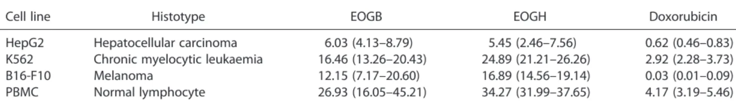
In the present study, both essential oils displayed potent cytotoxicity in different tumour cell lines. Table 1 shows the measured IC50 values. EOGB showed IC50 values from 6.03 to
16.46μg/ml for the HepG2 and K562 cell lines, and EOGH
showed IC50 values from 5.45 to 24.89μg/ml for the HepG2
and K562 cell lines, respectively. Doxorubicin, used as a positive control, showed IC50values from 0.03 to 2.92μg/ml for the
B16-F10 and K562 cell lines, respectively. According to criteria adopted by the American National Cancer Institute and our cyto-toxic screening programme, a crude extract/oil that shows IC50
values below 30μg/ml and a lead compound that shows IC50
values below 1μg/ml in tumour cell line assays are considered
promising for anticancer drug development.[10] Thus, EOGB and EOGH showed promising results. These essential oils have been previously reported as cytotoxic agents, with IC50 values
below 25μg/ml. The main compounds found in EOGB
(caryophyllene oxide) and EOGH (β-pinene, α-pinene and (E )-caryophyllene) were also previously tested on tumour cell lines, with onlyβ-pinene showing weak cytotoxic activity, suggesting that the minor compounds in the oils or associations among the major compounds in the oils are responsible for their cytotoxic activity.[6]In another study, (E)-caryophyllene showed cytotoxic activity, with an IC50 value of ~20μg/ml to tumour
cells, but was not cytotoxic to normal cells.[11,12]
Cancer therapies using cytotoxic agents commonly exhibit acute toxic side effects due to the non-selective activity of the cytotoxic agents, resulting in damage to normal (non-tumour) cells during normal replication. Therefore, the cytotoxicity of the essential oils was also evaluated in normal cells (PBMCs). The results, presented in Table 1, showed that EOGB and EOGH were also cytotoxic to normal cells, suggesting only a low selectively for tumour cells. Doxorubicin, a clinically useful chemotherapy agent, also presents cytotoxicity to normal cells.
All subsequent experiments were conducted on the HepG2 cell line to study the cytotoxic properties of the essential oils on this cell line. For this, cells were incubated for 24 h with the essential oils at concentrations of 2.5 and 5.0μg/ml. These
concentrations were chosen based on their IC50 values in this
cell line (6.03μg/ml for EOGB and 5.45μg/ml for EOGH).
EOGB and EOGH reduced the number of viable HepG2 cells in a concentration-dependent manner (p<0.05, Figure 1A), as
mea-sured by the trypan blue exclusion method. As examined by AO/ EB staining, an increasing number of apoptotic cells were observed (p<0.05, Figure 1B). No significant increase in the percentage of
necrotic cells was observed (p>0.05, date not shown). Neither
es-sential oil caused membrane disruption at any concentration tested (p>0.05, date not shown). EOGB and EOGH treatment significantly
increased the percentage of DNA fragmentation (p<0.05,
Figure 1C). These modifications are also compatible with apoptotic cells. In addition, a remarkable activation of caspase-3 was recorded in lysates from the HepG2 cells treated with EOGB and EOGH (p<0.05, Figure 1D), suggesting caspase-mediated apoptotic cell
death. In morphological analyses with haematoxylin–eosin staining, EOGB- and EOGH-treated cells also presented morphologies consis-tent with apoptosis, including abundant vacuoles, reductions in cell volume, chromatin condensation and fragmentation of the nuclei (Figure 2). Thus, this study presents for thefirst time the pro-apoptotic effects of EOGB and EOGH. Doxorubicin, used as a positive control, also showed apoptotic characteristics.
Some studies have also reported cytotoxic activity for plants belonging to the Guatteria genus, such as G. boliviana,[13] G. friesiana[14] and G. pogonopus[15]; however, the compounds isolated from other Guatteria genus are different from the compounds found in the leaf essential oils from G. blepharophylla and G. hispida. Mahiou et al.[13] demonstrated that puertogaline B, (+)-guatteboline, ( )-antioquine, tiliageine, funiferine, sepeerine and pangkorimine isolated from G. bolivianastem bark present potent cytotoxic activity in KB cells. The antitumour effects of the leaf essential oil ofG. friesianahas been reported in bothin vitro andin vivo models, and these effects appear to be assigned to the oil’s main components,
α-,β- andγ-eudesmol.[14]These eudesmol isomers have shown promising anticancer potential, with α- and γ-eudesmol exhibiting stronger cytotoxic and pro-apoptotic activity than
β-eudesmol.[16–19] The antitumour effects of the leaf essential oil of G. pogonopus have also been reported in both in vitro
andin vivomodels, but, unlike G. friesiana, these effects were not assigned to the main components, γ-patchoulene, (E )-caryophyllene andβ-pinene.[15]
As cited above, the cytotoxicity of the leaf essential oils from G. blepharophylla(caryophyllene oxide) andG. hispida(β-pinene,α-pinene and (E)-caryophyllene) was also not assigned to their main components.[6]
Table 1. Cytotoxic activity of the leaf essential oils ofGuatteria blepharophylla(EOGB) andGuatteria hispida(EOGH) on tumour cells and normal cells
Cell line Histotype EOGB EOGH Doxorubicin
HepG2 Hepatocellular carcinoma 6.03 (4.13–8.79) 5.45 (2.46–7.56) 0.62 (0.46–0.83) K562 Chronic myelocytic leukaemia 16.46 (13.26–20.43) 24.89 (21.21–26.26) 2.92 (2.28–3.73) B16-F10 Melanoma 12.15 (7.17–20.60) 16.89 (14.56–19.14) 0.03 (0.01–0.09) PBMC Normal lymphocyte 26.93 (16.05–45.21) 34.27 (31.99–37.65) 4.17 (3.19–5.46)
The data are presented as the IC50values, inμg/ml, and their 95% confidence interval obtained by non-linear regression from four or
six experiments by a methyl-[3H]thymidine incorporation assay after a 72 h incubation. Doxorubicin was used as the positive control.
In conclusion, both essential oils investigated were able to inhibit tumour cell proliferation and induce cell death by apoptosis pathways.
Acknowledgements
This work was financially supported by the Brazilian agencies CAPES, CNPq, FAPESB and FAPITEC/SE. The authors thank Elisalva T. Guimarães and Daniele Brustolim for their assistance
inflow cytometry data acquisition. The English was edited by American Journal Experts (key#75A5-1C0C-DEFD-E9DC-0971).
References
1. J. E. Richardson, L. W. Chatrou, J. B. Mols, R. H. J. Erkens, M. D. Pirie,
Philos. Trans. R. Soc. B2004,359, 1495.
2. M. O. Soladoye, N. A. Amusa, S. O. Raji-Esan, E. C. Chukwuma, A. A.
Taiwo,Ann. Biol. Res.2010,1, 261.
A B
C D
Figure 1. Effect of the leaf essential oils ofGuatteria blepharophylla(EOGB) andGuatteria hispida(EOGH) on the viability of HepG2 hepatocellular
car-cinoma cells after 24 h incubation. (A) Cell viability measured by the trypan blue dye exclusion method. (B) Cell viability measured byfluorescence
mi-croscopy using acridine orange/ethidium bromide; viable cells (white bar), apoptotic cells (grey bar) and necrotic cells (black bar). (C) Internucleosomal
DNA fragmentation determined byflow cytometry using propidium iodide and Triton X-100. (D) Caspase-3 activation measured by colorimetric assay.
The negative control was treated with the vehicle (0.1% DMSO) used for diluting the tested substance. Doxorubicin (Dox, 1μg/ml) was used as a
pos-itive control. The data are presented as the mean values ± SEM from four or six experiments. *p<0.05 compared to the negative control by ANOVA
followed by Student–Newman–Keuls test
A C E
B FD
25 µm
Figure 2. Effect of the leaf essential oils ofGuatteria blepharophylla(EOGB) andGuatteria hispida(EOGH) on the cell morphology of HepG2
hepato-cellular carcinoma cells. The cells were stained with haematoxylin–eosin and analysed by optical microscopy after 24 h incubation with EOGB at
con-centrations of 2.5 (C) and 5.0μg/ml (D) and EOGH at concentrations of 2.5 (E) and 5.0μg/ml (F). The negative control (A) was treated with the vehicle
(0.1% DMSO) used for diluting the tested substances. Doxorubicin (1μg/ml) was used as a positive control (B). Black arrows show nuclear DNA
frag-mentation or chromatin condensation
3. E. V. Costa, S. D. Teixeira, F. A. Marques, M. C. Duarte, C. Delarmelina,
M. L. Pinheiro, J. R. Trigo, B. H. Sales Maia,Phytochemistry2008,69, 1895.
4. S. D. G. Aciole, C. F. Piccoli, L. J. E. Duque, E. V. Costa, M. A. Navarro-Silva,
F. A. Marques, B. H. L. N. S. Maia, M. L. B. Pinheiro, M. T. Rebelo,Rev.
Colomb. Entomol.2011,37, 262.
5. E. V. Costa, F. A. Marques, M. L. B. Pinheiro, R. M. Braga, C. Delarmelina,
M. C. T. Duarte, A. L. T. G. Ruiz, J. E. Carvalho, B. H. L. N. S. Maia,J. Braz.
Chem. Soc.2011,22, 1111.
6. S. S. Ribeiro, A. M. Jesus, C. S. Anjos, T. B. Silva, A. D. C. Santos, J. R. Jesus, M. S. Andrade, T. S. Sampaio, W. F. Gomes, P. B. Alves, A. A. Carvalho, C. Pessoa, M. O. Moraes, M. L. B. Pinheiro, A. P. N. Prata, A. F. Blank, R. Silva-Mann, V. R. S. Moraes, E. V. Costa, P. C. L. Nogueira, D. P. Bezerra,
Planta Med.2012,78, 1601.
7. M. Griffiths, H. Sundaram,Methods Mol. Biol.2011,731, 451.
8. P. Brousseau, Y. Payette, H. Tryphonas, B. Blakley, D. Flipo, M. Fournier. InManual of Immunological Methods, M. Beudet, E. Kouassi, P. Lapierre,
I. Voccia (eds.). CRC Press LLC: Boca Raton,1999, pp. 28–29.
9. G. Nicolletti, M. C. Magliorati, F. Pagliacci, C. Grignani, C. Riccardi,
J. Immunol. Methods1997,139, 217.
10. M. Suffness, J. M. Pezzuto. InMethods in Plant Biochemistry: Assays for
Bio-activity, K. Hostettmann (ed.). Academic Press: London,1990, pp. 71–133.
11. R. Tundis, M. R. Loizzo, M. Bonesi, F. Menichini, D. Dodaro,
N. G. Passalacqua, G. Statti, F. Menichini,Nat. Prod. Res.2009,
23, 1707.
12. E. Amiel, R. Ofir, N. Dudai, E. Soloway, T. Rabinsky, S. Rachmilevitch,
Evid. Base Compl. Alt. Med.2012,2012, 872394.
13. V. Mahiou, F. Roblot, A. Fournet, R. Hocquemiller, Phytochemistry
2000,54, 709.
14. A. C. Britto, A. C. de Oliveira, R. M. Henriques, G. M. Cardoso, D. S. Bomfim,
A. A. Carvalho, M. O. Moraes, C. Pessoa, M. L. Pinheiro, E. V. Costa,
D. P. Bezerra,Planta Med.2012,78, 409.
15. J. E. N. Fontes, R. P. C. Ferraz, A. C. S. Britto, A. A. Carvalho, M. O.
Moraes, C. Pessoa, E. V. Costa, D. P. Bezerra,Chem. Biodivers.2013,
10, 722.
16. T. J. Hsieh, F. R. Chang, Y. C. Chia, C. Y. Chen, H. F. Chiu, Y. C. Wu,
J. Nat. Prod.2001,64, 616.
17. M. Ben Sghaier, I. Skandrani, N. Nasr, M. G. Franca, L. Chekir-Ghedira,
K. Ghedira,Environ. Toxicol. Pharmacol.2011,32, 336.
18. Y. Li, T. Li, , C. Miao, J. Li, W. Xiao, E. Ma,Phytother. Res.2013,27, 338.
19. D. S. Bomfim, R. P. C. Ferraz, N. C. Carvalho, M. B. P. Soares,
M. L. B. Pinheiro, E. V. Costa, D. P. Bezerra, Basic Clin.
Pharmacol. Toxicol.2013,113, 300.