Submitted17 April 2016 Accepted 10 June 2016 Published14 July 2016
Corresponding authors Han-Xiang An,
anhanxiang@yahoo.com Yun Zhang, zhangy@fjirsm.ac.cn
Academic editor Praveen Arany
Additional Information and Declarations can be found on page 11
DOI10.7717/peerj.2203
Copyright 2016 Zhou et al.
Distributed under
Creative Commons CC-BY 4.0
OPEN ACCESS
Association between aberrant APC
promoter methylation and breast cancer
pathogenesis: a meta-analysis of 35
observational studies
Dan Zhou1,2, Weiwei Tang3, Wenyi Wang3, Xiaoyan Pan3, Han-Xiang An3and
Yun Zhang1,2
1Department of Translational Medicine, Xiamen Institute of Rare Earth Materials, Xiamen, China 2Department of Translational Medicine, Key Laboratory of Design and Assembly of Functional
Nanostructures, Fujian Provincial Key Laboratory of Nanomaterials, Fujian Institute of Research on the Structure of Matter, Fuzhou, China
3Department of Medical Oncology, The First Affiliated Hospital of Xiamen University, Xiamen, China
ABSTRACT
Background. Adenomatous polyposis coli (APC) is widely known as an antagonist of
the Wnt signaling pathway via the inactivation ofβ-catenin. An increasing number of studies have reported that APC methylation contributes to the predisposition to breast cancer (BC). However, recent studies have yielded conflicting results.
Methods. Herein, we systematically carried out a meta-analysis to assess the correlation between APC methylation and BC risk. Based on searches of the Cochrane Library, PubMed, Web of Science and Embase databases, the odds ratio (OR) with 95% confidence interval (CI) values were pooled and summarized.
Results. A total of 31 articles involving 35 observational studies with 2,483 cases and 1,218 controls met the inclusion criteria. The results demonstrated that the frequency of APC methylation was significantly higher in BC cases than controls under a random effect model (OR=8.92, 95% CI [5.12–15.52]). Subgroup analysis further confirmed the reliable results, regardless of the sample types detected, methylation detection methods applied and different regions included. Interestingly, our results also showed that the frequency of APC methylation was significantly lower in early-stage BC patients than late-stage ones (OR=0.62, 95% CI [0.42–0.93]).
Conclusion. APC methylation might play an indispensable role in the pathogenesis of
BC and could be regarded as a potential biomarker for the diagnosis of BC.
SubjectsGenetics, Epidemiology, Oncology, Women’s Health Keywords Breast cancer, APC, Methylation, Meta-analysis
INTRODUCTION
reproductive, hormonal and environmental factors, have been associated with an increased incidence of BC (Harrison et al.,2015). Previous studies have reported that early detection using mammography is effective and can improve the overall survival rate (Brooks et al.,
2010). However, false positive mammograms always result in the diagnosis and over-treatment of developing BC. Therefore, no acknowledged biomarker has yet been proven to be sufficiently sensitive and specific for routine use in clinical diagnosis.
Epigenetic as well as genetic alterations are both stable and heritable and occur in tumor suppressor genes involved in tumourigenesis. The most common epigenetic alteration involving aberrant DNA methylation, a reliable and sensitive biomarker for nearly all types of cancer including breast cancer, often leads to the transcriptional silencing of tumor suppressor genes (Zmetakova et al.,2013). Several studies have demonstrated that tumor DNA derived from malignant cells can be detected in various bodily fluids and serum of BC patients and can potentially serve as a non-invasive diagnostic material (Martínez-Galán et al.,2014). A growing number of tumor suppressor genes has been shown to be directly involved in cell cycle regulation, DNA repair, cell signal transduction and angiogenesis (Dumitrescu,2012). Notably, the promoter methylation of genes involved in the canonical Wnt signaling pathway, which regulates cell differentiation, proliferation and homeostasis, are observed more often in BC patients compared with cancer-free controls (Klarmann, Decker & Farrar,2014).
The adenomatous polyposis coli (APC) gene is widely known as an antagonist of the Wnt signaling pathway via the inactivation ofβ-catenin, which is regarded as a transcriptional activator (Virmani et al.,2001). The APC gene, located at chromosome 5q21–5q22, was originally implicated in colorectal cancer (Van der Auwera et al.,2008). The inhibition or down-regulation of APC expression through APC promoter methylation contributes to the formation of colorectal cancer (Ashktorab et al.,2013). Similar to the findings in colorectal cancer, APC promoter methylation is associated with various early- or late-stage human malignancies, including BC (Matsuda et al.,2009). The promoter hypermethylation of APC is most often related to the nuclear accumulation ofβ-catenin, which may result in the loss of cell growth control (Sparks et al.,1998). Thus, APC promoter methylation, which acts as a non-invasive biomarker, can be used to distinguish BC patients from cancer-free controls. However, recent studies have yielded conflicting results with regard to the significant association between APC methylation and BC pathogenesis.Wojdacz et al.(2011a) reported that there was no significant difference in the frequency of APC methylation in peripheral blood leukocyte DNA between BC patients and cancer-free controls.Cho et al.(2010) also showed that the APC gene was rarely hypermethylated in blood DNA in BC patients.
Given these controversial results, we conducted this comprehensive meta-analysis of the current observational studies to evaluate the association between the aberrant methylation of the APC promoter and increased BC risk.
MATERIALS & METHODS
Search strategylower data limits were imposed; only abstracts, unpublished and incomplete studies were excluded. Titles, abstracts of potential references and reference lists from relevant studies were carefully checked. We performed the search strategy using the following search terms and their various combinations: ‘‘APC,’’ ‘‘Adenomatous polyposis coli,’’ ‘‘methylation,’’ ‘‘breast cancer,’’ ‘‘breast neoplasm’’ and ‘‘mammary carcinoma.’’
Selection criteria
The studies included in the present meta-analysis addressed the association between APC methylation and increased BC risk. Our inclusion criteria were as follows: (1) provided sufficient data on the frequency of APC methylation in BC patients and controls; (2) original observational studies in full-text form; and (3) when several studies overlapped, the most recent or large-scale article was selected. The following were exclusion criteria: (1) data based on reviews, animal models, case reports or cell line studies; (2) studies lacking key information necessary for calculations; (3) duplicated studies; and (4) studies including BC patients or controls who underwent radiotherapy and chemotherapy which may influence APC promoter methylation levels.
Data extraction
The relevant data were extracted from the eligible studies independently by two authors (D Zhou and WW Tang). Differing opinions, if any, were resolved by discussion in accordance with the original literature. The following information was extracted in a predefined table: the name of the first author, the year of publication, the country of origin, the sample type, the experimental methods used to detect APC methylation, sample size, tumor stage, tumor grade and APC methylation frequencies. Additionally, we classified stage 0, I and II as early-stage BC and stage III and IV as late-stage BC, as confirmed by the AJCC staging system. Furthermore, grades I and II were combined as low-grade BC; grade III was regarded as high-grade BC. This meta-analysis was performed following the statement of preferred reporting items set by the PRISMA Group (File S1) (Moher et al.,2009).
Statistical analysis
according to region, experimental methods for detecting APC methylation, and sample types in order to explore the potential origin of inter-study heterogeneity. In addition, we conducted a sensitivity analysis by removing a single study to examine the stability of the results. The funnel plot, Begg’s test and Egger’s test were investigated in order to determine the degree of publication bias. The treatment effect was plotted against a measure of study size in the funnel plot. When publication bias was present, the shape of the funnel plot was asymmetric. Trim and fill analysis was used to estimate the number of potential missing studies resulting from the asymmetry of the funnel plot.
RESULTS
Study selection and characteristics
The selection process is displayed as a flow chart inFig. 1based on the search strategies as previously described. After a careful initial search of the abstracts, 74 potentially relevant articles were identified excluding 1 duplicate and 93 irrelevant studies. Then, we reviewed the full text articles. Among these studies, 43 were excluded (21 articles did not design a control group; 9 articles focused on BC cell lines; 8 articles lacked available data; and 5 articles were reviews). Finally, 31 studies published from 2001 to 2016 involving 35 studies were included in this systematic meta-analysis (PubMed 19, Web of Science 10, Embase 2).
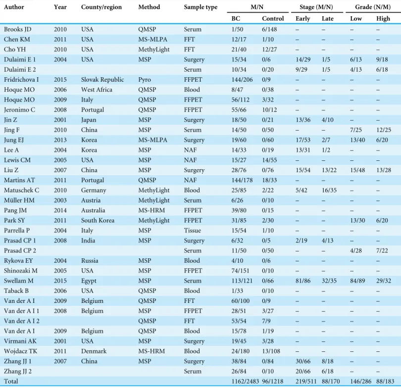
The general characteristics of eligible studies were summarized and displayed inTable 1. A total of 2,483 BC patients and 1,218 controls were employed in multiple countries or regions including Asia (n=10) (Jin et al.,2001;Jing et al.,2010;Jung et al.,2013;Lee et al.,2004;Liu et al.,2007;Park et al.,2011b;Prasad et al.,2008;Zhang et al.,2007), Europe (n=13) (Fridrichova et al.,2015;Hoque et al.,2009;Jeronimo et al.,2008;Martins et al.,
2011;Matuschek et al.,2010;Muller et al.,2003;Parrella et al.,2004;Rykova et al.,2004;Van der Auwera et al.,2009a;Van der Auwera et al.,2009b;Van der Auwera et al.,2008;Wojdacz et al.,2011b), Africa (n=2) (Hoque et al.,2006;Swellam et al.,2015), North America (n=9) (Brooks et al.,2010;Chen et al.,2011;Cho et al.,2010;Dulaimi et al.,2004;Lewis et al.,2005;Shinozaki et al.,2005;Taback et al.,2006;Virmani et al.,2001) and Oceania (n=1) (Pang et al.,2014). Furthermore, the methylated APC levels in BC patients and controls were examined with 6 methods. Of these methods, methylation specific PCR (MSP) was adopted in 17 studies, quantitative real-time MSP (QMSP) was used in 9 studies, methylation specific-multiplex ligation-dependent probe amplification (MethyLight) was used in 4 studies, methylation specific-multiplex ligation-dependent probe amplification (MS-MLPA) was employed in 2 studies, methylation-sensitive high-resolution melting analysis (MS-HRM) was used in 2 studies and pyrosequencing was used in only 1 study. Furthermore, BC tissues (i.e., fresh frozen tissues, formalin fixed paraffin-embedded tissues and tissues from surgery), samples derived from blood (i.e., blood cells and serum) and needle aspirated fluid (NAF) were enrolled to assess the methylation levels of the APC promoter.
Meta-analysis
Figure 1 Flow chart of the collection of studies for this meta-analysis.
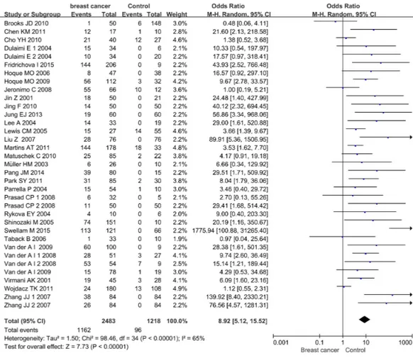
among the included studies (p<0.00001,I2=65%), the random effect model was adopted to evaluate the combined effects of APC promoter methylation. The overall analysis indicated that the frequency of APC promoter methylation was remarkably higher in BC patients than in cancer-free controls. The combined OR for 35 included relevant studies showed that APC methylation was significantly correlated with increased BC risk and the absence of APC expression played an important role in BC pathogenesis (OR=8.92, 95% CI [5.12–15.52]).
Sensitivity analysis
A sensitivity analysis was conducted by omitting one individual study every time to evaluate the stability of the pooled OR and to choose the heterogeneous study. As shown inFig. 3, the combined OR between APC methylation and increased BC risk was indeed reliable without heterogeneous studies.
Subgroup analysis
Table 1 General characteristics of eligible studies.
Author Year County/region Method Sample type M/N Stage (M/N) Grade (N/M)
BC Control Early Late Low High
Brooks JD 2010 USA QMSP Serum 1/50 6/148 – – – –
Chen KM 2011 USA MS-MLPA FFT 12/17 1/10 – – – –
Cho YH 2010 USA MethyLight FFT 21/40 12/27 – – – –
Dulaimi E 1 2004 USA MSP Surgery 15/34 0/6 14/29 1/5 6/13 9/18
Dulaimi E 2 Serum 10/34 0/20 9/29 1/5 4/13 6/18
Fridrichova I 2015 Slovak Republic Pyro FFPET 144/206 0/9 – – – –
Hoque MO 2006 West Africa QMSP Blood 8/47 0/38 – – – –
Hoque MO 2009 Italy QMSP FFPET 56/112 3/32 – – – –
Jeronimo C 2008 Portugal QMSP FFPET 55/66 10/12 – – – –
Jin Z 2001 Japan MSP Surgery 18/50 0/21 13/36 4/10 – –
Jing F 2010 China MSP Serum 14/50 0/50 – – 7/25 12/25
Jung EJ 2013 Korea MS-MLPA Surgery 19/60 0/60 17/53 2/7 13/40 6/20
Lee A 2004 Korea MSP NAF 14/33 0/19 13/31 1/2 – –
Lewis CM 2005 USA MSP NAF 15/27 14/55 – – – –
Liu Z 2007 China MSP Surgery 28/76 0/76 15/54 13/22 15/48 13/28
Martins AT 2011 Portugal QMSP NAF 144/178 18/33 – – – –
Matuschek C 2010 Germany MethyLight Blood 25/85 2/22 5/42 16/35 – – Müller HM 2003 Austria MethyLight Serum 6/26 0/10 – – – –
Pang JM 2014 Australia MS-HRM FFPET 39/80 0/15 – – – –
Park SY 2011 South Korea MethyLight FFPET 31/85 2/30 – – 13/30 6/20
Parrella P 2004 Italy MSP Tissue 15/54 1/10 – – – –
Prasad CP 1 2008 India MSP Surgery 6/32 0/5 2/19 4/13 – –
Prasad CP 2 Serum 11/50 0/50 – – 4/28 7/22
Rykova EY 2004 Russia MSP Blood 4/10 0/6 – – – –
Shinozaki M 2005 USA MSP FFPET 74/151 0/10 – – – –
Swellam M 2015 Egypt MSP Serum 113/121 0/66 81/86 32/35 84/89 29/32
Taback B 2006 USA QMSP Blood 1/33 0/10 – – – –
Van der A I 2009 Belgium QMSP FFT 60/100 0/9 – – – –
Van der A I 1 2008 Belgium MSP FFPET 28/51 3/27 – – – –
Van der A I 2 QMSP FFT 53/54 7/9 – – – –
Van der A I 2009 Belgium QMSP Blood 15/78 1/19 – – – –
Virmani AK 2001 USA MSP Surgery 19/45 3/28 – – – –
Wojdacz TK 2011 Denmark MS-HRM Blood 24/180 13/108 – – – – Zhang JJ 1 2007 China MSP Surgery 38/84 0/84 30/66 8/18 – –
Zhang JJ 2 Serum 26/84 0/10 20/66 6/18 – –
Total 1162/2483 96/1218 219/511 88/170 146/286 88/183
Notes.
Figure 2 Forest plot of APC promoter methylation and breast cancer risk based on the random effects model.The small squares and horizontal lines represent the OR and 95% CI of individual studies. If the 95% CI included 1, the difference in APC methylation between patients with breast cancer and controls was not significant. The centre of the diamond represents the combined treatment effect (calculated as a weighted average of individual ORs) and the horizontal tips represent the 95% CI. OR represents the odds ratio. 95% CI represents the 95% confidence interval.
Figure 3 Sensitive analysis of pooled OR based on the random effects model.The results were calcu-lated by omitting each study in turn. The circles represent the individual studies in this meta-analysis. The two ends of the dotted lines represent the 95% CI. OR represents the odds ratio. 95% CI represents the 95% confidence interval.
the methylation of the APC promoter, the combined OR value was 18.18 for MSP (95% CI [7.96–41.52]), 3.93 for QMSP (95% CI [1.78–8.69]), 3.29 for MethyLight (95% CI [1.27–8.52]) and 31.81 for MS-MLPA (95% CI [5.30–191.06]). Heterogeneity in the QMSP (I2=36%) and MS-MLPA (I2=0%) subgroups was far lower than that of the MethyLight and MS-HRM subgroups (I2=83%).
To assess the association between APC methylation and tumor stage, 11 studies comprising 681 BC patients were pooled to calculate the OR. The results showed that the frequency of APC promoter methylation was significantly lower in early-stage patients than in late-stage patients (OR=0.62, 95% CI [0.42–0.93],I2=34%). Meanwhile, the OR of 8 studies revealed that the association between APC methylation and tumor grade was not statistically significant (OR=0.78, 95% CI [0.51–1.21],I2=0%).
Publication bias
Table 2 Subgroup analysis for the relationship between APC promoter methylation and breast cancer.
Subgroup No BC
M/N
Control M/N
OR (95% CI) Heterogeneity test
I2 p Chi2
Sample types
Tissue 19 731/1397 42/480 9.93 [5.10, 19.34] 50% 0.006 36.34 Blood or Serum 13 258/848 22/631 9.44 [2.56, 34.83] 78% <0.00001 55.34
NAF 3 173/238 32/107 3.95 [2.10, 7.42] 6% 0.34 2.13
Region
Asia 10 205/604 2/479 24.48 [10.94, 54.74] 0% 0.53 8.07
Europe 13 629/1200 58/306 4.63 [2.44, 8.78] 50% 0.02 24.18
North America 9 168/430 36/314 3.79 [1.70, 8.44] 42% 0.09 13.76 Africa 2 121/168 0/104 172.05 [1.76, 16792.96] 80% 0.02 5.07
Oceania 1 39/80 0/15 29.51 [1.71, 509.92] NA NA NA
Methods
MSP 17 448/986 21/617 18.18 [7.96, 41.52] 54% 0.004 35.03
QMSP 9 393/718 45/310 3.93 [1.78, 8.69] 39% 0.11 13.20
MethyLight 4 83/236 16/89 3.29 [1.27, 8.52] 36% 0.20 4.66
MS-MLPA 2 31/77 1/70 31.81 [5.30, 191.06] 0% 0.58 0.31
MS-HRM 2 63/260 13/123 4.49 [0.14, 146.62] 83% 0.02 5.76
Pyro 1 144/206 0/9 43.93 [2.52, 766.48] NA NA NA
Notes.
NAF, Needle aspirate fluid; MSP, Methylation specific PCR; QMSP, Quantitative real-time MSP; Pyro, Pyrosequencing; MS-MLPA, Methylation specific-multiplex ligation-dependent probe amplification; MS-HRM, Methylation-sensitive high-resolution melting analysis; NA, Not available; M, Number of APC promoter methylated patients; N, Number of control.
methylation and BC risk (OR=1.444, 95% CI [1.081–1.965]), further proving the stability of our meta-analysis.
DISCUSSION
To the best of our knowledge, this is the first meta-analysis to systematically evaluate the association between APC promoter methylation and BC pathogenesis. BC is a significant clinical and public health problem and is mainly attributed to epigenetic and genetic changes. Epigenetic alternation involving DNA methylation is a relatively early event that serves as a tumor molecular biomarker candidate in BC and can be detected in all pathological tumor stages. The APC gene is considered to be a tumor suppressor gene, and the silencing of its expression may result in cell-to-cell adhesion disorders and the disruption of the Wnt signaling pathway. APC methylation, a contributing factor to the absence of APC expression, is often linked toβ-catenin accumulation and TCF/LEF-induced transcription (Klarmann, Decker & Farrar,2008). Numerous studies have reported that APC methylation is highly specific for BC and can be used as a biomarker in the diagnosis of BC (Dumitrescu,2012;
Figure 4 Publication bias analysis.(A) The funnel plot of APC methylation and breast cancer risk. The log of OR against the standard error of the log of the OR was plotted in this graph.(B) The Begg’s plot of APC methylation and breast cancer risk. The circles represent the individual studies in this meta-analysis. The line in the centre represents the pooled OR. (C) The Begg’s plot of publication bias after trim-and-fill analysis. The circles represent the included studies. The diamonds represent the presumed missing studies. OR represents the odds ratio.
To resolve these contradictory results, we gathered relevant studies and carried out this meta-analysis using systematic statistical methods. Herein, we included a total of 35 studies with 2,483 cases and 1,218 controls published from 2001 to 2016. Our results based on the pooled OR revealed that the level of APC methylation was observably higher in BC patients compared to cancer-free controls, which indicated that APC methylation could serve as a potential biomarker for BC diagnosis, regardless of the various sample types detected, APC methylation detection methods applied and cases employed in different regions.
correlation was still strong and stable. Therefore, an appropriate APC methylation detection method considering the regions and sample types employed is essential for routine clinical diagnosis. Additionally, we found that the status of APC methylation increased notably in late-stage patients compared with early-stage ones, which indicated that APC methylation might be closely related to the malignant evolution of BC.
As mentioned above,Wojdacz et al.(2011b) examined the use of methylation biomarkers as screening tools for BC diagnosis. They found no significant difference in the frequency between 180 BC patients and 108 healthy controls and a weak association between APC methylation and BC pathogenesis. This discrepancy mainly resulted from the methylation detection method. They used MS-HRM which may yield heterogeneous methylation values derived from the primer and cut-off values, and it tended to produce a lower evaluation of methylation when applying less methylated samples (Migheli et al.,2013).
Surprisingly, only Egger’s linear regression showed an obvious publication bias other than Begg’s test and funnel plots.Egger et al.(1997) suggested that Egger’s test was more sensitive than Begg’s test. The publication bias mainly resulted from the inclusion criteria. Only full-text published studies were collected in this meta-analysis. Therefore, unpublished studies and conference abstracts were not included. Additionally, other study characteristics including the source of funding and prevailing theories at the time of publication, can contribute to publication bias. However, we included a large number of BC patients (n=2,483) to ensure the reliability of the meta-analysis and minimize the potential publication bias.
Although the meta-analysis indeed confirmed the significance of a correlation between APC methylation and BC pathogenesis, several limitations should be considered. First, the sample sizes used in several studies were small, which may have increased the risk of publication bias and limited the results of the meta-analysis. Second, the quality of the selected studies varied, as we included high-quality and low-quality studies. Therefore, heterogeneity likely existed. Third, the cut-off points of APC methylation and the primers based on CPG islands were difficult to unify. Thus, we were unable to calculate the pooled sensitivity and specificity of APC methylation.
In conclusion, the results of our meta-analysis highlight the clinical significance and scien-tific value of APC promoter methylation in the diagnosis of BC. Consequently, APC methyla-tion is a potential biomarker for monitoring BC development. However, given the limitamethyla-tions listed above, high-quality studies with large-scale and consistent standards should be carried out. The guidelines for the reporting of tumor marker studies recommended by the National Cancer Institute are necessary for adaptation to high-quality studies (McShane et al.,2005).
ADDITIONAL INFORMATION AND DECLARATIONS
Funding
Grant Disclosures
The following grant information was disclosed by the authors: National Natural Science Foundation of China: 31400699.
Natural Science Foundation of Fujian Province of China: 2014J01142.
Competing Interests
The authors declare there are no competing interests.
Author Contributions
• Dan Zhou conceived and designed the experiments, performed the experiments, wrote the paper, prepared figures and/or tables.
• Weiwei Tang performed the experiments, prepared figures and/or tables. • Wenyi Wang analyzed the data.
• Xiaoyan Pan contributed reagents/materials/analysis tools.
• Han-Xiang An and Yun Zhang conceived and designed the experiments, reviewed drafts of the paper.
Data Availability
The following information was supplied regarding data availability: The raw data has been supplied as aFiles S1andS2.
Supplemental Information
Supplemental information for this article can be found online athttp://dx.doi.org/10.7717/ peerj.2203#supplemental-information.
REFERENCES
Anker P, Mulcahy H, Chen XQ, Stroun M. 1999.Detection of circulating tumour DNA
in the blood (plasma/serum) of cancer patients.Cancer and Metastasis Reviews 18:65–73DOI 10.1023/A:1006260319913.
Ashktorab H, Rahi H, Wansley D, Varma S, Shokrani B, Lee E, Daremipouran M,
Laiyemo A, Goel A, Carethers JM, Brim H. 2013.Toward a comprehensive and
systematic methylome signature in colorectal cancers.Epigenetics8:807–815 DOI 10.4161/epi.25497.
Brooks JD, Cairns P, Shore RE, Klein CB, Wirgin I, Afanasyeva Y, Zeleniuch-Jacquotte
A. 2010.DNA methylation in pre-diagnostic serum samples of breast cancer
cases: results of a nested case-control study.Cancer Epidemiology34:717–723 DOI 10.1016/j.canep.2010.05.006.
Chen KM, Stephen JK, Raju U, Worsham MJ. 2011.Delineating an epigenetic
contin-uum for initiation, transformation and progression to breast cancer.Cancers (Basel) 3:1580–1592DOI 10.3390/cancers3021580.
Cho YH, Yazici H, Wu HC, Terry MB, Gonzalez K, Qu M, Dalay N, Santella RM. 2010.
Dulaimi E, Hillinck J, Ibanez de Caceres I, Al-Saleem T, Cairns P. 2004.Tumor suppressor gene promoter hypermethylation in serum of breast cancer patients. Clinical Cancer Research10:6189–6193DOI 10.1158/1078-0432.CCR-04-0597.
Dumitrescu RG. 2012.Epigenetic markers of early tumor development.Methods in
Molecular Biology 863:3–14DOI 10.1007/978-1-61779-612-8_1.
Egger M, Davey Smith G, Schneider M, Minder C. 1997.Bias in meta-analysis detected
by a simple, graphical test.BMJ 315:629–634DOI 10.1136/bmj.315.7109.629.
Fridrichova I, Smolkova B, Kajabova V, Zmetakova I, Krivulcik T, Mego M, Cierna
Z, Karaba M, Benca J, Pindak D, Bohac M, Repiska V, Danihel L. 2015.CXCL12
and ADAM23 hypermethylation are associated with advanced breast cancers. Translational Research165:717–730DOI 10.1016/j.trsl.2014.12.006.
Harrison K, Hoad G, Scott P, Simpson L, Horgan GW, Smyth E, Heys SD, Haggarty P. 2015.Breast cancer risk and imprinting methylation in blood.Clin Epigenetics7: Article 92DOI 10.1186/s13148-015-0125-x.
Higgins JP, Thompson SG, Deeks JJ, Altman DG. 2003.Measuring inconsistency in
meta-analyses.BMJ327:557–560 DOI 10.1136/bmj.327.7414.557.
Hoque MO, Feng Q, Toure P, Dem A, Critchlow CW, Hawes SE, Wood T, Jeronimo C,
Rosenbaum E, Stern J, Yu M, Trink B, Kiviat NB, Sidransky D. 2006.Detection of
aberrant methylation of four genes in plasma DNA for the detection of breast cancer. Journal of Clinical Oncology24:4262–4269DOI 10.1200/JCO.2005.01.3516.
Hoque MO, Prencipe M, Poeta ML, Barbano R, Valori VM, Copetti M, Gallo AP, Brait M, Maiello E, Apicella A, Rossiello R, Zito F, Stefania T, Paradiso A, Carella M, Dallapiccola B, Murgo R, Carosi I, Bisceglia M, Fazio VM, Sidransky D, Parrella
P. 2009.Changes in CpG islands promoter methylation patterns during ductal
breast carcinoma progression.Cancer Epidemiology, Biomarkers & Prevention 18:2694–2700DOI 10.1158/1055-9965.EPI-08-0821.
Inoue K, Fry EA. 2015.Aberrant splicing of estrogen receptor, HER2, and CD44 genes in
breast cancer.Genetics and Epigenetics7:19–32DOI 10.4137/GEG.S35500.
Jeronimo C, Costa I, Martins MC, Monteiro P, Lisboa S, Palmeira C, Henrique R,
Teixeira MR, Lopes C. 2003.Detection of gene promoter hypermethylation in fine
needle washings from breast lesions.Clinical Cancer Research9:3413–3417.
Jeronimo C, Monteiro P, Henrique R, Dinis-Ribeiro M, Costa I, Costa VL, Filipe L, Carvalho AL, Hoque MO, Pais I, Leal C, Teixeira MR, Sidransky D. 2008.
Quantitative hypermethylation of a small panel of genes augments the diagnostic accuracy in fine-needle aspirate washings of breast lesions.Breast Cancer Research and Treatment 109:27–34DOI 10.1007/s10549-007-9620-x.
Jin Z, Tamura G, Tsuchiya T, Sakata K, Kashiwaba M, Osakabe M, Motoyama T. 2001.
Adenomatous polyposis coli (APC) gene promoter hypermethylation in primary breast cancers.British Journal of Cancer85:69–73DOI 10.1054/bjoc.2001.1853.
Jing F, Yuping W, Yong C, Jie L, Jun L, Xuanbing T, Lihua H. 2010.CpG island
Jung EJ, Kim IS, Lee EY, Kang JE, Lee SM, Kim DC, Kim JY, Park ST. 2013.Comparison of methylation profiling in cancerous and their corresponding normal tissues from korean patients with breast cancer.Annals of Laboratory Medicine33:431–440 DOI 10.3343/alm.2013.33.6.431.
Klarmann GJ, Decker A, Farrar WL. 2008.Epigenetic gene silencing in the Wnt pathway
in breast cancer.Epigenetics3:59–63DOI 10.4161/epi.3.2.5899.
Klarmann GJ, Decker A, Farrar WL. 2014.Epigenetic gene silencing in the Wnt pathway
in breast cancer.Epigenetics3:59–63DOI 10.4161/epi.3.2.5899.
Lee A, Kim Y, Han K, Kang CS, Jeon HM, Shim SI. 2004.Detection of tumor markers
including carcinoembryonic antigen, APC, and cyclin D2 in fine-needle aspiration fluid of breast.Archives of Pathology and Laboratory Medicine128:1251–1256 DOI 10.1043/1543-2165(2004)128<1251:DOTMIC>2.0.CO;2.
Lewis CM, Cler LR, Bu DW, Zochbauer-Muller S, Milchgrub S, Naftalis EZ, Leitch
AM, Minna JD, Euhus DM. 2005.Promoter hypermethylation in benign breast
epithelium in relation to predicted breast cancer risk.Clinical Cancer Research 11:166–172.
Li S, Zeng XT, Ruan XL, Weng H, Liu TZ, Wang X, Zhang C, Meng Z, Wang XH. 2014.
Holmium laser enucleation versus transurethral resection in patients with benign prostate hyperplasia: an updated systematic review with meta-analysis and trial sequential analysis.PLoS ONE9:e101615DOI 10.1371/journal.pone.0101615.
Liu Z, Yang L, Cui DX, Liu BL, Zhang XB, Ma WF, Zhang Q. 2007.Methylation status
and protein expression of adenomatous polyposis coli (APC) gene in breast cancer. Ai Zheng 26:586–590.
Martínez-Galán J, Torres B, Del Moral R, Muñnoz-Gámez JA, Martín-Oliva D, Villalobos M, Núñez MI, Luna Jde D, Oliver FJ, Ruiz de Almodóvar JM. 2014.
Quantitative detection of methylated ESR1 and 14-3-3-σgene promoters in serum as candidate biomarkers for diagnosis of breast cancer and evaluation of treatment efficacy.Cancer Biology and Therapy7:958–965DOI 10.4161/cbt.7.6.5966.
Martins AT, Monteiro P, Ramalho-Carvalho J, Costa VL, Dinis-Ribeiro M, Leal C,
Henrique R, Jeronimo C. 2011.High RASSF1A promoter methylation levels are
predictive of poor prognosis in fine-needle aspirate washings of breast cancer lesions. Breast Cancer Research and Treatment 129:1–9DOI 10.1007/s10549-010-1160-0.
Matsuda Y, Schlange T, Oakeley EJ, Boulay A, Hynes NE. 2009.WNT signaling
enhances breast cancer cell motility and blockade of the WNT pathway by sFRP1 suppresses MDA-MB-231 xenograft growth.Breast Cancer Research11:Article R32 DOI 10.1186/bcr2317.
Matuschek C, Bolke E, Lammering G, Gerber PA, Peiper M, Budach W, Taskin H,
Prisack HB, Schieren G, Orth K, Bojar H. 2010.Methylated APC and GSTP1 genes
McShane LM, Altman DG, Sauerbrei W, Taube SE, Gion M, Clark GM, Statistics
Subcommittee of the NCIEWGoCD. 2005.Reporting recommendations for tumor
marker prognostic studies (REMARK).Journal of the National Cancer Institute 97:1180–1184DOI 10.1093/jnci/dji237.
Migheli F, Stoccoro A, Coppede F, Wan Omar WA, Failli A, Consolini R, Seccia M,
Spisni R, Miccoli P, Mathers JC, Migliore L. 2013.Comparison study of MS-HRM
and pyrosequencing techniques for quantification of APC and CDKN2A gene methylation.PLoS ONE 8:e52501DOI 10.1371/journal.pone.0052501.
Moher D, Liberati A, Tetzlaff J, Altman DG, Group P. 2009.Preferred reporting items
for systematic reviews and meta-analyses: the PRISMA statement.Journal of Clinical Epidemiology 62:1006–1012DOI 10.1016/j.jclinepi.2009.06.005.
Muller HM, Widschwendter A, Fiegl H, Ivarsson L, Goebel G, Perkmann E, Marth C,
Widschwendter M. 2003.DNA methylation in serum of breast cancer patients: an
independent prognostic marker.Cancer Research63:7641–7645.
Pang JM, Deb S, Takano EA, Byrne DJ, Jene N, Boulghourjian A, Holliday A, Millar
E, Lee CS, O’Toole SA, Dobrovic A, Fox SB. 2014.Methylation profiling of ductal
carcinomain situand its relationship to histopathological features.Breast Cancer Research16:Article 423DOI 10.1186/s13058-014-0423-9.
Park CK, Kim J, Yim SY, Lee AR, Han JH, Kim CY, Park SH, Kim TM, Lee SH, Choi
SH, Kim SK, Kim DG, Jung HW. 2011a.Usefulness of MS-MLPAfor detection of
MGMT promoter methylation in the evaluation of pseudoprogression in glioblas-toma patients.Neuro-Oncology13:195–202DOI 10.1093/neuonc/noq162.
Park SY, Kwon HJ, Lee HE, Ryu HS, Kim SW, Kim JH, Kim IA, Jung N, Cho NY,
Kang GH. 2011b.Promoter CpG island hypermethylation during breast cancer
progression.Virchows Archiv458:73–84DOI 10.1007/s00428-010-1013-6.
Parrella P, Poeta ML, Gallo AP, Prencipe M, Scintu M, Apicella A, Rossiello R, Liguoro G, Seripa D, Gravina C, Rabitti C, Rinaldi M, Nicol T, Tommasi S, Paradiso A,
Schittulli F, Altomare V, Fazio VM. 2004.Nonrandom distribution of aberrant
promoter methylation of cancer-related genes in sporadic breast tumors.Clinical Cancer Research10:5349–5354DOI 10.1158/1078-0432.CCR-04-0555.
Prasad CP, Mirza S, Sharma G, Prashad R, DattaGupta S, Rath G, Ralhan R. 2008.
Epigenetic alterations of CDH1 and APC genes: relationship with activation of Wnt/beta-catenin pathway in invasive ductal carcinoma of breast.Life Sciences 83:318–325DOI 10.1016/j.lfs.2008.06.019.
Rykova EY, Skvortsova TE, Laktionov PP, Tamkovich SN, Bryzgunova OE, Starikov AV, Kuznetsova NP, Kolomiets SA, Sevostianova NV, Vlassov VV. 2004.
Investigation of tumor-derived extracellular DNA in blood of cancer patients by methylation-specific PCR.Nucleosides Nucleotides Nucleic Acids23:855–859 DOI 10.1081/NCN-200026031.
with sentinel lymph node metastasis.Clinical Cancer Research11:2156–2162 DOI 10.1158/1078-0432.CCR-04-1810.
Sparks AB, Morin PJ, Vogelstein B, Kinzler KW. 1998.Mutational analysis of the
APC/beta-catenin/Tcf pathway in colorectal cancer.Cancer Research58:1130–1134.
Swellam M, Abdelmaksoud MD, Sayed Mahmoud M, Ramadan A, Abdel-Moneem
W, Hefny MM. 2015.Aberrant methylation of APC and RARbeta2 genes in breast
cancer patients.IUBMB Life67:61–68DOI 10.1002/iub.1346.
Taback B, Giuliano AE, Lai R, Hansen N, Singer FR, Pantel K, Hoon DS. 2006.
Epi-genetic analysis of body fluids and tumor tissues: application of a comprehensive molecular assessment for early-stage breast cancer patients.Annals of the New York Academy of Sciences1075:211–221DOI 10.1196/annals.1368.029.
Torre LA, Bray F, Siegel RL, Ferlay J, Lortet-Tieulent J, Jemal A. 2015.Global cancer statistics, 2012.CA: A Cancer Journal for Clinicians65:87–108
DOI 10.3322/caac.21262.
Van der Auwera I, Bovie C, Svensson C, Limame R, Trinh XB, Van Dam P, Van
Laere SJ, Marck EV, Vermeulen PB, Dirix LY. 2009a.Quantitative assessment of
DNA hypermethylation in the inflammatory and non-inflammatory breast cancer phenotypes.Cancer Biology & Therapy8:2252–2259.
Van der Auwera I, Elst HJ, Van Laere SJ, Maes H, Huget P, Van Dam P, Van Marck EA,
Vermeulen PB, Dirix LY. 2009b.The presence of circulating total DNA and
methy-lated genes is associated with circulating tumour cells in blood from breast cancer patients.British Journal of Cancer100:1277–1286DOI 10.1038/sj.bjc.6605013.
Van der Auwera I, Van Laere SJ, Van den Bosch SM, Van den Eynden GG, Trinh BX, Van Dam PA, Colpaert CG, Van Engeland M, Van Marck EA, Vermeulen PB,
Dirix LY. 2008.Aberrant methylation of the Adenomatous Polyposis Coli (APC)
gene promoter is associated with the inflammatory breast cancer phenotype.British Journal of Cancer99:1735–1742DOI 10.1038/sj.bjc.6604705.
Virmani AK, Rathi A, Sathyanarayana UG, Padar A, Huang CX, Cunnigham HT, Farinas AJ, Milchgrub S, Euhus DM, Gilcrease M, Herman J, Minna JD, Gazdar
AF. 2001.Aberrant methylation of the adenomatous polyposis coli (APC) gene
promoter 1A in breast and lung carcinomas.Clinical Cancer Research7:1998–2004.
Wojdacz TK, Thestrup BB, Cold S, Overgaard J, Hansen LL. 2011a.No difference in
the frequency of locus-specific methylation in the peripheral blood DNA of women diagnosed with breast cancer and age-matched controls.Future Oncol 7:1451–1455 DOI 10.2217/fon.11.123.
Wojdacz TK, Thestrup BB, Overgaard J, Hansen LL. 2011b.Methylation of
cancer related genes in tumor and peripheral blood DNA from the same breast cancer patient as two independent events.Diagn Pathol6:Article 116 DOI 10.1186/1746-1596-6-116.
Zhang DP, Li XW, Lang JH. 2015.Prognostic value of beta-catenin expression in
Zhang JJ, Ouyang T, Wan WH, Xu GW, Deng GR. 2007.Detection and significance of APC gene promoter hypermethylation in serum of breast cancer patients.Ai Zheng 26:44–47.
Zmetakova I, Danihel L, Smolkova B, Mego M, Kajabova V, Krivulcik T, Rusnak I, Rychly B, Danis D, Repiska V, Blasko P, Karaba M, Benca J, Pechan J,
Fridri-chova I. 2013.Evaluation of protein expression and DNA methylation profiles