CLINICAL SCIENCE
Correlation of
MGMT
promoter methylation status
with gene and protein expression levels in
glioblastoma
Miyuki Uno,I Sueli Mieko Oba-Shinjo,I Anamaria Aranha Camargo,III Ricardo Pereira Moura,IIIPaulo Henrique de Aguiar,IHector Navarro Cabrera,I Marcos Begnami,IVSe´rgio Rosemberg,IIManoel Jacobsen Teixeira,I Suely Kazue Nagahashi MarieI
IDepartment of Neurology, Faculdade de Medicina da Universidade de Sa˜o Paulo, Sa˜o Paulo, SP/Brazil.IIDepartment of Pathology, Faculdade de Medicina da Universidade de Sa˜o Paulo, Sa˜o Paulo, SP/Brazil. IIILudwig Institute for Cancer Research, Hospital Alema˜o Oswaldo Cruz, Sa˜o Paulo, SP/Brazil. IVDiagnostika – Patologia Ciru´rgica e Citologia, Sa˜o Paulo, SP/Brazil.
OBJECTIVES: 1) To correlate the methylation status of the O6-methylguanine-DNA-methyltransferase (MGMT)
promoter to its gene and protein expression levels in glioblastoma and 2) to determine the most reliable method for usingMGMTto predict the response to adjuvant therapy in patients with glioblastoma.
BACKGROUND: The MGMT gene is epigenetically silenced by promoter hypermethylation in gliomas, and this modification has emerged as a relevant predictor of therapeutic response.
METHODS:Fifty-one cases of glioblastoma were analyzed forMGMTpromoter methylation by methylation-specific PCR and pyrosequencing, gene expression by real time polymerase chain reaction, and protein expression by immunohistochemistry.
RESULTS:MGMTpromoter methylation was found in 43.1% of glioblastoma by methylation-specific PCR and 38.8% by pyrosequencing. A low level of MGMT gene expression was correlated with positive MGMT promoter methylation (p= 0.001). However, no correlation was found between promoter methylation and MGMT protein expression (p= 0.297). The mean survival time of glioblastoma patients submitted to adjuvant therapy was significantly higher among patients withMGMTpromoter methylation (log rank = 0.025 by methylation-specific PCR and 0.004 by pyrosequencing), and methylation was an independent predictive factor that was associated with improved prognosis by multivariate analysis.
DISCUSSION AND CONCLUSION:MGMTpromoter methylation status was a more reliable predictor of susceptibility to adjuvant therapy and prognosis of glioblastoma than were MGMT protein or gene expression levels. Methylation-specific polymerase chain reaction and pyrosequencing methods were both sensitive methods for determiningMGMTpromoter methylation status using DNA extracted from frozen tissue.
KEYWORDS: Glioblastoma;MGMTpromoter methylation;MGMTgene; MGMT protein; Prognosis.
Uno M, Oba-Shinjo SM, Camargo AA, Moura RP, Aguiar PH, Cabrera HN, et al. Correlation ofMGMTpromoter methylation status with gene and protein expression levels in glioblastoma. Clinics. 2011;66(10):1747-1755.
Received for publication onJune 2, 2011;First review completed onJune 10, 2011;Accepted for publication onJune 30, 2011 E-mail: [email protected] / [email protected]
Tel.: 55 11 3061-7458
INTRODUCTION
Gliomas are the most common primary brain tumors in adults.1Glioblastomas (GBMs, World Health Organization Grade IV) are the most frequent and malignant of these gliomas, with tumorigenicity demonstrated even in xeno-graft models.2 The median survival of GBM patients
rarely exceeds 12 months.3,4 GBMs are divided into two subgroups: primary GBMs that emergede novoand second-ary GBMs that are formed from lower-grade astrocytomas.5-7 Radiotherapy, either alone or in association with chemother-apy, is a frequent complementary treatment to surgical resection in GBM. Recent clinical trials have demonstrated that the combined use of radiotherapy and alkylating agents, particularly temozolamide, improves overall survival.7,8 Nonetheless, only one third of GBM patients seem to benefit from these therapies. The epigenetic silencing of the O6 -methylguanine-DNA-methyltransferase (MGMT) gene by promoter hypermethylation is emerging as a clinically relevant predictor of response to treatment in glioma patients; this predictive value may be limited to GBM.9 Copyrightß2011CLINICS– This is an Open Access article distributed under
the terms of the Creative Commons Attribution Non-Commercial License (http:// creativecommons.org/licenses/by-nc/3.0/) which permits unrestricted non-commercial use, distribution, and reproduction in any medium, provided the original work is properly cited.
MGMT promoter hypermethylation can be detected in approximately half of gliomas and is associated with longer overall survival (OS) in patients who receive alkylating chemotherapy in association to radiotherapy.10,11Alkylating agents, most commonly chloroethylnitrosoureas (carmus-tine [BCNU], lomus(carmus-tine, and fotemus(carmus-tine), procarbazine, and temozolomide, induce cell death by forming crosslinks between adjacent DNA strands through alkylation of the O6 position of guanine. Transcriptionally active MGMT rapidly removes the alkyl adducts, preventing the formation of crosslinks and thereby causing resistance to alkylating drugs.11,12Hypermethylation of theMGMTpromoter with consequent loss of MGMT protein expression reduces the DNA repair activity of glioma cells, overcoming their resistance to alkylating agents.11
To translate this finding into a molecular diagnosis, MGMT promoter methylation assessment must be reliable and applicable to clinical practice. Several different meth-odologies are available for assessing the methylation status: 1) direct study of MGMT promoter methylation or 2) indirect assessment of its mRNA or protein expression levels.
Various assays have been reported for determining the MGMTpromoter methylation status,13but the most widely used technique is methylation-specific polymerase chain reaction (MSP) analysis after bisulfite treatment.14 MSP detects CpG island methylation with high sensitivity and specificity, particularly when high-quality DNA extracted from frozen tissue is analyzed. Significant risks of false-positive or false-negative results have been reported, especially when the DNA quality and/or quantity is low as in cases of DNA extracted from paraffin-embedded material.15Although MSP is a non-quantitative method, the methylatedMGMT allele is attributed solely to neoplastic cells16by bisulfite treatment; MSP is, therefore, considered a cost-effective method for determining theMGMTpromoter methylation status in tumor samples. Pyrosequencing (PyroS) was recently introduced as an alternative method based on sequencing by the synthesis principle to yield quantitative results for each individual CpG position,17-19 including an internal control to check the efficacy of the bisulfite treatment. Because of these characteristics, PyroS has been reported to be the most accurate, robust, and high-throughput method for determining MGMT methylation status.20,21
Alternatively, MGMT methylation status may also be inferred indirectly from MGMT gene expression levels determined by real-time PCR or from MGMT protein expression level detected by immunohistochemistry (IHC), a well-established method that is available in the majority of histopathology laboratories.22 Recently, the MGMT mRNA expression level has been associated with malig-nant glioma outcome independently ofMGMTmethylation status.23
With the availability of these various methodologies for exploringMGMTas a predictor of outcome or response to therapy, it is important to determine which method presents the best combination of sensitivity, specificity, and favorable cost-benefit ratio using the same set of samples.
Therefore, the objective of the present study was to compare these various methods using the same set of GBM samples and to correlate the results with the clinical end-point of overall survival of the GBM patients.
MATERIALS AND METHODS
Tissue samples
GBM specimens were obtained during therapeutic surgical management of patients by the neurosurgery group at Hospital das Clı´nicas, Department of Neurology, School of Medicine, University of Sa˜o Paulo, Sa˜o Paulo, Brazil. The specimens were examined by a neuropathologist at the Department of Pathology of the same institution. GBM cases were all primary and were diagnosed within three months of the initial appearance of symptoms. This study was approved by the local research ethics committee (#691/05), and informed consent was obtained from each patient. Fresh GBM samples and non-neoplastic brain tissue from temporal lobectomy for epilepsy23,24were macrodissected and imme-diately snap-frozen in liquid nitrogen upon surgical removal. A 4mm-thick cryosection of each sample was analyzed under a light microscope after hematoxylin-eosin staining for assessing necrosis and the presence of cellular debris and non-neoplastic areas; following removal from the frozen block, samples were microdissected prior to DNA and RNA extractions. Fifty-one GBM samples from 17 female and 34 male patients with a mean age of 50.2 (SD¡14.6) years, and
19 non-neoplastic tissue samples (mean age: 37 years) were included in the present study. Twenty-nine out of 51 GBM patients were submitted to adjuvant radiotherapy (fractio-nated focal irradiation in daily fractions of 2 Gy given five days per week for six weeks for a total of 60 Gy) and/or chemotherapy (carmustine). The degree of tumor resection was classified as gross total resection (GTR) when more than 90% of the tumor was resected or partial resection (PR) when less than 90% of the tumor was resected. Demographic and clinical findings are presented in Table 1.
DNA extraction and bisulfite treatment
DNA was extracted from frozen tissue using standard phenol/chloroform methods. To evaluate the DNA concen-tration and purity, we measured the absorbances at 260 and 280 nm. A260/A280 ratios in the range of 1.8–2.0 were considered satisfactory for purity standards. Bisulfite treat-ment of up to 800 ng of DNA was performed using EpiTect Bisulfite Kits (Qiagen, Hilden, Germany).
Total RNA extraction and cDNA synthesis
Total RNA was isolated from tissues using RNeasy Mini Kits (Qiagen). A conventional reverse transcription reaction was performed to yield single-stranded cDNA. The first strand of cDNA was synthesized from 1mg of total RNA previously treated with 1 unit of DNase I (FPLC-pure, GE Healthcare, Uppsala, Sweden) using random and oligo (dT) primers, RNase inhibitor, and SuperScript III reverse transcriptase according to the manufacturer’s recommenda-tions (Invitrogen, Carlsbad, CA). The resulting cDNA was subsequently treated with 1 unit of RNase H (GE Healthcare), diluted with TE buffer, and stored at -20
˚
C until later use.Quantitative real time PCR
was used for relative expression analysis. The primer sequences were as follows (59 to 39): MGMT F: GCTGA-ATGCCTATTTCCACCA, MGMT R: CACAACCTTCA-GCAGCTTCCA, HPRT F: TGAGGATTTGGAAAGGGT-GT, HPRT R: GAGCACACAGAGGGCTACAA; GUSB F: AAAATACGTGGTTGGAGAGCTCATT, GUSB R: CCGA-GTGAAGATCCCCTTTTTA;TBPF: AGGATAAGAGAGC-CACGAACCA and TBP R: CTTGCTGCCAGTCTGGAC-TGT.
SYBR Green I amplification mixtures (12ml) contained 3ml of cDNA, 6ml of 2X Power SYBR Green I Master Mix (Applied Biosystems, Foster City, CA) and forward and reverse primers. PCR reactions were run on an ABI Prism 7500 sequence detector (Applied Biosystems) as follows: 2 min at 50
˚
C, 10 min of polymerase activation at 95˚
C, and 40 cycles of 15 s at 95˚
C and 1 min at 60˚
C. All of the primers were synthesized by IDT (Integrated DNA Technologies, Coralville, IA). The minimum primer concentrations neces-sary were determined to be those concentrations that gave the lowest threshold cycle (Ct) and maximum amplification efficiency while minimizing non-specific amplification; primer concentrations used were 200 nM for MGMT, HPRT and TBP and 400 nM for GUSB. Analysis of the DNA melting curves demonstrated a single peak for all primers. Standard curves were analyzed for all genes to check the efficiency of amplification of each gene. Additionally, agarose gel electrophoresis was employed to check the size of the PCR product amplified.The 2-DDCtequation was applied to calculate the relative MGMTexpression in tumor samples compared to the mean of the non-neoplastic tissues whereDCt = Ct (MGMTgene) – Ct (geometric mean of housekeeping genes) andDDCt =DCt (tumor) – meanDCt (non-neoplastic tissues).25For statistical analysis, theMGMT expression status was scored as high-or low-expression acchigh-ording to the median of the GBM relative expression values.
Methylation-specific PCR
MSP analyzed positions 118-137 and 174-195 with the following specific primers designed to distinguish methy-lated (MetMGMT) from unmethymethy-lated DNA (Unmet-MGMT)26 (59-39): UnmetMGMT F: TTTGTGTTTTGATGT-TTGTAGGTTTTTGT, UnmetMGMT R: AACTCCACACTC-TTCCAAAAACAAAACA, MetMGMT F: TTTCGACGTTC-GTAGGTTTTCGC-39and MetMGMT R:GCACTCTTCCGA-AAACGAAACG.
MSP using SYBR Green I was performed using PCR Core Reagents (Applied Biosystems) with 20 ng of bisulfite-treated DNA. Final concentrations in a volume of 10ml were 1X SYBR Green PCR Buffer, 1 mM of each dNTP, 200 nM of primers and 0.3 units of AmpliTaq DNA polymerase. PCR was carried out on an ABI Prism 7500 sequence detector (Applied Biosystems) with the following amplification program: 10 min at 95
˚
C followed by 40 cycles of 95˚
C for 15 s and 60˚
C for 1 min. Universal unmethylated and universal poly-methylated DNA (EpiTect Control DNA Set, Qiagen) were included as controls in each set of reactions in addition to a negative control sample without DNA. Methylated and unmethylated MGMT promoter sequences were analyzed by comparing the melting curves of control DNAs. Additionally, the reactions were checked by 3% agarose gel electrophoresis to verify the presence of methylated and unmethylated MGMT promoter PCR products of lengths 81 bp and 93 bp, respectively.Pyrosequencing analysis
PyroS analysis was carried out for 5 CpG sites in exon 1 (positions 17 to 39, Ensembl ID: OTTHUMT00000051009) of theMGMTpromoter using a PyroMark Q24 System (Biotage, Sweden). Primers (PyroMark Assay Database, Biotage) were designed to hybridize with CpG-free regions to secure methylation-independent amplification as an internal con-trol. PCR was performed with 20 ng bisulfite-treated DNA,
Table 1 -Demographic characteristics of and clinical data from the GBM patients analyzed in this study.
Case no. Age at dx (years)1 Gender2 Surgical resection3 Treatment4
1 71 M GTR RT&CT
2 65 F GTR SR
3 47 F GTR RT
4 78 F GTR RT
5 45 M PR RT&CT
6 54 F PR RT
7 68 M PR SR
8 67 M GTR RT&CT
9 57 F PR SR
10 17 F PR CT
11 59 F PR RT
12 41 F PR RT&CT
13 55 M GTR SR
14 42 M PR RT
15 56 F GTR SR
16 45 M GTR RT&CT
17 62 F PR SR
18 52 M PR SR
19 51 M GTR SR
20 35 M PR SR
21 39 M PR RT&CT
22 60 M PR SR
23 46 M PR RT&CT
24 35 M PR SR
25 49 M PR SR
26 52 F GTR RT&CT
27 57 M PR RT
28 16 M PR SR
29 55 M GTR RT
30 40 M GTR SR
31 26 M GTR RT
32 40 M PR RT&CT
33 68 F PR SR
34 28 F PR SR
35 38 F PR RT
36 32 M GTR RT&CT
37 55 M PR RT&CT
38 54 M PR RT&CT
39 61 F PR SR
40 52 M PR RT&CT
41 63 M PR SR
42 52 M PR SR
43 76 M PR SR
44 39 M PR RT&CT
45 68 F PR RT
46 58 F PR RT
47 26 M PR SR
48 69 M PR RT
49 31 M PR SR
50 47 M PR RT&CT
51 63 M PR RT&CT
1Age at diagnosis was calculated from date of birth to date of surgery. 2M, male; F, female.
3GTR, gross total resection; PR, partial resection.
200 mM of each primer, 12.5ml PyroMark 2X PCR master mix, 2.5ml CoralLoad Concentrate 10X (provided in the PyroMark PCR Master Mix, Qiagen), and HotStar Taq polymerase. PCR conditions were 95
˚
C for 15 min; 45 cycles of 94˚
C for 30 s, 53˚
C for 30 s, and 72˚
C for 30 s; and 72˚
C for 10 min. PCR products of 104 bp were checked by 2% agarose gel electrophoresis. Subsequent quantification of the methy-lation density of five CpG sites was performed using the PyroMark Q24 software. Subsets of GBM cases were reanalyzed with repeated PCR and PyroS reactions to test the reproducibility of the findings. Two subgroups were defined for statistical analysis according to an average of CpG residue methylation: 1) an unmethylated group, for which the mean range was ,10%, and 2) a methylated group, including those samples with an intermediate status (mean range of 10-26%) or a methylated status (mean range $27%).Immunohistochemistry
Four-micrometer sections were prepared from paraffin-embedded tumor blocks for IHC analysis. Briefly, antigen retrieval was performed in 10 mM citrate buffer (pH 6.0) at 122
˚
C for 3 min using an electric pressure cooker (BioCare Medical, Walnut Creek, CA). Specimens were subsequently blocked and incubated further with a mouse monoclonal antibody raised against human MGMT (Clone MT 3.1, Neomarkers Inc., Fremont, CA) at a final dilution of 1:500 at 4˚
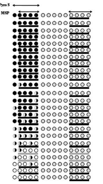
C overnight. Antigen-antibody reactions were revealed using a commercial kit (Novolink, Novocastra, Newcastle, UK) at room temperature; diaminobenzidine was used to detect the signal, and Harris hematoxylin was used as a nuclear stain. A positive control tissue (colon adenocarci-noma) was used to confirm the consistency of the immunostaining, and all samples were stained in a single batch.Figure 1 -Comparison of theMGMTpromoter methylation status of GBM cases analyzed using MSP and PyroS. Five CpG sites were analyzed using PyroS, and 8 CpG sites were analyzed using MSP. For PyroS, theMGMTmethylation status was scored according to the average percentage of specifically methylated CpG sites as unmethylated (U) when,10% CpG sites were methylated, intermediate (I) when 10 to 26% CpG sites were methylated, or methylated (M) when$27% of CpG sites were methylated. For MSP, theMGMT
MGMT expression was assessed and scored in tumor cells by two independent observers who were blinded to tumor methylation status and clinical data according to the
following semi-quantitative classification method based on percent of nuclei that were positive: 0 (no staining), 1 (10– 25%), 2 (26–50%), or 3 (.50%). Endothelial staining was
used as an internal control for MGMT immunostaining. In accordance with previous reports, MGMT staining was considered positive when uniform MGMT staining was detected in the cell nuclei.27Reactivity that was restricted to the cytosol and granular nuclear reactivity were considered negative.27Endothelial cells and perivascular lymphocytes were excluded from positive cell counts. For statistical analysis, scores of 0 were defined as the absence of protein expression, and scores of 1 to 3 were defined as positive for protein expression. The x2 test was used to evaluate the association between the MGMT promoter methylation status (negative versus positive) and protein expression levels (absenceversuspositive).
Statistical analysis
Thex2test was used to evaluate the associations between the MGMT promoter methylation status (negative versus positive) and the gene and protein expression levels. The Mann-Whitney test was used to analyze the differences in the relative expression levels between the two groups (methy-latedversus unmethylated) as determined by MSP and PyroS. Overall survival (OS) was calculated from the day of surgery to the day of death and was expressed in months. The Kaplan-Meier survival curve was analyzed using thelog rank(Mantel Cox) test and multivariate analysis using the Cox proportional hazards model. The logistic regression model included the following parameters: age at diagnosis, gender (femaleversus male), degree of tumor surgical resection (gross total resection, GTR versus partial resection, PR) and MGMT promoter methylation status (methylated versus unmethy-lated) assessed using PyroS and MSP. Calculations were performed using STATA version 7 (STATA Corp., College Station, TX) and SPSS 15.0 (SPSS, Chicago, IL). Also,p-values lower than 0.05 were considered statistically significant.
RESULTS
MGMT promoter methylation status
MGMTpromoter methylation was detected in 43.1% (22 out of 51) of the GBM samples by MSP and in 38.8% (4 intermediate and 15 methylated out of 49) of the samples by PyroS. Two of the 51 GBM cases were excluded from the PyroS method analysis, due to unsuccessful PCR amplifica-tion. MGMT methylation status was determined with 91% concordance for the two methods. Although PyroS revealed two cases of intermediate methylation that went undetected by MSP, the former method failed to detect three methy-lated cases that were detected by MSP. These three cases were methylated at a CpG site revealed by the primer set for PyroS, as shown in Figure 1.
MGMT gene expression
MGMT gene expression was determined by qRT-PCR. The median expression levels in GBM did not differ
significantly from those of non-neoplastic brain tissues (1.16 versus 0.91, respectively, p =0.597). However, when MGMT expression levels were analyzed in the two subgroups (methylatedversus unmethylated), a significant difference was observed with higher expression levels found in the methylated subgroup using either MSP (p,0.0001, Figure 2A) or PyroS (p,0.001, Figure 2B). Furthermore, as revealed by qualitative analysis, the correlation between the methylation status of the MGMT promoter and the relative MGMT expression level (either positive promoter methylation and low expression or negative promoter methylation and high expression) was statistically significant (p =0.001, 72.5% concordance for MSP;p =0.002, 71.4% concordance for PyroS).
MGMT protein expression
Positive expression of MGMT protein in tumor cell nuclei (as analyzed by IHC) was observed in 38 out of 51 (74.5%) GBM cases; there were 33 cases with a positivity score of 1+positivity and only five cases with a score of 2+(Figure 3). Among the 38
positive cases, 47.4% (by MSP) and 57.1% (by PyroS) were positive forMGMT promoter methylation. In contrast, only 31% (4 out of 13) of cases showing no staining on IHC were positive forMGMTpromoter methylation. Consequently, no significant correlation between MGMT protein expression and MGMT promoter methylation status was found. The con-cordances between IHC and either MSP or PyroS were only 47% and 57.1%, respectively. Other comparative analyses of the IHC protein expression scores and methylation status (con-sidering 0 and 1+staining as negative protein expression or 2+
as positive protein expression) demonstrated a similarly low concordance between the two methods.
An overview of the results of the present study is shown in Figure 3 as a heatmap and includes MGMT promoter methylation status and gene and protein expression findings.
Influence ofMGMTpromoter methylation status
and gene and protein expression on prognosis
The mean OS of the GBM cases was 13¡14.0 months. MGMT gene and protein expression levels were not correlated with OS (p.0.05, data not shown). Similarly, MGMT promoter methylation status (as determined by MSP) did not appear to affect OS, which was 17.2 months for the methylated group and 9.5 months for the unmethy-lated group (p =0.297). However, when only the 29 GBM cases submitted to adjuvant chemotherapy with radio-therapy and/or chemoradio-therapy were considered, the mean OS times differed significantly between the two groups (27.4 months for the methylated group and 12 months for the unmethylated group, p =0.025), indicating a stronger ther-apeutic response in the methylated group (Figure 4A). The mean OS was also significantly longer in the methylated group (31.7 months) than in the unmethylated group (11.8 months) (p =0.004) determined by PyroS (Figure 4B). A
multivariate Cox regression model (which considered age at diagnosis, gender, degree of tumor surgical resection, and MGMT promoter methylation status) showed that only MGMT promoter methylation status assessed either by MSP or PyroS was an independent prognostic factor (hazard ratio = 0.342, p= 0.023 and hazard ratio = 0.218, p= 0.005, respectively) as indicated in Table 2. Comparison of the methylation levels of the five CpG sites individually (above versus below the median level) yielded similar results (p =0.004 for CpG1, CpG2 and CpG5 and p =0.002 for CpG3 and CpG4, data not shown).
DISCUSSION
In the present study, MGMT promoter methylation as determined by two distinct methods had a significant impact on overall survival among patients treated with radiotherapy and/or chemotherapy as reported by other researchers in clinical trials and meta-analyses.10,11,28-31The frequency ofMGMTpromoter methylation in our study was in agreement with previously described results in primary GBM, which range from 36-45%.10,15,32-33Using one set of GBM samples, we were able to predict the therapeutic
Figure 4 -Kaplan-Meier curves showing the overall survival of GBM patients submitted to adjuvant therapy (radiotherapy and/or chemotherapy) and grouped according toMGMTpromoter methylation status as determined by MSP (A) and PyroS (B). The difference in overall survival times between the methylated and unmethylated groups was statistically significant for both methods (log-ranktest:
response using either a qualitative (MSP) or a quantitative (PyroS) method to assess the methylation status of the MGMT promoter. A high level of concordance (91%) between the two methods was observed. MSP provided slightly higher sensitivity while covering more CpG sites in the analysis than PyroS (8 versus 5 CpG sites). However, PyroS detected an intermediate methylation state that was not revealed by MSP. Additionally, the analysis of the five CpG sites together or separately predicted response to therapy in our set of GBM cases. In fact, the status of only a few CpG sites may be an adequate predictor of OS if high quality DNA is extracted from frozen tissue, which improves the sensitivity of methylation assessment,32 and adequate internal controls to detect incomplete bisulfite conversion and false priming are used. The accurate and robust results for MGMT promoter methylation status achieved by assessment of only four CpG sites20 located immediately downstream of those investigated in the present study corroborate this notion. Moreover, the methylation status of CpG4 alone has also proved to be a good predictor of OS.31
Another important issue in bringing a result from the laboratory bench to bedside practice is the cost-benefit ratio. To this end, we analyzed the reliability of two low cost methods for indirectly assessing MGMT methylation status: 1) MGMT gene expression level by straightforward qRT-PCR method and 2) MGMT protein expression level by IHC, a routine method widely available in histopathology laboratories. We compared the results of both methodolo-gies to theMGMTmethylation status. The positive correla-tion observed between the presence of MGMT promoter methylation and low expression of this gene may be explained by the use of the same microdissected frozen tumor fragment for DNA and RNA extractions and also by normalization of qRT-PCR results using three housekeeping genes instead of one.34,35 Nevertheless, the concordance between theMGMTgene expression level and methylation status determined by MSP was 72.5%, and this discrepancy may be due to the difficulty of determining the cut-off level
for dividing the cases into groups with high or lowMGMT gene expression. In the present study, the cut-off was arbitrarily determined as the median average value of the gene expression level in all GBM cases (1.16), although other criteria may also be acceptable. Recently, MGMT mRNA expression has been shown to play a direct role in mediating tumor sensitivity to alkylating agents independently of MGMT promoter methylation.23 Some methodological differences, such as the housekeeping genes used in that study compared to the present one, may explain the discrepancy, as may the fact that some CpG sites better reflect the MGMT gene expression level than others.36 Therefore, the reliability of assessing MGMT methylation status indirectly by MGMT qRT-PCR remains an open question.
No significant correlation between MGMT protein expres-sion (as determined by IHC) and methylation status was found, and concordances of only 47.4% (MSP) and 57.1% (PyroS) were observed. The presence of sampling bias in the methods with the smaller frozen fragments needed for the methylation study compared to the typically larger paraffin-embedded sections used for IHC, and the inclusion of endothelial cells, tumor-infiltrating lymphocytes or a variety of normal resident cells preserved within tumors may represent confounding factors for evaluation by IHC.21,37,38 Heterogeneity of the cell subpopulation comprising GBM tumors is an additional factor that could explain the discrepancy between the results of these two methods. The concomitant detection of both methylated and unmethylated status in the majority of GBM specimens analyzed in the present study corroborates the existence of this heterogene-ity, as described previously.39,40Furthermore, other factors, such as the p53 status, may also influence the final level of MGMT protein expression. Tumors with normal p53 status are more likely to have low or absent MGMT expression independently of theMGMTpromoter methylation status.41 Therefore, although IHC is a more accessible method than MSP or PyroS, MGMT protein expression is not a reliable method for inferring MGMT methylation status.
CONCLUSION
In summary, in one set of samples, theMGMTpromoter methylation status but not the MGMT mRNA or protein expression levels was confirmed as a factor predicting the response to adjuvant therapy in GBM patients.
ACKNOWLEDGMENTS
We are grateful to Vivian Minami Bertola at Qiagen Biotecnologia Brasil for technical assistance with pyrosequencing. We also thank the Psychiatry Institute for help with logistics in surgical therapy.
Grant: Funding was provided by FAPESP (04/12133-6), the Ludwig
Institute for Cancer Research, and CNPq.
REFERENCES
1. Wrensch M, Minn Y, Chew T, Bondy M, Berger MS. Epidemiology of primary brain tumors: current concepts and review of the literature. Neuro-oncology. 2002;4:278–99.
2. Miura FK, Alves MJF, Rocha MC, da Silva R, Oba-Shinjo SM, Marie SKN. Xenograft transplantation of human malignant astrocytoma cells into immunodeficient rats: an experimental model of glioblastoma. Clinics. 2010;65:305–9.
3. Korshunov A, Sycheva R, Golanov A. The prognostic relevance of molecular alterations in glioblastomas for patients age ,50 years. Cancer. 2005;104:825–32, doi: 10.1002/cncr.21221.
Table 2 -Multivariate proportional hazards analysis (Cox model) of age, gender, degree of surgical tumor resection, andMGMTmethylation status (by MSP and PyroS) of patients with GBM who underwent adjuvant therapy.
Variable HR (95% CI) p-value MSP
MGMTpromoter methylation status (by MSP)1
0.342 (0.13-0.86) 0.023
Age at diagnosis 1.00 (0.97-1.04) 0.783 Gender2 1.54 (0.67-3.55) 0.311 GTR3 0.54 (0.21-1.40) 0.205
PyroS
MGMTpromoter methylation status (by PyroS)1
0.218 (0.08-0.63) 0.005
Age at diagnosis 1.02 (0.98-1.06) 0.230 Gender2 1.30 (0.54-3.13) 0.553 GTR3 0.55 (0.21-1.42) 0.216
HR, hazard ratio; CI, confidence interval; GTR: gross total resection, PR: partial resection.
Age at diagnosis (from date of birth to date of surgery). 1MGMT methylated compared to MGMT unmethylated. 2Compared to male.
4. Louis DN, Ohgaki H, Wiestler OD, Cavenee WK, Burger PC, Jouvet A, et al. The 2007 WHO classification of tumours of the central nervous system. Acta Neuropathol. 2007;114:97–109, doi: 10.1007/s00401-007-0243-4. 5. Fults D, Brockmeyer D, Tullous MW, Pedone CA, Cawthon RM. p53
mutation and loss of heterozygosity on chromosomes 17 and 10 during human astrocytoma progression. Cancer Res. 1992;52:674–9.
6. Ishii N, Tada M, Hamou MF, Janzer RC, Meagher-Villemure K, Wiestler OD, et al. Cells with TP53 mutations in low grade astrocytic tumors evolve clonally to malignancy and are an unfavorable prognostic factor. Oncogene. 1999;18:5870–8, doi: 10.1038/sj.onc.1203241.
7. Stupp R, Hegi ME, Diserens A-C, Godard S, Dietrich P-Y, Regli L, et al. Clinical trial substantiates the predictive value of O-6-methylguanine-DNA methyltransferase promoter methylation in glioblastoma patients treated with temozolomide. Clin Cancer Res. 2004;10:1871–4, doi: 10. 1158/1078-0432.CCR-03-0384.
8. Cairncross G, Berkey B, Shaw E, Jenkins R, Scheithauer B, Brachman D, et al. Phase III trial of chemotherapy plus radiotherapy compared with radiotherapy alone for pure and mixed anaplastic oligodendroglioma: Intergroup Radiation Therapy Oncology Group Trial 9402. J Clin Oncol. 2006;24:2707–14, doi: 10.1200/JCO.2005.04.3414.
9. Weller M, Wick W. Are we ready to demystify age in glioblastoma? Or does older age matter in glioblastoma? Neuro-oncology. 2011;13:365–6. 10. Esteller M, Garcia-Foncillas J, Andion E, Goodman SN, Hidalgo OF,
Vanaclocha V, et al. Inactivation of the DNA-repair gene MGMT and the clinical response of gliomas to alkylating agents. N Engl J Med. 2000;343:1350–4, doi: 10.1056/NEJM200011093431901.
11. Hegi ME, Diserens A-C, Gorlia T, Hamou M-F, de Tribolet N, Weller M, et al. MGMT gene silencing and benefit from temozolomide in glioblastoma. N Engl J Med. 2005;352:997–1003, doi: 10.1056/NEJMoa043331.
12. Gerson SL. MGMT: its role in cancer aetiology and cancer therapeutics. Nat Rev Cancer. 2004;4:296–307, doi: 10.1038/nrc1319.
13. Singer-Sam J, LeBon JM, Tanguay RL, Riggs AD. A quantitative HpaII-PCR assay to measure methylation of DNA from a small number of cells. Nucleic Acids Res. 1990;18:687, doi: 10.1093/nar/18.3.687.
14. Cankovic M, Mikkelsen T, Rosenblum ML, Zarbo RJ. A simplified laboratory validated assay for MGMT promoter hypermethylation analysis of glioma specimens from formalin-fixed paraffin-embedded tissue. Lab Invest. 2007;87:392–7.
15. Hamilton MG, Rolda´n G, Magliocco A, McIntyre JB, Parney I, Easaw JC. Determination of the methylation status of MGMT in different regions within glioblastoma multiforme. J Neurooncol. 2011;102:255–60, doi: 10. 1007/s11060-010-0307-5.
16. Shen L, Kondo Y, Rosner GL, Xiao L, Hernandez NS, Vilaythong J, et al. MGMT promoter methylation and field defect in sporadic colorectal cancer. J Natl Cancer Inst. 2005;97:1330–8, doi: 10.1093/jnci/dji275. 17. Colella S, Shen L, Baggerly KA, Issa JP, Krahe R. Sensitive and
quantitative universal Pyrosequencing methylation analysis of CpG sites. BioTechniques. 2003;35:146–50.
18. Tost J, Dunker J, Gut IG. Analysis and quantification of multiple methylation variable positions in CpG islands by Pyrosequencing. BioTechniques. 2003;35:152–6.
19. Dupont J-M, Tost J, Jammes H, Gut IG. De novo quantitative bisulfite sequencing using the pyrosequencing technology. Anal Biochem. 2004;333:119–27, doi: 10.1016/j.ab.2004.05.007.
20. Mikeska T, Bock C, El-Maarri O, Hu¨bner A, Ehrentraut D, Schramm J, et al. Optimization of quantitative MGMT promoter methylation analysis using pyrosequencing and combined bisulfite restriction analysis. J Mol Diagn. 2007;9:368–81, doi: 10.2353/jmoldx.2007.060167.
21. Shaw RJ, Hall GL, Lowe D, Liloglou T, Field JK, Sloan P, et al. The role of pyrosequencing in head and neck cancer epigenetics: correlation of quantitative methylation data with gene expression. Arch Otolaryngol Head Neck Surg. 2008;134:251–6, doi: 10.1001/archoto.2007.50.
22. Preusser M, Charles Janzer R, Felsberg J, Reifenberger G, Hamou M-F, Diserens A-C, et al. Anti-O6-methylguanine-methyltransferase (MGMT) immunohistochemistry in glioblastoma multiforme: observer variability and lack of association with patient survival impede its use as clinical biomarker. Brain Pathol. 2008;18:520–32.
23. Kreth S, Thon N, Eigenbrod S, Lutz J, Ledderose C, Egensperger R, et al. O-methylguanine-DNA methyltransferase (MGMT) mRNA expression predicts outcome in malignant glioma independent of MGMT promoter methylation. PLoS ONE. 2011;6:e17156, doi: 10.1371/journal.pone. 0017156.
24. Marie SKN, Okamoto OK, Uno M, Hasegawa APG, Oba-Shinjo SM, Cohen T, et al. Maternal embryonic leucine zipper kinase transcript abundance correlates with malignancy grade in human astrocytomas. Int J Cancer. 2008;122:807–15, doi: 10.1002/ijc.23189.
25. Livak KJ, Schmittgen TD. Analysis of relative gene expression data using real-time quantitative PCR and the 2(-Delta Delta C(T)) Method. Methods. 2001;25:402–8, doi: 10.1006/meth.2001.1262.
26. Esteller M, Hamilton SR, Burger PC, Baylin SB, Herman JG. Inactivation of the DNA repair gene O6-methylguanine-DNA methyltransferase by promoter hypermethylation is a common event in primary human neoplasia. Cancer Res. 1999;59:793–7.
27. McLendon RE, Cleveland L, Pegram C, Bigner SH, Bigner DD, Friedman HS. Immunohistochemical detection of the DNA repair enzyme O6-methylguanine-DNA methyltransferase in formalin-fixed, paraffin-embedded astrocytomas. Lab Invest. 1998;78:643–4.
28. Kamiryo T, Tada K, Shiraishi S, Shinojima N, Kochi M, Ushio Y. Correlation between promoter hypermethylation of the O6-methylgua-nine-deoxyribonucleic acid methyltransferase gene and prognosis in patients with high-grade astrocytic tumors treated with surgery, radio-therapy, and 1-(4-amino-2-methyl-5-pyrimidinyl)methyl-3-(2-chlor-oethyl)-3-nitrosourea-based chemotherapy. Neurosurgery. 2004;54:349– 57, doi: 10.1227/01.NEU.0000103422.51382.99.
29. Cao VT, Jung T-Y, Jung S, Jin S-G, Moon K-S, Kim I-Y, et al. The correlation and prognostic significance of MGMT promoter methylation and MGMT protein in glioblastomas. Neurosurgery. 2009;65:866–75, doi: 10.1227/01.NEU.0000357325.90347.A1.
30. Weller M, Stupp R, Reifenberger G, Brandes AA, van den Bent MJ, Wick W, et al. MGMT promoter methylation in malignant gliomas: ready for personalized medicine? Nat Rev Neurol. 2010;6:39–51, doi: 10.1038/ nrneurol.2009.197.
31. Karayan-Tapon L, Quillien V, Guilhot J, Wager M, Fromont G, Saikali S, et al. Prognostic value of O6-methylguanine-DNA methyltransferase status in glioblastoma patients, assessed by five different methods. J Neurooncol. 2010;97:311–22, doi: 10.1007/s11060-009-0031-1. 32. Nakamura M, Watanabe T, Yonekawa Y, Kleihues P, Ohgaki H. Promoter
methylation of the DNA repair gene MGMT in astrocytomas is frequently associated with G:C –.A:T mutations of the TP53 tumor suppressor gene. Carcinogenesis. 2001;22:1715–9, doi: 10.1093/carcin/22.10.1715. 33. Hegi ME, Diserens A-C, Godard S, Dietrich P-Y, Regli L, Ostermann S,
et al. Clinical trial substantiates the predictive value of O-6-methylgua-nine-DNA methyltransferase promoter methylation in glioblastoma patients treated with temozolomide. Clin Cancer Res. 2004;10:1871–4, doi: 10.1158/1078-0432.CCR-03-0384.
34. Nakagawa T, Ido K, Sakuma T, Takeuchi H, Sato K, Kubota T. Prognostic significance of the immunohistochemical expression of O6-methylgua-nine-DNA methyltransferase, P-glycoprotein, and multidrug resistance protein-1 in glioblastomas. Neuropathology. 2009;29:379–88, doi: 10. 1111/j.1440-1789.2008.00983.x.
35. Valente V, Teixeira SA, Neder L, Okamoto OK, Oba-Shinjo SM, Marie SKN, et al. Selection of suitable housekeeping genes for expression analysis in glioblastoma using quantitative RT-PCR. BMC Mol Biol. 2009;10:17, doi: 10.1186/1471-2199-10-17.
36. Everhard S, Tost J, El Abdalaoui H, Crinie`re E, Busato F, Marie Y, et al. Identification of regions correlating MGMT promoter methylation and gene expression in glioblastomas. Neuro-oncology. 2009;11:348–56. 37. Rodriguez FJ, Thibodeau SN, Jenkins RB, Schowalter KV, Caron BL, O’neill
BP, et al. MGMT immunohistochemical expression and promoter methyla-tion in human glioblastoma. Appl Immunohistochem Mol Morphol. 2008;16:59–65.
38. Anda T, Shabani HK, Tsunoda K, Tokunaga Y, Kaminogo M, Shibata S, et al. Relationship between expression of O6-methylguanine-DNA methyltransferase, glutathione-S-transferase pi in glioblastoma and the survival of the patients treated with nimustine hydrochloride: an immunohistochemical analysis. Neurol Res. 2003;25:241–8, doi: 10. 1179/016164103101201445.
39. Brell M, Tortosa A, Verger E, Gil JM, Vin˜olas N, Villa´ S, et al. Prognostic significance of O6-methylguanine-DNA methyltransferase determined by promoter hypermethylation and immunohistochemical expression in anaplastic gliomas. Clin Cancer Res. 2005;11:5167–74, doi: 10.1158/1078-0432.CCR-05-0230.
40. Natsume A, Kondo Y, Ito M, Motomura K, Wakabayashi T, Yoshida J. Epigenetic aberrations and therapeutic implications in gliomas. Cancer Sci 2010;101:1331–6, doi: 10.1111/j.1349-7006.2010.01545.x. 41. Srivenugopal KS, Shou J, Mullapudi SR, Lang FF, Rao JS, Ali-Osman F.