Shell microstructure and its inheritance in the calcitic helcionellid
Mackinnonia
Michael J. Vendrasco
a,band Antonio G. Checa
aa
Departamento de Estratigrafía y Paleontología, Avenida Fuentenueva s/n, Universidad de Granada, 18071, Spain; [email protected]
b
Department of Biological Science, California State University, Fullerton, 92834, USA
Received 2 July 2014, accepted 16 December 2014
Abstract.Mackinnonia davidi from the Cambrian (Series 2) of Australia has a prismatic outer shell layer and, as newly described here, a calcitic semi-nacre inner layer. The pattern is the same as in stenothecids such as Mellopegma, providing more evidence for a strong phylogenetic signal in the shell microstructure of Cambrian molluscs. In addition, calcite now appears to have been common in helcionellids and other molluscs during the early and middle Cambrian, with many species exhibiting foliated calcite. This is surprising given the dominance of aragonite in molluscs, both modern and from post-Cambrian fossil deposits with exceptional shell microstructure preservation, including localities from the Ordovician of the Cincinnati region, USA.
Key words: Cambrian, helcionellid, Mollusca, Mackinnonia, Australia, shell microstructure, biomineralization.
INTRODUCTION
Fine microstructural detail can be preserved in calcium phosphate internal moulds and replacements of the shells of Cambrian molluscs (Runnegar 1985). While some common molluscan shell microstructures such as prismatic, crossed-lamellar and foliated calcite occur in Cambrian molluscs (Runnegar 1985), the textures amongst early molluscs are diverse and often unusual (Kouchinsky 2000). Stenothecids are a group of laterally-compressed helcionellids whose inner shell micro-structure in at least some forms consists of calcitic semi-nacre (Vendrasco et al. 2011). This texture is rare in modern molluscs but is common in lophophorates (Taylor et al. 2010).
Mackinnonia Runnegar in Bengtson et al. 1990 is a
widespread early Cambrian helcionellid mollusc known from China, Australia, Siberia, Greenland and North America (Parkhaev 2001, 2005; Skovsted & Peel 2007). The genus is characterized by a shell with a strongly undulating inner surface and straight outer wall (Runnegar 1990). The polygonal textures on the convex parts of the internal phosphate mould (corresponding to depressions in the inner surface of the shell; see Fig. 1) consist of a polygonal texture that represents prismatic shell microstructure (Runnegar 1990). Imprints of original shell crystals from concave parts of the internal moulds were previously unknown.
The occurrence of shell microstructure data in molluscs from both the Cambrian (e.g. Runnegar 1985) as well as Ordovician (e.g. Mutvei 1997) potentially allows the inference of broad-scale evolutionary patterns. Recent data from the Ordovician suggest that the aragonitic shell microstructure nacre was fairly wide-spread by this time (Vendrasco et al. 2013; MJV, personal observation).
Here we describe the nature of the inner shell layer
in Mackinnonia davidi from the early Cambrian of
Australia. Then we discuss the significance of this type of helcionellid microstructure pattern as it relates to phylogenetic analysis and seawater control over shell mineralogy.
MATERIALS AND METHODS
(CIC) of the University of Granada (Spain). Measure-ments were made from digital SEM photographs using ImageJ (Rasband 1997–2014). Specimens are deposited at the South Australian Museum (SAMA), Adelaide, South Australia.
RESULTS
Most internal moulds of Mackinnonia davidi have at least faint polygonal texture along comarginal ridges and sometimes at the apex. The walls of polygons are typically 2–3.5 µm in thickness (Fig. 2D), while the centres of polygons range from about 5–8.5 µm in width/diameter with outlines that vary from sub-circular to rounded-polygonal. Most of the polygonal imprints are concave, with the walls standing proud relative to the central region. The internal structure of polygons is
unclear, although the phosphate seems to be arranged in thin layers here (Fig. 2D). In transitional zones between ridges and troughs in the internal moulds, the regular polygonal network in some cases gives way to thin, roughly comarginal ridges made up of segments of polygonal walls (Fig. 2B).
Most regions of the comarginal troughs in internal moulds of Mackinnonia davidi lack identifiable regular texture. However, in some cases imprints of the whole or portions of flat, roughly equidimensional structures can be seen. These tend to vary within about 1–3 µm in width and the outlines range through a variety of polygonal shapes (e.g., square, rectangular, hexagonal; Fig. 2E–H). There tends to be a high degree of orientation of these tablets and in many cases one or two of the corresponding sides of the tablets are consistently preserved in one region (Fig. 2E, G).
Fig. 1. Shell microstructure patterns in Mackinnonia and stenothecids. A, cross-sectional view of Mackinnonia fossils, showing how troughs in the internal mould express the inner shell layer while ridges express the outer shell layer. B, general microstructure pattern of stenothecids and Mackinnonia with a prismatic outer layer and calcitic semi-nacre inner layer. Outer shell surface is at the bottom. C, sectional view of Mellopegma and other molluscs where the shell margin thins and expresses the outer shell layer on the inner shell surface. Outer shell surface is at the bottom.
________________________________________________________________________________________________________
DISCUSSION
Inner shell microstructure of calcitic semi-nacre
Thin sections reveal that Mackinnonia had a straight shell wall. Thus the comarginal ridges in internal moulds represent regions where the shell thins and expresses the outer shell microstructure on the inner shell surface (Runnegar 1990, fig. 159), while troughs in the inner phosphate coating represent regions where the inner shell microstructure is imprinted (Fig. 1).
The inner shell microstructure of Mackinnonia is clearly laminar and composed of tablets. Thus it can be classified either as nacre (where tablets are aragonitic) or calcitic semi-nacre (with calcitic tablets). Tablets occurring in troughs of internal moulds of Mackinnonia
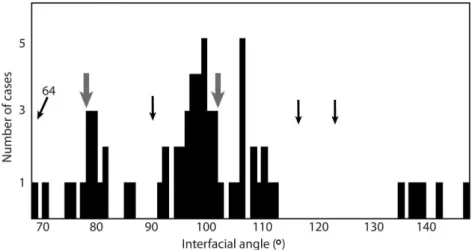
typically are straight-sided and occasionally have a rhomboidal shape. Some have an outline that looks similar to tablets of nacre (Fig. 2F), but we interpret the texture instead as calcitic because the interfacial angles of tablets generally match those expected for rhombic calcitic semi-nacre, 78° and 102° (Taylor & Weedon 2000), and are far from those expected for nacre (Fig. 3).
Calcitic(?) prismatic outer shell microstructure
The polygonal texture on the comarginal ridges of internal moulds represents the imprint of prismatic shell microstructure where prisms are about 10 µm in diameter (Runnegar 1990). The dimensions of the polygons are consistent with prismatic shell microstructure in modern molluscs, although prism diameters are at the small end of the range. Thin sections of Mellopegma show a tall
and narrow prismatic shell microstructure, although the prisms seen in thin section are 3 µm diameter, about a third the size of the prisms imprinted on internal moulds (Runnegar 1990, fig. 159e).
The mineralogy of the prisms is more likely calcite, although there is some doubt. In favour of calcite are the observations that prisms are remarkably well-preserved in calcitic specimens (Runnegar 1990), and no modern mollusc has an aragonitic outer shell layer and calcitic inner shell layer – those with calcitic inner layers have calcitic outer layers as well (although oysters with such a configuration can have an aragonitic myostracum). To explain the difference in size estimates of inferred prisms noted above, it is possible that each prism contained 2–3 columnar calcite crystals. Such a con-figuration, however, is unusual in molluscs.
Prismatic is by far the most common outer shell microstructure in modern molluscs and many early Cambrian molluscs show polygonal imprints (e.g., Kouchinsky 2000; Parkhaev 2004). Thus it seems likely that the earliest molluscan shells had a prismatic outer layer. The organic scaffolding around prisms is often thick in Cambrian molluscs (Vendrasco et al. 2010), similar to the modern bivalve Neotrigonia, indicating that the earliest mollusc shells tended to be more flexible in parts than in most modern molluscs.
Phylogenetic signal to shell microstructure data
A shell that consists of a prismatic outer layer and rhombic calcitic semi-nacre inner layer characterizes
Mackinnonia davidi as well as many stenothecid
molluscs (Vendrasco et al. 2010, 2011). Stenothecids
and Mackinnonia share many general morphological features, including lateral compression, an undulating inner shell surface, a prominent pegma-like structure (Fig. 2B), a convex ventral margin and preservation of outer shell microstructure where the shell thins. The correspondence between shell microstructure and inferences of phylogenetic relationship based on general morphology has been noted for other groups of Cambrian molluscs (Runnegar & Pojeta 1992; Kouchinsky 1999; Vendrasco et al. 2010). The additional evidence provided herein for the correlation in Mackinnonia and stenothecids strengthens the argument that shell microstructure has a strong phylogenetic signal in early molluscs and thus has utility in inferring phylogenetic relationships within early molluscs and mollusc-like Problematica.
Commonality of calcite in shells of Cambrian molluscs
A growing number of early to middle Cambrian molluscs have been found to have had at least a partly calcitic shell. This includes foliated calcite in
Pseudomyona and Eotebenna (Runnegar & Jell 1976; Runnegar 1985) and calcitic semi-nacre in the stenothecids Mellopegma, Acanthotheca and possibly
Stenotheca (Vendrasco et al. 2011). In addition, calcitic textures similar to foliated calcite can be inferred from photographs of Calyptroconus, Anuliconus, Aequiconus
and another helcionellid (Parkhaev 2001, pl. xxiv). Furthermore, a recent examination of fossils from the early Cambrian of Australia revealed calcitic semi-nacre in the Mackinnonia-like Parailsanella and foliated calcite in the paragastropod Pelagiella and other helcionellids tentatively identified as Bemella, Marocella,
Figurina and Microconulus (to be treated in a
sub-sequent publication). In fact, a majority of Cambrian molluscs whose inner shell microstructure is known in detail appear to have had a calcitic shell.
These calcitic molluscs range from just after the first switch to calcite seas that shelled metazoans experienced (Porter 2007). Thus perhaps seawater chemistry influenced the shell mineralogy of early molluscs. Temperature (Carter et al. 1998) and various biological factors such as predation (Carter 1980) clearly contribute to the relative fitness of calcite versus aragonite, but the relative importance of these selective pressures is at present unclear. Predatory pressure increased during the Great Ordovician Biodiversifi-cation Event, and this correlates with the apparent independent evolution of nacre in many groups of molluscs (Vendrasco et al. 2013). Perhaps at this time selective pressure via predation outweighed the push of seawater chemistry.
Helcionellids as a mixture of modern mollusc and other lophotrochozoan character states
Calcitic semi-nacre might occur in other molluscs, for example platyceratids including Platyceras spinigerum
from the Carboniferous of the USA (Carter & Clark 1985) and possibly older representatives dating back to the Ordovician. The ‘nacre’, identified by Batten (1984) from both groups of the diphyletic Platyceratidae (Frýda et al. 2009), may likewise be calcitic semi-nacre. But this shell microstructure otherwise is unknown in post-Cambrian molluscs. Calcitic semi-nacre is much more common in lophophorates (Taylor & Weedon 2000).
Other traits of helcionellids likewise reflect their close relationship to the ancestors of modern non-molluscan lophotrochozoans. For example, exceptional fossils of the coiled helcionellid Pelagiella preserve chaetae (Thomas et al. 2010), a character more common in annelids and brachiopods than in molluscs. In addition, many helcionellids had a densely porous shell (Parkhaev 2006; Vendrasco et al. 2011); pores occur in the shells of some molluscs but are much more common in brachiopods. As with many early members of stem lineages, helcionellids share a mixture of character states of the crown group (Mollusca) with states of closely related phyla (Brachiopoda, Bryozoa, Polychaeta).
Acknowledgements. We thank Bruce Runnegar for unsorted residues from acid maceration, and Maggie Cusack and Alexander Gubanov for helpful discussions. Jiri Frýda and Kathleen Histon improved the manuscript with helpful reviews. The work was funded by a Marie Curie Postdoctoral Fellowship (IIF 301668) from the European Commission. This research is a contribution to IGCP Project 591.
REFERENCES
Batten, R. L. 1984. The calcitic wall in the Paleozoic families Euomphalidae and Platyceratidae (Archeogastropoda).
Journal of Paleontology, 58, 1186–1192.
Bengtson, S., Conway Morris, S., Cooper, B. J., Jell, P. A. & Runnegar, B. 1990. Early Cambrian Fossils from South Australia. Memoir 9. Association of Australasian Palae-ontologists, Brisbane, 364 pp.
Carter, J. G. 1980. Environmental and biological controls of bivalve shell mineralogy and microstructure. In Skeletal Growth of Aquatic Organisms (Rhoads, D. C. & Lutz, R. A., eds), pp. 69–113. Plenum Press, New York. Carter, J. G. & Clark, G. R. 1985. Classification and
Carter, J. G., Barrera, E. & Tevesz, M. J. S. 1998. Thermal potentiation and mineralogical evolution in the Bivalvia (Mollusca). Journal of Paleontology, 72, 991–1010. Frýda, J., Racheboeuf, P. R., Frýdová, B., Ferrová, L., Mergl, M.
& Berkyová, S. 2009. Platyceratid gastropods – stem group of patellogastropods, neritimorphs or something else? Bulletin of Geosciences, 84, 107–120.
Kouchinsky, A. V. 1999. Shell microstructures of the Early Cambrian Anabarella and Watsonella as new evidence on the origin of the Rostroconchia. Lethaia, 32, 173–180. Kouchinsky, A. V. 2000. Skeletal microstructures of hyoliths
from the Early Cambrian of Siberia. Alcheringa, 24, 65–81.
Mutvei, H. 1997. Siphuncular structure in Ordovician endocerid cephalopods. Acta Palaeontologica Polonica, 42, 375– 390.
Parkhaev, P. Y. 2001. Molluscs. In The Cambrian Bio-stratigraphy of the Stansbury Basin, South Australia
(Alexander, E. M., Jago, J. B., Rozanov, A. Yu. & Zhuravlev, A. Yu., eds), Transactions of the Palae-ontological Institute of the Russian Academy of Sciences, 282, 113–203.
Parkhaev, P. Y. 2004. New data on the morphology of shell muscles in Cambrian helcionelloid mollusks. Pale-ontological Journal, 38, 254–256.
Parkhaev, P. Y. 2005. Two new species of the Cambrian helcionelloid mollusks from the northern part of the Siberian Platform. Paleontological Journal, 39, 615–619. Parkhaev, P. Y. 2006. On the Genus Auricullina Vassiljeva,
1998 and shell pores of the Cambrian helcionelloid mollusks. Paleontological Journal, 40, 20–33.
Porter, S. M. 2007. Seawater chemistry and early carbonate biomineralization. Science, 316, 1302.
Rasband, W. S. 1997–2014. ImageJ, U. S. National Institutes of Health, Bethesda, Maryland, USA, http://imagej.nih.gov/ij/, 1997–2014 [accessed 14 December 2014].
Runnegar, B. 1985. Shell microstructures of Cambrian molluscs replicated by phosphate. Alcheringa, 9, 245–257.
Runnegar, B. 1990. Mollusca. In Early Cambrian Fossils from South Australia (Bengtson, S., Conway Morris, S., Cooper, B. J., Jell, P. A. & Runnegar, B.), Memoir of the Association of Australasian Palaeontologists, 9, 232–257.
Runnegar, B. & Jell, P. A. 1976. Australian Middle Cambrian molluscs and their bearing on early molluscan evolution.
Alcheringa, 1, 109–138.
Runnegar, B. & Pojeta, J. 1992. The earliest bivalves and their Ordovician descendents. American Malacological Bulletin, 9, 117–122.
Skovsted, C. B. & Peel, J. S. 2007. Small shelly fossils from the argillaceous facies of the Lower Cambrian Forteau Formation of western Newfoundland. Acta Palae-ontologica Polonica, 52, 729–748.
Taylor, P. D. & Weedon, M. J. 2000. Skeletal ultrastructure and phylogeny of cyclostome bryozoans. Zoological Journal of the Linnean Society, 128, 337–399.
Taylor, P. D., Vinn, O. & Wilson, M. A. 2010. Evolution of biomineralization in ‘Lophophorates’. Special Papers in Palaeontology, 84, 317–333.
Thomas, R. D. K., Vinther, J. & Kerry, M. 2010. Paired chaetae associated with spiral shells of the late Early Cambrian mollusc Pelagiella from the Kinzers Formation; taphonomy, functional morphology, and potential evolutionary relationships. Geological Society of America Abstracts with Programs, 42, 633.
Vendrasco, M. J., Porter, S. M., Kouchinsky, A. V., Li, G. & Fernandez, C. Z. 2010. New data on molluscs and their shell microstructures from the Middle Cambrian Gowers Formation, Australia. Palaeontology, 53, 97–135. Vendrasco, M. J., Kouchinsky, A. V., Porter, S. M. &
Fernandez, C. Z. 2011. Phylogeny and escalation in
Mellopegma and other Cambrian molluscs. Palaeontologia Electronica, 14, 1–44.