SYSTEMATICS, MORPHOLOGY AND PHYSIOLOGY
Morphological Study of the Hindgut in Larvae of
Anticarsia gemmatalis
Hübner
(Lepidoptera: Noctuidae)
S
HEILAM. L
EVY1, Â
NGELAM.F. F
ALLEIROS2, F
LÁVIOM
OSCARDI3, E
LISAA. G
REGÓRIO1 EL
UISA. T
OLEDO11Depto. Morfologia, Instituto de Biociências, Universidade Estadual Paulista - UNESP, Campus de Botucatu, 18618-000
Botucatu, SP, e-mail: [email protected]
2Depto. Histologia, Centro de Ciências Biológicas, Universidade Estadual de Londrina - UEL, Londrina, PR 3Centro Nacional de Pesquisa da Soja - Embrapa, Londrina, PR
Neotropical Entomology 33(4): 427-431 (2004)
Estudo Morfológico do Intestino Posterior em Larvas de Anticarsia gemmatalis Hübner (Lepidoptera: Noctuidae)
RESUMO - A lagarta da soja (Anticarsia gemmatalisHübner) tem grande interesse econômico, pois
afeta significativamente a cultura da soja em todo o mundo. Este trabalho descreve a morfologia do intestino posterior de larvas de A. gemmatalis, com ênfase nos seus aspectos histológicos. O intestino
posterior é constituído por regiões morfologicamente distintas, identificadas como piloro, íleo, cólon e reto. Independente da região, a parede do intestino posterior é constituída por fina cutícula, epitélio simples e camada muscular. A íntima cuticular apresenta espículas no anel intersticial posterior, entre o intestino médio e o posterior, e na região posterior do piloro. A musculatura do reto é formada por camada única de largas fibras circulares, diferindo das demais regiões do intestino posterior que apresentam duas camadas de fibras musculares. As extremidades distais dos túbulos de Malpighi atravessam as paredes do reto, constituindo o sistema criptonefridial característico de Lepidoptera.
PALAVRAS-CHAVE: Tubo digestivo, histologia, lagarta da soja, inseto
ABSTRACT - The velvetbean caterpillar (Anticarsia gemmatalis Hübner) has great economical interest
as it affects the soybean crop worldwide. This work describes the morphology of the hindgut in A. gemmatalis larvae emphasizing their histological aspects. Distinct morphological regions, identified as
pylorus, ileum, colon and rectum, constitute the hindgut of A.gemmatalis. A thin cuticular intima, a simple
epithelium and muscular layer compose the hindgut wall, independent of the region. Microspines project from the cuticular intima in the posterior interstitial ring, between the midgut and the hindgut, and the posterior pyloric region. A single circular layer of large fibers, differing from the other hindgut regions that present two layers of muscular fibers, forms the rectal musculature. The distal ends of Malpighian tubules cross the rectum wall and constitute the cryptonephric excretory system typical in Lepidoptera larvae.
KEY WORDS: Digestive tube, histology, velvetbean caterpillar, insect
The digestive tract of insects is considered an effective physical and chemical barrier against the potentially invasive pathogens that are ingested with the feeding. Three main regions constitute the digestive tract: foregut, midgut and hindgut (Eaton 1988, Terra & Ferreira 1994, Chapman 1998).
It is well known that the insect midgut is responsible for food digestion and nutrient absorption (Santos et al. 1984,
Billingsley & Lehane 1996, Cristofoletti et al. 2001). The
undigested material goes straight to the hindgut where the water absorption and feces formation and elimination occur. The hindgut of most Lepidoptera larvae may be morphologically subdivided in pylorus, ileum, colon and rectum (Drecktrah et al. 1966, Judy & Gilbert 1970, Chi et al.
1975). The Malpighian tubules open into the pylorus; their distal ends are associated with the rectal wall and form the
Lepidopteran characteristic cryptonephric excretory system (Wigglesworth 1984) that conserves water by absorbing it actively from the feces either to the hemolymph or to the lumen of the Malpighian tubules (Maddrell & O´Donnell 1992).
The larvae of Anticarsia gemmatalis Hübner are
considered one of the most serious pests of soybean crop, and they are known as velvetbean caterpillar. Although there is an effective program for the biological control of A. gemmatalis using virus that get into the insect through the
digestive tract (Moscardi & Carvalho 1993, Flipsen et al.
in larvae of A. gemmatalis, under light and scanning electron
microscopy.
Material and Methods
A. gemmatalis larvae were obtained from the Laboratório
Entomológico of the Centro Nacional de Pesquisa da Soja (CNPSo)/Embrapa, Londrina - PR, Brazil. The larvae were maintained in the laboratory with artificial diet (Hoffmann-Campo et al.1985), under controlled temperature (25-27oC),
photoperiod (14h light/10h dark) and 80% relative humidity. Larvae of the 4th and 5th instars (12-16 days old), after a
rapid rinsing in 70% alcohol, were dissected in insect saline solution (ISS- 1.8 g de NaCl; 1.88 g de KCl; 0.16 g de CaCl; 0.004 g de NaHCO3; distillated water - q.s.p. 100 ml) under stereomicroscope.
For studies with light microscope, the hindgut was isolated and fixed in glutaraldehyde (2.5%) and paraformaldehyde (4%) solution in phosphate buffer (0.1 M, pH 7.3). After dehydration in graded ethanol series, the material was embedded in JB4 hystoresin. The 3 µm sections
were stained with hematoxylin-eosin (H.E.), analyzed and
photographed under an Axiophot (Zeiss) photomicroscope. For scanning electron microscope studies, the hindgut was fixed in 2.5% glutaraldehyde in 0.1 M phosphate buffer (pH 7.3), post-fixed in 1% osmium tetroxide solution in the same buffer, dehydrated in graded ethanol solutions, critical point dried and gold coated in a sputtering device. The materials were analyzed and photographed using SEM 515 (Phillips) scanning electron microscope.
Results and Discussion
The hindgut in A. gemmatalis larvae is the most complex
portion of the digestive tract, being constituted by the pylorus, ileum, colon and rectum. It is internally lined by a continuous epithelium covered by a thin chitinous cuticular intima (Figs. 1A, 1C, 2B and 2D), as described for other Lepidoptera species (Drecktrah et al. 1966, Mathur 1966, Chi et al. 1975). According to Gillott (1995) the cuticle of the
hindgut is thinner than the one of the foregut because this region is responsible for some water and nutrient absorption. A posterior interstitial ring lined by small cuboidal epithelial cells with central spherical nuclei marks the junction
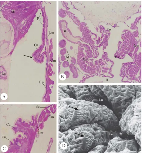
Figure 1. Morphology of the pylorus in A. gemmatalis hindgut. A - Junction between the midgut (Mi) and hindgut: the
posterior interstitial ring (Pr) with small spines (arrow). H.E. 320x. B - General view of the anterior and posterior (Po) pyloric portions. H.E. 62x. C - Detail of the transition between anterior and posterior pyloric portions, showing the squamous (Se) and the cubic (Cc) epithelial cells. H.E. 250x. D - Scanning electron micrograph of the hindgut lumen surface showing groups of spines (arrow) near to pyloric valve (Pv). 720x. Epithelium (Ep), Lumen (Lu), longitudinal (Lm) and circular (Cm) muscular layers, cuticular intima (Cu), pyloric valve (Pv), Malpighian tubules (Mt), common ducts of the Malpighian tubules (★), and ileum (IL).
C D
A
B
Cu Cc
Se
Cm
L m Lu
Mi
P r
Cm L m
Ep Cu
M t
Cm
IL P v
L m P o
Lu
of mid-hindgut, and the cuticule intima at this region presents small spines (Fig. 1A); the longitudinal (external) and circular (internal) muscles cover the ring (Fig. 1A).
Posterior from this ring, the epithelial cells become increasingly flatten at the pyloric region (Figs. 1A, 1B and 1C). The pylorus of A. gemmatalis is the most complex
histological region comparing with the other hindgut regions. Two distinct pyloric portions were recognized in A. gemmatalis (Fig. 1B), as described for Hyalophora cecropia
L. (Judy & Gilbert 1970). The anterior pyloric portionis lined with a simple basophilic squamous epithelium and the chitinous intima has no spines; the muscle layer presents inner circular and outer longitudinal fibers (Figs. 1A, 1B and 1C). The posterior pylorus has a smaller lumen; irregular shaped cuboidal epithelial cells (Fig. 1C) and microspines project from the chitinous intima (Fig. 1D). Similar spinner cuticle lining the pyloric posterior region was also observed in H. cecropia (Judy & Gilbert 1970), Heliothis zea Boddie, Heliothis virescens Fabricius and Spodoptera frugiperda
Smith (Chi et al. 1975). According to Barth (1972), the presence
of this chitinous structure can be correlated with the insects feeding habits: if they feed with fluid material, like the phytophagous and hematophagous insects, the internal wall is smooth; in solid feeding species, the cuticular layer showed chitinous microspines that helps in the undigested material transport toward the other portions of the hindgut. However, microspines were found in fluid-feeding insects, as described in different species of ants (Caetano et al. 2002). Elzinga (1998)
suggested that the microspines might be correlated with the presence of well-developed hindgut subdivisions and valves between these sections. Such chambers permit regulation of food movement and retention of digested food remnants to be acted upon by symbionts, as observed in crickets, cockroaches and termites; the microspines appear to aid in retaining bacteria and protozoan (Elzinga & Hopkins 1995, Elzinga 1996).
The posterior pyloric region has a thick musculature constituted by an inner circular layer and an outer longitudinal layer, which characterized the pyloric valve (Figs. 1B and 1C). The outer longitudinal muscle fibers penetrate among the circular muscle fibers (Fig. 1B). The circular muscle layer helps in the pyloric valve contraction, while the longitudinal muscle layer is responsible for the pyloric valve relaxes after the food movements toward the ileum (Gillott 1995).
A pair of common Malpighian ducts, which are ampulla-shaped, inserted through the musculature on the ventrolateral sides of the pyloric valve (Fig. 1B), characterizing the end of pyloric region in A. gemmatalis. The same aspect was
described for other Lepidoptera (Standlea & Yonke 1968, Mathur 1972, Eaton 1988), Coleoptera (Areekul 1957, Vasques 1988), Diptera (Patil & Govindan 1984) and Hymenoptera (Caetano & Overal 1984, Arab & Caetano 2002).
The ileum epithelium in A. gemmatalis has acidophilic
flattened polygonal epithelial cells with elongated nuclei and apical cytoplasm exhibit brush border-like structure lined by cuticular intima (Figs. 2A and 2B). The brush border structure represents the apical plasma membrane invaginations associated with mitochondria described under transmission electron microscopy in the ileum cell of Schistocerca gregaria
Forskal, which allows the water absorption that occurs in this region (Irvine et al. 1988). This absorption is necessary
to maintain the insect metabolism as well to promote the feces dehydration that starts into the ileum (Barth 1972). The outer circular muscles are more developed than the inner longitudinal fibers (Figs. 2A and 2B); similar aspects were observed in Lepidoptera species (Mathur 1966, Judy & Gilbert 1970, Chi et al. 1975) and in some Coleoptera (Areekul 1957).
However, Vasques (1988) described a different muscular arrangement in Pyrearinus termitilluminans Costa, with inner
circular and outer longitudinal muscular layer.
The histological characteristics of the colon in A. gemmatalis (Figs. 2A and 2C) are similar to those described
in other insets (Judy & Gilbert 1970, Gonçalves 1980). The colon epithelium is composed by a simple squamous epithelial cells, and the thicker cuticular intima is thrown into longitudinal folds; because of this folding, the lumen is very restricted and has a compound tubular appearance (Fig. 2C), giving to the lumen a potential for expansion when large amount of food pass through (Chi et al. 1975). The outer
circular muscular layer is more developed than the inner longitudinal layer (Fig. 2C).
A fold rectal wall penetrates into the distal colon regional and constitutes the rectal valve in A. gemmatalis (Fig. 2C).
Well-developed bounds of dilator muscles detected in this region (Fig. 2C) allow the valve contraction and dilatation, so helping feces transport to the rectum; besides, the rectal valve does not allow the feces reflux to the colon when the rectum contracts to defection. This structure was described in most of insects, but it is less developed in fluid feeding species (Barth 1972).
The rectum wall in A. gemmatalis has large squamous
epithelial cells with polymorphic nuclei and the chitinous cuticular intima is smooth (Figs. 2C and 2D), as described for other Lepidoptera (Judy & Gilbert 1970, Chi et al. 1975).
Malpighian tubules are visualized between the epithelium and the thin rectal cellular membranes (Figs. 2C and 2D) and constitute the rectal complex that characterizes the cryptonephric excretory system typically found in Lepidoptera larvae (Drecktrah et al. 1966, Eaton 1988). The ability of the
Malpighian tubules to excrete uric acid with very little water loss is an important factor for the success of insects in terrestrial environments as uric acid is the principal nitrogenous end product of protein metabolism in insects (Hegner & Engeman 1968, Maddrell & O’Donnell 1992). A thin sheet-like layer of muscle is found in the rectum wall, formed by large flattened muscle cells (Figs. 2C and 2D) that make this hindgut region different from the others concerning the musculature.
The rectal pads are absent in A. gemmatalis, as well as in
other Lepidoptera larvae as H. cecropia (Judy & Gilbert 1970), H. zea, H. virescens and S. frugiperda (Chi et al. 1975). The
histological analysis of the anus in A. gemmatalis was not
possible to be a complished, as its strong junction to the exoskeleton did not allow the dissection.
Acknowledgements
larvae and the Departamento de Morfologia, UNESP, Botucatu, SP, for helping in the histological procedures. This work was supported by the Conselho Nacional de Desenvolvimento Científico e Tecnológico (CNPq) and Fundação de Amparo à Pesquisa do Estado de São Paulo (FAPESP).
Literature Cited
Arab, A. & F.H. Caetano. 2002. Segmental specializations in the Malpighian tubules of the fire ant Solenopsis saevissima Forel 1904 (Myrmicinae): an electron
microscopical study. Arthropod Struc. Develop. 30: 281-292.
Areekul, S. 1957. The comparative internal larval anatomy of several genera of Scarabaeidae (Coleoptera). Ann. Entomol. Soc. Am. 50: 562-577.
Barth, R. 1972. (eds.) Entomologia geral. Rio de Janeiro, Fundação Instituto Oswaldo Cruz, 374p.
Billingsley, P.F. & M.J. Lehane. 1996. Structure and ultrastructure of the insect midgut, p. 3-30. In M.J. Lehane & P.F. Billingsley (eds.), Biology of the insect midgut. London, Chapman and Hall, 486p.
Caetano, F.H, K. Jaffé & F.J. Zara. 2002. (eds.) Formigas: Biologia e anatomia. Rio Claro, FHC, 131p.
Caetano, F.H. & W.L. Overal. 1984. Estudos do trato digestivo de vespas (Hymenoptera, Vespidae) e estruturas excretoras associadas. I. Morfologia. Rev. Bras. Entomol. 28: 403-408.
Chapman, R.F. 1998. (ed.) The insects: Structure and function. Harvard, Cambridge University Press, 412p. Figure 2. Morphology of the A. gemmatalis hindgut. A - Ileum (IL) with flattened polygonal epithelial cells (Pe). H.E.
62x. B - Detail of ileum wall: epithelial cells with elongated nuclei (Nu), and apical brush border-like structure (arrow head). H.E. 250x. C - Transition between the colon (Co) and rectum (Re): longitudinal folds (arrow) and rectal valve (Rv). H.E. 62x. D - Detail of the cryptonephric excretory system: rectum epithelial cells with polymorphic nuclei (Nu), rectal cellular membranes (Rm), and sheet muscle (S). H.E. 250x. Cuticular intima (Cu), colon epithelial cells (Ce), rectum epithelial cells (Rc), circular (Cm) muscular layer, Malpighian tubules (Mt), lumen (Lu) and cryptonephric excretory system (Ex).
Re
Rv
Rc
IL P e
Co
L m
Ce Co
Ex Cm
M t Rm Cu
Lu
Cu
S
A B
B B
Nu Cm
M t
Chi, C., W.A. Drew, J.H. Young & M.R. Curd. 1975. Comparative morphology and histology of the larval digestive system of two genera of Noctuidae (Lepidoptera): Heliothis and Spodoptera. Ann. Entomol.
Soc. Am. 68: 371-380.
Cristofoletti, P.T., A.F. Ribeiro & W.R. Terra. 2001. Apocrine secretion of amylase and exocytosis of trypsin along the midgut of Tenebriomolitor larvae. J. Insect Physiol. 47:
143-155.
Drecktrah, H.G., K.L. Knight & T.A. Brindley. 1966. Morphological investigations of the internal anatomy of the fifth larval instar of the european corn borer. Iowa St. J. Sci. 40: 57-86.
Eaton, J.L. 1988. (ed.) Lepidopteran anatomy. New York, Wiley-Interscience Publication, 257p.
Elzinga, R.J. 1996. A comparative study of microspines in the alimentary canal of five families of Orthoptera (Saltatoria). Int. J. Insect Morphol. Embriol. 25: 249-260.
Elzinga, R.J. 1998. Microspines in the alimentary canal of arthropoda, onychophora, annelida. Int. J. Insect Morphol. Embriol. 27: 341-349.
Elzinga, R.J. & T.L. Hopkins. 1995. Microspines in the hindgut regions of four families of cockroaches (Blattaria). Int. J. Insect Morphol. Embriol. 24: 203-211.
Flipsen, J.T.M., J.W.M. Martens, M.M. Van Oers, J.M. Vlak & W.M. Van Lent. 1995. Passage of Autographa californica nuclear polyhedrosis virus through the
midgut epithelium of Spodoptera exigua larvae. Virology
208: 328-335.
Gillott, C. 1995. (ed.) Entomology. New York, Plenum Press, 798p. Gonçalves, I.S. 1980. Histologia do tubo digestivo de
Rugitermes niger Oliveira, 1979, (Isoptera,
Kalotermitidae). Rev. Bras. Entomol. 24: 215-216.
Hegner, R.W. & J.G. Engeman. 1968. (eds.) Invertebrate zoology. London, Macmillan Company, 619p.
Hoffmann-Campo, C.B.H., E.B. Oliveira & F. Moscardi. 1985. Criação massal da lagarta da soja (Anticarsia gemmatalis). Londrina, EMBRAPA/CNPSo, 23p.
Irvine, B., N. Audsley, R. Lechleitner, J. Meredith, B. Thomson & J. Phillips. 1988. Transport properties of
locust ileum in vitro: Effects of cyclic AMP. J. Exp. Biol.
137: 361-385.
Judy, K.J. & L.I. Gilbert. 1970. Histology of the alimentary canal during the metamorphosis of Hyalophora cecropia
(L). J. Morphol. 131: 277-300.
Maddrell, S.H.P. & M.J. O’Donnell. 1992. Insect Malpighian tubules: V.ATPase action in ion and fluid transport.J. Exp. Biol. 172: 417-429.
Mathur, L.M.L. 1966. Morphology of the alimentary canal and ciated glands of the mature larva of Achaea janata L.
(Lepidoptera: Noctuidae). Indian J. Entomol. 28: 318-331.
Mathur, L.M.L. 1972. Developmental changes in the alimentary canal of Prodenia litura Fabr. (Lepidoptera). J. Nat. Hist. 6:
39-46.
Moscardi, F. & R.C.Z. Carvalho. 1993. Consumo e utilização de soja por Anticarsia gemmatalis Hüb. (Lepidoptera:
Noctuidae) infectada, em diferentes estádios larvais, por seu vírus de poliedrose nuclear. An. Soc. Entomol. Bras. 22: 267-280.
Patil, G.M. & R. Govindan. 1984. Internal anatomy of silkworm Uzi-Fly, Exorista sorbillans (Wiedemann) (Diptera:
Tachinidae). Indian J. Seric. 23: 22-31.
Santos, C.D., A.F. Ribeiro, C. Ferreira & W.R. Terra. 1984. The larval midgut of cassava hornworm (Erinnyisello):
Ultrastructure, fluid flux and the secretory activity in relation to the organization of digestion. Cell Tissue Res. 237: 565-574.
Standlea, P.P. & T.R. Yonke. 1968. Clarification of the description of the digestive system of Heliothis zea. Ann.
Entomol. Soc. Am. 61: 1478-1481.
Terra, W.R. & C. Ferreira. 1994. Insect digestive enzymes: Properties, compartmentalization and function. Comp. Biochem. Physiol. 109B: 1-62.
Vasques, A.M. 1988. Estudo morfológico e histológico do tubo digestivo de larvas de Pyrearinus termitilluminans Costa,
1982 (Coleoptera, Elateridae, Pyrophorini). Rev. Bras. Entomol. 32: 253-258.
Wigglesworth, V.B. 1984. (ed.) Insect physiology. London, Chapman and Hall, 191p.