Divergence at the Gr39a Locus in Drosophila
Anastasia Gardiner1*, Daniel Barker1, Roger K. Butlin2, William C. Jordan3, Michael G. Ritchie1
1School of Biology, University of St. Andrews, St. Andrews, Scotland, United Kingdom,2Department of Animal and Plant Sciences, University of Sheffield, Sheffield, United Kingdom,3Institute of Zoology, Zoological Society of London, London, United Kingdom
Background.Gene families typically evolve by gene duplication followed by the adoption of new or altered gene functions. A different way to evolve new but related functions is alternative splicing of existing exons of a complex gene. The chemosensory gene families of animals are characterised by numerous loci of related function. Alternative splicing has only rarely been reported in chemosensory loci, for example in 5 out of around 120 loci inDrosophila melanogaster. The gustatory receptor geneGr39ahas four large exons that are alternatively spliced with three small conserved exons. Recently the genome sequences of eleven additional species of Drosophila have become available allowing us to examine variation in the structure of theGr39alocus across a wide phylogenetic range of fly species.Methodology/Principal Findings.We describe a fifth exon and show that the locus has a complex evolutionary history with several duplications, pseudogenisations and losses of exons. PAML analyses suggested that the whole gene has a history of purifying selection, although this was less strong in exons which underwent duplication. Conclusions/Significance. Estimates of functional divergence between exons were similar in magnitude to functional divergence between duplicated genes, suggesting that exon divergence is broadly equivalent to gene duplication.
Citation: Gardiner A, Barker D, Butlin RK, Jordan WC, Ritchie MG (2008) Evolution of a Complex Locus: Exon Gain, Loss and Divergence at the Gr39a Locus in Drosophila. PLoS ONE 3(1): e1513. doi:10.1371/journal.pone.0001513
INTRODUCTION
Gustatory receptor (Gr) genes comprise a large fraction (,50%) of the Drosophila chemosensory receptor gene superfamily [1], encoding 7-transmembrane (7TM) proteins involved in taste and smell. Most DrosophilaGrsare very divergent, sometimes showing as little as 8% amino acid identity to each other [1]. Much of the diversity of the chemoreceptor family has evolved through widespread and repeated whole-gene duplications, followed by functional divergence of those duplicates that do not degrade to pseudogenes [2,3]. Another mechanism that enlarges the eukary-otic protein repertoire in general is alternative splicing. Although this is currently thought to be rare among chemosensory receptor loci [4,5], threeD. melanogaster Grgenes (Gr23a, Gr28b,Gr39a) are notable in that they have been shown to undergo alternative splicing, together coding for 11 proteins, or 16% of all gustatory receptors in the species [1,6]. Analysis of theGr repertoire in 12 Drosophila species allowed us to identify the orthologues of the Gr39a genes in these species [7]. This locus showed an unusual pattern of structural changes compared with otherGrs. Here we investigate in detail the evolution of the Gr39a gene using a comparative, bioinformatic approach.
Located on the left arm of the second chromosome of D. melanogaster, theGr39agene has four large exons (A,B,CandD), each including coding sequences for six transmembrane domains, followed by three small exons that together encode the seventh transmembrane domain and COOH-terminus [6]. Any one of the large exons may be spliced to the smaller exons, generating four different 7TM protein products. These are expressed in the main taste organs of D. melanogaster, the labellum (Gr39aA, Gr39aB, Gr39aC, Gr39aD), though some are also expressed in the thorax (Gr39aC, Gr39aD), abdomen (Gr39aC) and wings (Gr39aD) [6]. The function ofGr39ais unknown, but its close phylogenetic affinity with theD. melanogastermale specific pheromone receptorGr68a[8] suggests a possible involvement in pheromone recognition [9].
We have annotated the orthologs of D. melanogaster Gr39a in eleven other recently sequenced Drosophila species [10], repre-senting a wide range of phylogenetic divergence from D.
melanogaster(Figure 1A, [10]). Of these species, nine (D. melanogaster, D. simulans, D. sechellia, D. yakuba, D. erecta, D. ananassae, D. pseudoobscura, D. persimilis and D. willistoni) are in the subgenus Sophophora, and the remaining three (D. mojavensis,D. virilisandD. grimshawi) are within the subgenus Drosophila. The two subgenera are estimated to have diverged from each other 40–60 million years ago [11,12]. Lower level groups and subgroups have been identified within the subgenera (Figure 1A). We examined the structural and potentially functional differences between theGr39a genes across these twelve species. We identified a new large exon, exonE, found in most species but lost in the melanogaster lineage. After an analysis in which we employ phylogenomic approaches more usually used to examine gene evolution, we propose a model of the evolution of Gr39a. We conclude that, despite strong purifying constraints on theGr39alocus overall, the exons that are prone to duplication or pseudogenisation show evidence of relaxed selection which probably facilitated ‘‘subfunctionalization’’ of the duplicated exon copies. Evidence of positive selection also suggests ‘‘specialization’’ and/or neofunctionalization of tandemly dupli-cated exons has occurred, though potential new functions are currently unknown.
Academic Editor:Richard Copley, Wellcome Trust Centre for Human Genetics, United Kingdom
ReceivedOctober 5, 2007;AcceptedJanuary 2, 2008;PublishedJanuary 30, 2008
Copyright:ß2008 Gardiner et al. This is an open-access article distributed under the terms of the Creative Commons Attribution License, which permits unrestricted use, distribution, and reproduction in any medium, provided the original author and source are credited.
Funding:This work was supported by the Natural Environment Research Council UK, grant NE/C003187/1. DB was supported by a Research Councils UK Academic Fellowship.
Competing Interests:The authors have declared that no competing interests exist.
RESULTS
Chemosensory receptor repertoires among different insects show great divergence, for instance only a few orthologous groups were identified when the complete olfactory and gustatory receptor repertoires of the fruit fly, honeybee and mosquito were compared [4,5,13]. Robertson and Wanner [5] suggest that the exons Gr39aA-DofDrosophila melanogasterform an orthologous group with seventeenGrgenes ofAnopheles gambiae(AgGr9a-n,AgGr10,AgGr11, andAgGr12), but the evidence of the orthology is lacking due to the very weak bootstrap support for this clade. In Apis mellifera, the orthologs ofGr39awere not identified [5]. TheDrosophilaexons are related among themselves by duplications, which seem to have occurred after theDrosophila-Anophelessplit.
We were able to uncover complex structural changes that have occurred to Gr39a since the subgeneraDrosophila and Sophophora diverged. The gene structure ofGr39adescribed forD. melanogaster, with four large exons A, B, C, and D followed by three small constitutive exons [6], is peculiar to the species of the melanogaster subgroup only (Figure 1A). In this subgroup, the first exonAhas either accumulated frame shift mutations or significantly degraded in two species: D. sechelliaand D. erecta, respectively. Each large exon contains its own start codon and 59splice signal allowing the locus to encode several protein products independent of the mutational alteration of the ORF in one of the exons, so we would expect the exonsB, CandDto be expressed inD. sechelliaandD. erecta. Species of the melanogaster subgroup have also lost an ancestral exonE,first described here and identified as a degraded copy inD. ananassae, an intact exon in the species of the obscura group,D. willistoni, D. mojavensisandD. grimshawi, and an exon with two frame-shift mutations inD. virilis(Figure 1A). In support of this interpretation, a search for evidence of exonEin the melanogaster subgroup revealed the presence of short sequences that code for about 100 amino acids alignable with the truncated exonEofD. ananassaeand intact exonEofD. pseudoobscura.
The Bayesian-MCMC phylogeny of GR39a exons and their homologues is summarized in Figure 2. The posterior probabilities shown in Figure 2 appear to lack significant bias as estimates of the probabilities of the clade, conditional on the data, model and priors: average standard deviation of split frequencies was 0.0060 at the start of the final MCMC sample (close to the ideal value of zero), and lag-1 ACF for lnL in the two MrBayes runs was 0.014 (P.0.05) and 0.036 (P.0.05).
ExonEoccupies a basal position within a clade including exons C and D (Figure 2). The reconciliation of the species tree and phylogenetic tree of exonsE, CandDby the Notung program [14] showed two events of exon duplication (duplication of exonEand subsequent origin of exons D and C before the subgenera Sophophora and Drosophila split) and three independent events of exon loss (Figure 3). This prediction supports the loss of exonEin the melanogaster group, and suggests the loss of exon C in D. willistoni and the subgenus Drosophila, but we did not find any evidence of the presence of exonCin these species.
accumulation of frame shifts, premature stop codons and large deletions (for example,Gr28bAinD. sechellia,Gr28bDandEinD. grimshawii, Or69aCinD. sechelliaandD. melanogaster).
The presence of sequence motifs that specify six transmembrane domains was detected in all the large exons ofGr39a; the last three small exons are present in all species and encode for the seventh transmembrane domain (Figure 1A). We analysed the splicing structure of Gr39a in all species. The location of splice sites is conserved throughout the genes examined (Figure 1B). All large exons (except the pseudo-exonsAinD. sechelliaandD. erecta) and two conserved small exons contain the 59 donor-splice motif [AG]qGT(NNGT), while the first 39 acceptor-splice motif (CnTn)NCAGq[GC] appears at the beginning of the block of
three conserved small exons (Figure 1B). This structure supports a model of mutually exclusive alternative splicing, when a single large exon is spliced with the small conserved exons and the other large exons are excluded as part of an intron.
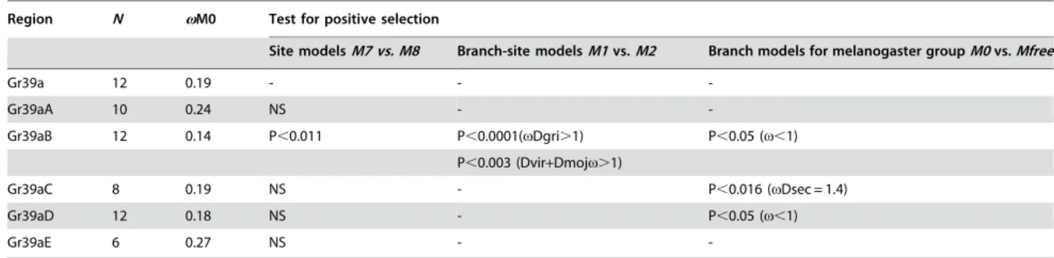
The estimatedvvalues for the whole gene and its large exons were all substantially lower than 1 (Table 1) suggesting that all parts of the gene are subject to strong purifying selection, however weaker selective constraints act on exonEand the duplicated exon A(vE= 0.27 andvA= 0.24; c.f.,0.17).
Evidence of expression of Gr39a inD. melanogaster [6], strong selective constraints, and preservation of the transmembrane domain structure and splice signals all suggest that Gr39a is a functionally active gene. The estimates of the coefficient of Figure 2.Gr39a‘‘exons’’ tree for 12Drosophilaspecies.Species: Dmel (D. melanogaster), Dsec (D. sechellia), Dsim (D. simulans), Dyak (D. yakuba), Dere (D. erecta), Dana (D. ananassae), Dpse (D. pseudoobscura), Dwil (D. willistoni), Dmoj (D. mojavensis), Dvir (D. virilis), Dgri (D. grimshawi). Exons with nonsense mutations are indicated by symbol ‘‘P’’. Numbers show clade posterior probabilities.
functional divergence (h)[15–17] between the protein isoforms of Gr39a are generally high (0.4936–0.6432) (Table 2) and compa-rable with estimates of h for highly functionally diverged duplicated genes [15] and duplicated Grs in Drosophila species (Table S2). These results suggest functional divergence between exonsB, C, D,and E. Interestingly, when we analysed functional divergence between isoformsBandA, including the single copies ofA from each species that were considered to be conserved orthologs of AofD. melanogaster, we detected very low values ofhbetweenAandB (h= 0.1408, not significantly different from zero), providing no evidence that these exons have diverged in their function, but rather suggesting that replacement mutations are due to neutral evolution. However, the inclusion of the duplicate isoforms ofAofD. mojavensis,
D. virilisandD. grimshawiiincreasedhto 0.28–0.38 (P,0.0001), when theA and B clusters were compared. The comparison of the A isoforms ofSophophoraversus theAisoforms ofD. mojavensis, D. virilis andD. grimsshawiialso provided evidence of functional divergence between them (hranged from 0.27 to 0.49, P,0.004–0.002). We also observed low divergence between exons C and D, possibly indicating similar functions.
We tested the main regions of the Gr39a gene for signs of positive selection using several models in PAML. Application of the site and branch-site models detected positive selection on exon B(P,0.011) in the subgenusDrosophiladuring the diversification of D. grimshawi,D. mojavensisandD. virilis(Table 1). Only 1.2% of sites of exonB underwent positive selection with v= 2.4; sites 28T, 60T, 68Sand193D. We applied branch models to test variation in
v in exons B, C and D on phylogenetic lineages within the melanogaster group (we excluded exonA, because it degraded in two melanogaster group species). v exceeded 1 (the neutral expectation) in exonCofD. sechellia(v= 1.43). Analyzing exonC, we also found an increase invinD. simulans(v=0.8138) andD. melanogaster (v=0.7554). To examine this further, pairwise comparisons of thedN(nonsynonymous substitution rate) and dS
(synonymous substitution rate) of the exons C, B, D (excluding exonA, because it degraded in D. sechellia) and small conserved exons were carried out inD. sechellia vs. D. melanogaster, and D. simulansvs. D. melanogaster (Figure 4A) (comparing closely related species excludes potential problems of accounting for multiple hits at the synonymous sites and saturation). At the conserved small exons thedSrates exceeded thedNrates approximately ten times, Figure 3. Predictions of duplication (in blue) and loss (in red) events
by parsimony.
doi:10.1371/journal.pone.0001513.g003
Table 1.PAML analysis of selection on theGr39agene and its tandemly duplicated large exons (A, B, C, D, E).
. . . .
Region N vM0 Test for positive selection
Site modelsM7 vs. M8 Branch-site modelsM1 vs. M2 Branch models for melanogaster groupM0 vs. Mfree
Gr39a 12 0.19 - -
-Gr39aA 10 0.24 NS -
-Gr39aB 12 0.14 P,0.011 P,0.0001(vDgri.1) P,0.05 (v,1)
P,0.003 (Dvir+Dmojv.1)
Gr39aC 8 0.19 NS - P,0.016 (vDsec = 1.4)
Gr39aD 12 0.18 NS - P,0.05 (v,1)
Gr39aE 6 0.27 NS -
-N, number of sequences tested
vM0, estimates of the overall ratio (v) of nonsynonymous substitution rate to the synonymous substitution rate
NS–not significant
Species abbreviation: Dgri–D. grimshawii; Dvir–D. virilis; Dmoj–D. mojavensis; Dsec–D. sechellia
doi:10.1371/journal.pone.0001513.t001
...
....
...
...
....
...
...
....
...
...
....
...
...
....
...
...
....
Table 2.Estimates of functional divergence between the large exons (A,B, C, D, E) of theGr39agene.
. . . .
A/B A/E A/C A/D B/E B/C B/D E/C E/D C/D
h 0.1408 0.6352 0.5360 0.6432 0.5575 0.5312 0.5944 0.5235 0.4936 0.2928
a 2.1806 3.4014 2.9970 2.6127 1.9484 1.6205 1.6260 2.8306 2.3156 2.0193
SEh 0.0873 0.1439 0.1348 0.1016 0.1097 0.1073 0.0812 0.1677 0.0984 0.1091
LRh 2.5998 19.460 15.795 40.038 25.790 24.479 53.458 9.7441 25.144 7.2000
P NS 0.0001 0.0001 0.0001 0.0001 0.0001 0.0001 0.002 0.0001 0.007
h-maximum likelihood estimate for the coefficient of functional divergence
a-maximum likelihood estimate for the gamma shape parameter for rate variation among sites
SEh-standard error of the estimate ofh
LRh-likelihood-ratio statistic for comparison of alternative hypothesish.0 against the null hypothesis ofh= 0
P–probability (NS–not significant) doi:10.1371/journal.pone.0001513.t002
....
...
....
...
...
....
...
...
....
...
...
....
...
...
indicating very strong negative selection against nonsynonymous mutations (Figure 4A). We found signs of a slight increase in thedS
anddNrates in exonDofD. sechellia, and also an increase in thedN
rates of exonCinD. sechelliaandD. simulans, while thedSrates of
exonCwere relatively stable compared with thedSrates in exonsB
orD. Thus, thedSrates of exonsBandDwere nearly three times
higher than theirdNrates, while in the exonCthe substitution rate at
the synonymous sites was about twice higher than that at the nonsynonymous sites (Figure 4A). For comparison, we also analysed thedS, dNrates and thevratio(dN/dS)amongst the exonsA, B, C, D
and small conserved exons between D. melanogaster and D. yakuba (Figure 4B). Along with the increase of thevin the exonC, we found similar patterns in exonA, in both cases changes ofvhappened due to an increased rate of nonsynonymous substitution.
DISCUSSION
About 10% of Drosophila genes contain tandemly duplicated exons, many of which are believed to be involved in mutually exclusive alternative splicing events [18]. Of the chemosensory receptor gene family, which contains around 120 genes, only three gustatory (Gr23a, Gr28b, Gr39a) and two olfactory (Or46a, Or69a) receptor genes have tandemly duplicated exons, which have been shown to undergo alternative splicing [1]. Among these genes, Gr39ais a unique case of rapid evolution through exon duplication and divergence, and in some cases exon loss.
Several structural changes have occurred to Gr39a, including the loss of exon C in D. willistoni and the species of Drosophila subgenus, and multiple duplications of exonAinD. pseudoobscura,
D. persimilis, D. willistoni, D. mojavensis, D. virilisand D. grimshawii, around the time of theDrosophila-Sophophoradivision. The species of the melanogaster group have lost the ancestral exon E, also pseudogenisation of exon A has occurred in D. sechellia and D. erecta. McBride and Arguello [19] also reported pseudogenisation of parts of theGrrepertoire in these last two species and related this to the ecological specialisation shown by these species [7]. Evidence for strong purifying constraints onGr39a supports its status as a functional gene. We found evidence of functional divergence between exonsB, E, CandD. However, based on the low level of functional divergence between the exonsCandD, it is likely that they have similar functions, even though both exons have persisted over a long evolutionary period. InD. melanogaster, both exons are expressed in the labellum and thorax, but also show spatial delimition with Gr39aC being expressed in the abdomen and Gr39aD in the wings [6]. The increase of the nonsynonymous substitution rate we detected inGr39aCofD. simulansand, especially, in the specialistD. sechelliamust indicate exon diversification.
We found little evidence of functional divergence between exons Aand B. Curiously, exonAis multiply duplicated in all lineages except the melanogaster group, where it was lost in the two specialist species. The duplicates of exonApresent inD. mojavensis, D. virilisandD. grimshawiishow evidence of functional divergence from exonB, as well as from exonAof theSophophorasubgenus. We also detected positive selection acting on Gr39aB in the Drosophilasubgenus. The extensive creation of new copies of exon Aand signs of positive selection acting onGr39aBin theDrosophila subgenus could indicate functional diversification of theGr39aA andGr39aBin these species.
Increased functional diversity results from extensive whole gene duplication in some cases, creating gene families, whereas in other cases it evolves through alternative splicing. Curiously, although the results of these two processes are similar, they are inversely correlated at the genomic level and it is unclear what conditions lead to one rather than the other [20]. There are several hypotheses proposed to explain the functional diversification of duplicated genes which presumably also apply to exon duplication. According to Ohno’s hypothesis, most gene duplicates, being functionally redundant, are eliminated from the genome (non-functionalization), except for rare occasions when beneficial mutations could lead to a new gene function (neofunctionalization) [21]. Another model predicts dividing the functions between the duplicate and the original copy (subfunctionalization) [22]. Both models assume that relaxation or a lack of selection on the duplicate allows the acquisition of novel replacement mutations and/or changes in expression pattern. According to Hughes [23], ‘‘specialization’’ of duplicates to different functions of the bi-functional ancestral gene can occur through positive selection. This model assumes a bi-functional nature of the ancestral gene, though in the case of already extensive gene families, different loci will already have similar functions but a degree of specialization (such as detecting related ligands). It has been shown that many chemoreceptors can recognize more than one ligand [24], suggesting their bi- or multi-functional nature. Duplication of such genes can facilitate the specialization of daughter genes and changes in expression, for instance in more restricted sets of tissues, can further drive specialization [23]. For tandemly duplicated exons, Kondrashov and Koonin [25] favour the ‘‘specialization’’ model of the duplicated exons, assuming that if both duplicated exons are translated immediately after duplica-tion, both will be subject to stabilizing selecduplica-tion, which excludes the possibility of short-term relaxation or lack of selection required to allow accumulation of replacement mutations. Alternatively, adaptation of alleles to different functions or sub-functions might Figure 4. Comparisons of substitution rates and v. A. Pairwise
comparison of the dS and dN rates of exons B, C, D and three conservative small exons (symbol 3 on the figure) inD. simulansvs.D. melanogasterandD. sechelliavs.D. melanogaster. B. Results of pairwise comparison of thedS, dNrates and of thevratio of the exonsA, B, C, D and three conservative small exons (symbol 3 on the figure) in the pair of species:D. melanogastervs.D. yakuba.
start even before duplication [26], thereby facilitating neofunctio-nalisation after duplication.
Despite our expectations that large alternatively spliced exons of the Gr39a would experience similar selective constraints and stabilizing selection, we found evidence of relaxation in the selective constraints on exonA(which is prone to duplication) and exonE. ExonEis either present as a single copy or lost in some lineages, while exonAhas multiply duplicated in most species. We found evidence of functional divergence between the copies ofA amongst the species of two subgenera, and we suggest that the exon divergence probably occurred through relaxation of selective constrains on this exon, which implies subfunctionalization of the duplicates according to Lynch and Force [22] or neofunctionaliza-tion according to Ohno’s hypothesis [21]. The selective constraints that act on other large exons (B, CandD) are relatively constant. We detected signals of positive selection on theGr39aBin theDrosophila subgenus and possibly on Gr39aC in some species of the melanogaster group. Exon B is present as a single copy in all Drosophila species examined; the signature of positive selection which we detected on this exon indicates an adaptive mode of its evolution. The ancient duplicates Gr39aC and Gr39aD have functionally diverged, but experience similar selective constrains. We suggest the participation of positive selection in their divergence. We found some evidence of an increase of the nonsynonymous substitution rate in several species of the melanogaster group with the strongest signal in the specialistD. sechellia. This observation might support Hughes’s model of ‘‘specialization’’ of duplicates [23] or the idea that positive selection can lead to a completely new function (neofunctionalization).
MATERIALS AND METHODS
Annotation of the orthologs of D. melanogaster Gr39a genes was performed for the other 11 Drosophila species whose genome sequences are publicly available [10] using a combination of Blast [27], GeneWise [28] and manual curation [7]. Annotations (Table S1, Figure S1) and proteins alignment (Figure S2) are available as Supplemental data. Figure S1 contains sequences of genes from the start codon to stop including introns, and also contains coding sequences (open reading frames, ORFs) in which introns are represented as gaps. The resulting sets ofGr39a sequences were multiply aligned at the codon level using ClustalW [29] on translations, followed by Protal2dna (K. Schuerer, C. Letondal; http://bioweb.pasteur.fr). Coding sequences identified were tested for the presence of transmembrane domains in their product using the TMHMM2.0 program [30]. For comparison of the Gr39a locus with other Drosophila gustatory (Gr23a, Gr28b, Gr39a)and olfactory (Or46a, Or69a) receptor genes that are known to undergo alternative splicing, we also identified their orthologs in the same set of species following the same procedure.
The phylogeny ofGr39anucleotide sequences, withGr68aDmel and Gr32aDmel as outgroups, was reconstructed by Bayesian Markov chain Monte Carlo (MCMC) analysis using MrBayes-3.1.2 [31] with the HKY+4c model; default priors on branch lengths, rate parameters and tree topology; and two runs, each with one chain of 30,000,000 generations sampled every 50,000 generations. The first 250 trees sampled in each run were discarded as burn-in, leaving a final MCMC sample of 702 trees. Convergence was assessed using the average standard deviation of split frequencies, output by MrBayes. Independence within the sample was assessed using autocorrelation in tree log-likelihoods of the 351 trees from each run [32], obtained with the ACF function of Minitab v14.20 (Minitab, inc.). The majority-rule consensus of the final MCMC sample was taken to be the phylogeny ofGr39a.
We used Notung 2.1 [14] to reconcile the ‘‘gene’’ tree (in our case, exon tree) and the species tree. Notung maps duplication and loss events onto branches of the species tree by reconstructing ancestral states according to parsimony rules. The topology of the species tree (Figure 1A) was obtained from [10].
The structural and potentially functional divergence of the large exons ofGr39a was explored using DIVERGE v1.04 [15]. The approach applied in DIVERGE was developed for the analysis of the functional divergence of duplicated genes which, from an evolu-tionary perspective, we assume also applies to duplicated exons. This approach was also suggested to be useful for studying functional divergence after speciation events, domain shuffling, lateral gene transfer etc. [15]. Briefly, after gene (or exon) duplication, the evolutionary rate (l) at an amino acid site may increase, leading to a functional divergence in the early stage after duplication, followed later by purifying constraints acting to maintain the novel function(s) [15]. If the evolutionary rates between the original (l1) and duplicate
(l2) stay the same or change proportionally over time, the coefficient
of rate correlation (rl) between them will be 1. A decrease of therl indicates differences in the evolutionary rates between the original and the duplicate copy, and a measure of such divergence is assigned ash= 12rl, wherehis a coefficient of functional divergence [15]. h= 0 indicates no functional divergence, and an increase inhfrom 0 to 1 shows increasing functional divergence from weak to extremely strong [15]. The significance ofhis assessed using the likelihood ratio statistic,LR, defined asLR~{2( lnH0{lnH1)whereH0is the likelihood of the model representing the null hypothesis (here, a model in which h is constrained to equal zero) and H1 is the likelihood of a more general model, representing the alternative hypothesis (his allowed to vary).LRwas converted to a P-value on the assumption that the null distribution ofLRisx2with degrees of freedom (d.f.) equal to the difference in the number of free parameters betweenH1andH0(here, d.f. = 1) [15,33]. We estimated hbetween the encoded protein isoforms Gr39aA, Gr39aB, Gr39aC, Gr39aD and Gr39aE, excluding the last transmembrane domain specified by three conserved exons. Because exonAduplicated in most lineages, we performed several comparisons of the Gr39aA isoforms. Initially, we compared the Gr39aA isoforms with Gr39aB, Gr39aC, Gr39aD and Gr39aE, by including only single copies ofA from each species that were considered to be conserved orthologues to theAofD. melanogasteron the basis of the highest similarity score calculated by GeneWise, then repeated this test by excludingD. grimshawiwhose copies of A scored similar values in GeneWise. We then repeated the analysis including duplicates of Gr39aA of one of the species where exon A duplicated (D. pseudoobscura,D. persimilis,D. willistoni, D. mojavensis,D. virilisandD. grimshawi) and compared these with Gr39aB. We also compared the Gr39aA of the Drosophila subgenus with the Gr39aA isoforms of theSophophorasubgenus.
The ‘‘M0’’ model ofcodemlin the PAML computer package [34] was used to determine the average selective constraint on the whole gene, and also separately for its exons (B, C, D, Eand the set of exons A, which comprised from the copies of A that were considered to be conserved orthologues to theAofD. melanogaster), through estimation of the ratio of the normalized nonsynonymous substitution rate (dN) to normalized synonymous substitution rate
(dS), or v= dN/dS.v.1 is considered strong evidence of positive
branch and branch-site models was assessed by comparingcodeml models ‘‘M8’’ with ‘‘M7’’, ‘‘Mfree’’ with ‘‘M0’’, and ‘‘M2’’ with ‘‘M1’’, respectively. Model M7 allows several site classes with v
drawn from abdistribution and fixed between 0 and 1, while M8 adds another class of sites with v.1 [34]. Branch models allow testing variation in v among different branches of a phylogeny [34]. The simplest model, M0, assumes onev for all branches, while the ‘‘free-ratio’’ or Mfree model allows differentv for all branches [34]. We used M0 and Mfree to estimatevfor the exons B, C and D in the melanogaster group (we excluded exon A because it had degraded in two species of the melanogaster group). Finally, branch-site models can detect episodic events of adaptive evolution on specific sites in different branches of a phylogeny [34]. In the M1 model, there are four classes of sites withvfixed below 1, while M2 allows a fraction of sites to havev.1 on user-selected (‘‘foreground’’) branches [34]. For convertingLRto a P-value, for M8 vs M7, d.f. = 2; for Mfree vs M0 in this study, d.f. = 8; for M1 vs M0, d.f. = 1. Finally, the pairwise comparison of the dS and dN rates was performed using codeml option
runmode =22 [34].
SUPPORTING INFORMATION
Table S1 Annotation and description of the identified Gr39a genes
Found at: doi:10.1371/journal.pone.0001513.s001 (0.02 MB XLS)
Table S2 The estimates of the functional divergence amongst Drosophila gustatory receptors
Found at: doi:10.1371/journal.pone.0001513.s002 (0.02 MB XLS)
Figure S1 The nucleotide sequences of the identified Gr39a genes
Found at: doi:10.1371/journal.pone.0001513.s003 (0.23 MB TXT)
Figure S2 The Gr39a multiple protein sequence alignment Found at: doi:10.1371/journal.pone.0001513.s004 (10.06 MB TIF)
ACKNOWLEDGMENTS
The genomic sequences ofD. erecta, D. ananassae, D. mojavensis, D. virilisand D.grimshawi were provided by Agencourt Bioscience Corporation. The genomes of D. simulans and D. yakuba were sequenced by Washington University (St. Louis), while D. sechellia and D. persimilis by the Broad Institute. Baylor University provided the data for the D. pseudoobscura genome.D. melanogastergenome was provided by the Berkeley Drosophila Genome Project and Celera. The authors are indebted to Christoph Echtermeyer for assistance with graphics and an anonymous reviewer for useful comments.
Author Contributions
Conceived and designed the experiments: RB WJ MR. Performed the experiments: AG. Analyzed the data: DB AG. Wrote the paper: RB DB AG WJ MR.
REFERENCES
1. Robertson HM, Warr CG, Carlson JR (2003) Molecular evolution of the insect chemoreceptor gene superfamily in Drosophila melanogaster. Proc Natl Acad Sci U S A 100: 14537–14542.
2. Nozawa M, Nei M (2007) Evolutionary dynamics of olfactory receptor genes in Drosophila species. Proc Natl Acad Sci U S A 104: 7122–7127.
3. Guo S, Kim J (2007) Molecular evolution of Drosophila odorant receptor genes. Mol Biol Evol 24: 1198–1207.
4. Hill CA, Fox AN, Pitts RJ, Kent LB, Tan PL, et al. (2002) G protein-coupled receptors inAnopheles gambiae. Science 298: 176–178.
5. Robertson HM, Wanner KW (2006) The chemoreceptor superfamily in the honey beeApis mellifera: expansion of the odorant, but not gustatory, receptor family. Genome Res 16: 1395–1403.
6. Clyne PJ, Warr CG, Carlson JR (2000) Candidate taste receptors in Drosophila. Science 287: 1830–1834.
7. Gardiner A, Barker D, Butlin RK, Jordan WC, Ritchie MG (2007) Drosophila Chemoreceptor Gene Evolution: Selection, Specialisation and Genome Size. Molecular Ecology, submitted.
8. Bray S, Amrein H (2003) A putative Drosophila pheromone receptor expressed in male-specific taste neurons is required for efficient courtship. Neuron 39: 1019–1029.
9. Amrein H (2004) Pheromone perception and behavior in Drosophila. Current Opinion in Neurobiology 14: 435–442.
10. Drosophila12 Genomes Consortium (2007) Evolution of genes and genomes on the Drosophila phylogeny. Nature 450: 203–218.
11. Russo CA, Takezaki N, Nei M (1995) Molecular phylogeny and divergence times of drosophilid species. Mol Biol Evol 12: 391–404.
12. Tamura K, Subramanian S, Kumar S (2004) Temporal Patterns of Fruit Fly (Drosophila) Evolution Revealed by Mutation Clocks. Mol Biol Evol 21: 36–44. 13. Fox AN, Pitts RJ, Robertson HM, Carlson JR, Zwiebel LJ (2001) Candidate odorant receptors from the malaria vector mosquito Anopheles gambiae and evidence of down-regulation in response to blood feeding. Proceedings of the National Academy of Sciences 98: 14693–14697.
14. Durand D, Halldorsson BH, Vernot B (2005) A Hybrid Micro-Macroevolu-tionary Approach to Gene Tree Reconstruction. J Comput Biol 13: 320–335. 15. Gu X (1999) Statistical methods for testing functional divergence after gene
duplication. Mol Biol Evol 16: 1664–1674.
16. Gu X (2001) Mathematical Modeling for Functional Divergence after Gene Duplication. Journal of Computational Biology 8: 221–234.
17. Gu X (2003) Functional divergence in protein (family) sequence evolution. Genetica 118: 133–141.
18. Letunic I, Copley RR, Bork P (2002) Common exon duplication in animals and its role in alternative splicing. Hum Mol Genet 11: 1561–1567.
19. McBride CS, Arguello RJ (2007) Five Drosophila Genomes Reveal Non-Neutral Evolution and the Signature of Host Specialization in the Chemoreceptor Superfamily. Genetics 177: 1395–1416.
20. Talavera D, Vogel C, Orozco M, Teichmann SA, de la Cruz X (2007) The (In)dependence of Alternative Splicing and Gene Duplication. PLoS Comput Biol 3: e33.
21. Ohno S (1970) Evolution by gene duplication. Heidelberg: Springer-Verlag. 160 p.
22. Lynch M, Force A (2000) The probability of duplicate gene preservation by subfunctionalization. Genetics 154: 459–473.
23. Hughes AL (1994) The evolution of functionally novel proteins after gene duplication. Proc R Soc Lond B 256: 119–124.
24. Hallem EA, Carlson JR (2004) The odor coding system of Drosophila. Trends in Genetics 20: 453–459.
25. Kondrashov FA, Koonin EV (2001) Origin of alternative splicing by tandem exon duplication. Hum Mol Genet 10: 2661–2669.
26. Proulx SR, Phillips PC (2006) Allelic divergence precedes and promotes gene duplication. Evolution 60: 881–892.
27. Altschul SF, Madden TL, Schaffer AA, Zhang J, Zhang Z, et al. (1997) Gapped BLAST and PSI-BLAST: a new generation of protein database search programs. Nucl Acids Res 25: 3389–3402.
28. Birney E, Clamp M, Durbin R (2004) GeneWise and Genomewise. Genome Res 14: 988–995.
29. Thompson JD, Higgins DG, Gibson TJ (1994) CLUSTAL W: improving the sensitivity of progressive multiple sequence alignment through sequence weighting, position-specific gap penalties and weight matrix choice. Nucl Acids Res 22: 4673–4680.
30. Krogh A, Larsson B, von Heijne G, Sonnhammer ELL (2001) Predicting transmembrane protein topology with a hidden Markov model: Application to complete genomes. J Mol Biol 305: 567–580.
31. Ronquist F, Huelsenbeck J (2003) MrBayes 3: Bayesian phylogenetic inference under mixed models. Bioinformatics 19: 1572–1574.
32. Pagel M, Meade A, Barker D (2004) Bayesian Estimation of Ancestral Character States on Phylogenies. System Biol 53: 673–684.
33. Cox DR (1962) Further Results on Tests of Separate Families of Hypotheses. Journal of the Royal Statistical Society B 24: 406–424.
34. Yang Z (1997) PAML: a program package for phylogenetic analysis by maximum likelihood. Computer Applications in the Biosciences 13: 555–556. 35. Yang Z, Bielawski JP (2000) Statistical methods for detecting molecular