Differential of Patients Presenting with Depressed
Immunity
Nuha Nuwayri-Salti1,2*, Khouzama Knio3, Adham Jammoul4, Rajaa Fakhoury5, Karim A. Sarhane6,
Hania Nakkash-Chmaisse7
1Department of Human Morphology, Faculty of Medicine, American University of Beirut, Beirut, Lebanon,2Department of Pediatrics, Rafic Hariri University Hospital, Beirut, Lebanon,3Department of Biology, Faculty of Arts and Sciences, American University of Beirut, Beirut, Lebanon,4Department of Neurology, Neurological Institute, Cleveland Clinic Foundation, Cleveland, Ohio, United States of America,5Biological and Environmental Science Department, Faculty of Science, Beirut Arab University, Beirut, Lebanon, 6Department of Biochemistry and Molecular Genetics, Faculty of Medicine, American University of Beirut, Beirut, Lebanon,7Department of Pharmacology, Faculty of Pharmacy, Beirut Arab University, Beirut, Lebanon
Abstract
Background:Systemic leishmaniasis has been known to present with prolonged fever, hepatosplenomegaly and wasting. Beside this classical form, a sub-clinical form has been identified. It is described with either one or two of the above symptoms missing; other findings have been reported instead, such as lymphadenopathy and anemia. In this report, we reveal a third unsuspected form which we are referring to as ‘‘atypical’’.
Methodology/Principal Findings:Patients suspected to be immune-deficient were referred to our immunology specialized laboratory to study some aspects of their immune functions (not normally covered in the general laboratory). Multiple specialized tests were performed, including microscopic examinations using appropriate stains, and mainly cultures of biopsies on several types of specialized media. 19?4% of 160 patients were found to have close to normal laboratory
profiles, but exhibited dysfunctional macrophages laden withLeishmaniaparasites.
Conclusions/Significance:Findings such as the ones we obtained allowed us to uncover the presence of patients with an atypical form of systemic leishmaniasis. It presents with symptoms masquarading a condition in which the immune system is non functional. This predisposes patients to recurrent secondary infections resulting in clinical pictures with a great variety of signs and symptoms. These findings alerted us to the fact that systemic leishmaniasis presents with a much wider spectrum of signs and symptoms than so far suspected and is far more common than diagnosed to date. Furthermore, among these 31 patients was a number of adults. This proved that in our area systemic leishmaniasis is surely not limited to the pediatric age group. Our recommendation is to entertain the diagnosis of atypical systemic leishmaniasis in any patient with an unexplained depressed immunity state and in whom no obvious immunologic defect can be identified.
Citation:Nuwayri-Salti N, Knio K, Jammoul A, Fakhoury R, Sarhane KA, et al. (2012) Atypical Systemic Leishmaniasis to Be Considered in the Differential of Patients Presenting with Depressed Immunity. PLoS Negl Trop Dis 6(8): e1782. doi:10.1371/journal.pntd.0001782
Editor:Carlos Franco-Paredes, Emory University, United States of America
ReceivedMarch 10, 2012;AcceptedJuly 3, 2012;PublishedAugust 21, 2012
Copyright:ß2012 Nuwayri-Salti et al. This is an open-access article distributed under the terms of the Creative Commons Attribution License, which permits unrestricted use, distribution, and reproduction in any medium, provided the original author and source are credited.
Funding:This work was supported by a grant from the National Institutes of Health (contract number AI 045187) Bethesda, Maryland, United States of America, and from the Diana Tamari Sabbagh Foundation (American University of Beirut). The funders had no role in study design, data collection and analysis, decision to publish, or preparation of the manuscript.
Competing Interests:The authors have declared that no competing interests exist.
* E-mail: [email protected]
Introduction
Visceral Leishmaniasis (VL) or Kala-Azar was first described by Leishman in 1906. It is characterized by fever (periodic or continuous), cachexia, organomegaly (liver and spleen) causing the abdomen to protrude, severe wasting of the limbs and trunk, and pancytopenia [1–4]. Due to systemic parasite dissemination, the disease is in general fatal if untreated [5].
Reviewing the literature, there seems to exist two types of systemic leishmaniasis. A form presenting with the above described signs and symptoms i.e. a classical form, and another type of the disorder referred to as the sub-clinical. The latter was first alluded to by Leishman in 1906; then it was followed by several groups, starting from the late 1950s with Manson-Bahr [6], who
recognized the presence of infected subjects carrying the parasite yet presenting with symptoms far milder. All of these groups based their diagnosis on positive immunological responses to the parasite [7–16].
In all cases, diagnosis of systemic leishmaniasis was dependent principally on the immune reaction to Leishmania antigens, especially that tissue examination does not always successfully demonstrate the parasite, neither do cultures of specimens from affected tissues.
As to our area, Bitar and Nachman reported 72 pediatric cases of leishmaniasis from five hospitals in Beirut (Lebanon). They were discovered between 1926 and 1964 (about 2 cases per year) [18]. Visceral Leishmaniasis presented then with symptoms and signs consistent with the typical textbook picture. This pathology invariably ended by the lymphatic system’s insufficiency, hence total immune paralysis and death of the patient.
Over the last 13 years, 160 patients diagnosed with depressed immunity, and who underwent exhaustive laboratory investiga-tions to no avail, were referred to our specialized laboratory for further evaluation of their immune system function.
Hence our objective in this report is to disclose to scientists in the field that systemic leishmaniasis has a clinical picture with far more protean signs and diverse symptoms.
Materials and Methods Ethics Statement
This project received approval from the Institutional Review Board of the American University of Beirut Medical Centre (AUB–MC) as part of our epidemiologic surveillance study initiated in 1994, when theLeishmaniastudy group was established. The project objectives were re-approved in 1999. We obtained written informed consent from all patients or from their parents/ guardians (when they were less than 18 years of age). It was agreed not to reveal the identity of the patients should the results of their laboratory tests be used in any research study.
Patients and Study Design
We reviewed all the cases referred to our laboratory situated at AUB–MC with the diagnosis of depressed immunity. Part of our routine in these cases was a questionnaire answered by each patient. We extracted the biodata with special attention to the
major clinical features in the symptomatology. We also included the geographic area where the patients lived and live, as well as their personal and family histories. Travel in and outside the area was recorded as well.
As for the battery of laboratory tests carried out on every patient, these consisted of a complete blood count (CBC), immunoglobulin titers (total and subclasses), complement activity and titers (total, C3 and C4). In addition, we tested chemotactic activity using a Skin Window. That was performed at 2 time intervals, one slide at 4 hours for the acute response, and the other slide at 18 hours for the chronic response [19]. In suspicious cases, the window was modified to detect the presence of monocytes with leishmaniaorganisms (report in preparation). Whenever the cellular response on this test was deficient and in the absence of any infectious agents affecting the cells, a Boyden chamber assay was performed to analyze the defect revealed by the Skin Window.
In addition, blood cultures on classical media (bacteria and fungi) and on Novy Nicolle McNeal medium (NNN medium) were also carried out.
The majority of patients had sections of biopsies from either a lymph node or a bone marrow, or had an aspirate of bone marrow and less often of the spleen. All of these samples were obtained in the course of the general investigation of their ‘‘defective’’ immunity and often their anemia. Available sections and/or aspirates were stained with leishman and acid fast stains for microscopic examination. Immunofluorescence for Leishmania parasites was used for confirmation in equivocal cases. For the latter, a positive control from axenic parasite cultures was always included.
Results
Patients’ Analysis
31 of the 160 referred patients (19?4%) were found to suffer from an infection withLeishmaniaparasites. Presentation of these subjects and behavior of the parasite in this group constitute the focus of the current report.
The majority of the infected patients were from the North of Lebanon (48%), Akkar District, which was identified as a focus in
Figure 1. Geographical distribution of the presenting patients. doi:10.1371/journal.pntd.0001782.g001
Author Summary
1999–2000 [20]. This locale was followed by Beirut area (23%) then by the South (16%) and last the Beqaa Valley (13%) (Figure 1).
The age at presentation of these cases ranged from a few months to 45 years. The majority by far (84?8%) were children in the age group between 2 months and 12 years, with a bimodal peak around 2 and 6 years. Worth stressing that there were a few adults. The gender was equally distributed, 16 males and 15 females. Duration of the presenting illness spanned from a few months to 11 years, in most cases lasting between 2 and 3 years. The symptoms varied in severity and nature of the targeted organ. This biodata, including the geographic locale, age at presenta-tion, gender, duration of illness in addition to the major clinical problems that brought the patient, is summarized in table 1.
The most frequent complaint was recurrent infections (74?2%) expressed by pathology either in the skin, the gastrointestinal tract, or the respiratory system. Skin infections (subcutaneous boils and/
Table 1.Salient biodata of the 31 patients.
Patients Age at Presentation (years) Gender Major Clinical Problem Duration Geographic Locale
1 7 F Failure to thrive 4 years North
2 5 M Recurrent infections (different systems) 2 years North
3 8 M Recurrent diarrhea 5 years Beirut
4 28 F Recurrent subcutaneous boils 1 year North
5 2 F Recurrent diarrhea and failure to thrive 17 months Beirut
6 8 F Pallor with poor energy 3 years Beirut
7 3 F Failure to thrive 3 years South
8 41 F High grade fever on and off with poor energy 2 years South
9 7 M Low grade fever with splenomegaly and
hypogammaglobulinemia
18 months North
10 6 M Recurrent upper respiratory tract infections 3 years North
11 8 F Recurrent subcutaneous boils 4 years North
12 8 M Failure to thrive 3 years North
13 10 F Low grade fever on and off, then continuous
over several weeks
6 months Beqaa
14 9 M Recurrent subcutaneous boils 1 year Beqaa
15 12 F Growth arrest at age 7 5 years North
16 7 M Recurrent subcutaneous boils 10 months Beirut
17 7 M Recurrent subcutaneous boils 3 years North
18 5 M Recurrent infections (different systems) 3 years Beirut
19 2 M Recurrent infections (different systems) 23 months Beqaa
20 45 F Low grade fever on and off, then continuous
over the last 3 months
11 months South
21 8 M Recurrent diarrhea 14 months South
22 3 M Recurrent subcutaneous boils 20 months Beqaa
23 10 M Recurrent subcutaneous boils 4 years South
24 6 M Recurrent diarrhea 3 years North
25 9 F Slow growth and poor energy 6 years North
26 8 M Recurrent subcutaneous boils 2 years Beirut
27 36 F Recurrent subcutaneous boils 16 months Beirut
28 13 M Low grade fever on and off 16 months North
29 5 F Recurrent subcutaneous boils 2 years North
30 12 F Low grade fever on and off with recurrent
diarrhea
11 years North
31 36 F Low grade fever with splenomegaly 4 months North
doi:10.1371/journal.pntd.0001782.t001
Table 2.Rates of occurrence of various symptoms.
Presenting symptoms #of cases Rate %
Frequent Infections 23/31 74?2
Skin (boils/deep abscesses) 12/23 52?2
Gastrointestinal tract (diarrheas) 10/23 43?5
Respiratory system (upper tract) 11/23 47?8
Failure to thrive 7/31 22?6
Fever of Unknown Origin (FUO) 7/31 22?6
Weakness and poor energy 3/31 9?7
Splenomegaly 2/31 6?4
or deep abscesses), occurring anywhere on the body, were in the lead. To note, these were devoid of any detectable insect bite site. Failure to thrive and Fever of Unknown Origin (FUO) came second in frequency. Splenomegaly was found in the minority of cases (Table 2).
Laboratory Results
Reviewing the results of the laboratory studies on these 31 patients revealed that the CBC parameters were all borderline normal except in one case. The immunologic investigation by and large was normal (notably, negativeLeishmaniaserology). IgG titers were upper normal in a few cases; only in one case, IgE levels were remarkably elevated with an eosinophil count ranging between 6 and 13% (on repeated testing). Worth mentioning that none of the
31 patients had positive skin test forLeishmaniaand tuberculosis. As for the organ biopsy, it did demonstrate the parasite but its efficacy depended on the sampled tissue. Figure 2 depicts two photomi-crographs illustrating intracellular parasites on a section of lymph node (A), and on a splenic aspirate (B).
The modified Skin Window was very efficient in demonstrating through its chronic phase the presence of the parasite within and without the monocytes in all 31 patients (Figure 3).
Microscopy/Culture Results
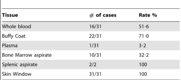
Every time we obtained a biopsy for microscopic examination, part of it went into culture on NNN medium. The efficiency of demonstrating the parasite using microscopy and/or culture is summarized in table 3.
Figure 2.Leishmaniaparasites shown in lymphoid organs.(A) Giemsa stained section 406mag. of a lymph node from patient 11 showing intracellular leishman bodies (purple in color) in the center of the photograph (white arrow). (B) Indirect immunohistochemistry using Fluorescein-labeled secondary antibody (FITC) and anti-leishmania primary antibody on a splenic aspirate from patient 9.Leishmaniaparasites are evident as
bright green entities (red arrows, 406mag.). doi:10.1371/journal.pntd.0001782.g002
Figure 3.Leishmaniaparasites shown on Skin Windows.(A) A normal Skin Window showing intracellular (white star) and extracellular clumps (doublets) of Leishmania parasites, oil immersion lens 1006mag. (B) A normal Skin Window showing four monocytes in motion with many
The best test for detecting the parasite microscopically was the Skin Window. The next in efficiency (culture and/or microscopy) was Buffy Coat, which revealed the presence of parasites in 71% of the cases. Whole blood followed. Bone marrow aspirate’s yield was positive only in one third of the cases which could be a reflection of its depressed activity. Although obtaining a splenic aspirate entails a serious risk (the reason why we limited it to 2 patients), we still think it is a very efficient tissue in yielding parasites.
Discussion
Kala-Azar has been established to present with a well defined clinical picture, but for the last few decades suspicion of having systemic leishmaniasis with a symptomatology different from the classically described, has been gaining greater and greater support. Such cases with a variable degree of illness were detected on the basis of positive serology [7–16].
In our series, the patients presented with symptoms typical of immune deficiency in the presence of a normal immune profile. Still they were found to harborLeishmaniaorganisms. In contrast to the reported sub-clinical cases among whom some subjects were totally symptom free while others had a milder form of the classical disorder, our cases presented with depressed immunity exhibiting symptoms pertaining to one or two target systems or organs.
A novel clinical picture of Visceral Leishmaniasis started to unfold. It consists of depressed immunity predisposing the subjects to infections with a wide spectrum of organisms (bacterial, viral and fungal). They presented with symptoms varying in type and severity. Although these secondary infections tend to resolve (with or without treatment), yet they invariably recurr. Thus in most patients and before referral, systemic anti-bacterial treatment was extensively used, sometimes in association with surgical interven-tions especially for skin abscesses and other tumorous growths. Anti-fungal agents were mostly added in cases with gastrointestinal complaints. Improvement or partial relief of symptoms arising from the superimposed infections was reported in most of our cases, but no complete cure was ever attained. Furthermore, it is important to note that none of the patients evolved to display the full fledged picture of Kala-Azar.
This form of atypical presentation is distinct from what has been described so far as sub-clinical illness meaning that it is not a lighter classical disease. This entity is novel in the sense that all patients were suspected to be immune-compromised and hence were sent to us for further immune function studies. Incidentally while performing the Skin Window to test the acute and chronic immune cellular responses to injury, we detected leishman bodies within and without the monocytes and some of the lymphocytes constituting the chronic phase of this reaction.
Whereas other investigators relied on immunological tests (purely serological) for proving the presence of the parasite in the candidate patients, we favored demonstrating it microscopi-cally within any of the appropriate tissues involved. Although the spleen when enlarged seemed to be the ideal organ to reveal the
parasite, yet the difficulty and the high risk implicated in its aspiration discourages most investigators from sampling it. Buffy Coat examination has obviously several advantages and was efficient in a good number of patients; however its efficacy drops the more the patient is anemic.
Besides, this set of data support the fact that systemic leishmaniasis in Lebanon and probably the area exists to a much greater extent than what we reported in our previous surveillance study [20]. Furthermore, the cases were detected in referrals from only two medical centers. Admitting the fact that the number of patients we are reporting is small for solid statistical analysis, still we think it may not be futile to detect a trend that could characterize such patients. So far the skin seems to be the organ most frequently targeted by these parasites. This is not surprising since these preferentially reside in cells of the hair follicles. Other systems may represent the shock organs being frequently affected by infections as happens in defective body defenses. As expected, it is the younger subjects (infants and children) who are the population at high risk.
Our major recommendation is to entertain the diagnosis of atypical leishmaniasis irrespective of age or country of patients presenting with depressed immunity, especially so whenever thorough investigation covering a large spectrum of diseases fails and multiple shotgun style therapies are tried without any success.
Acknowledgments
We thank the patients and their parents/guardians who gave consent to participate, and the staff at participating centers.
Author Contributions
Conceived and designed the experiments: NNS KK AJ RF KAS HNC. Performed the experiments: NNS KAS AJ RF HNC KK. Analyzed the data: NNS KK AJ RF KAS HNC. Contributed reagents/materials/ analysis tools: NNS KK AJ RF KAS HNC. Wrote the paper: NNS KK AJ RF KAS HNC.
References
1. Neva FA (1990) Immunotherapy for parasitic disease. N Engl J Med 322: 55–7. 2. Badaro´ R, Jones TC, Lorenc¸o R, Cerf BJ, Sampaio D, et al. (1986) A prospective study of visceral leishmaniasis in an endemic area of Brazil. J Infect Dis 154: 639–49.
3. Lysenko AJ, Beljaev AE (1987) Quantitative approaches to epidemiology. In: Peters W, Killick Kendrick R, eds. The leishmaniases in biology and medicine. Vol I. Biology and epidemiology. London: Academic Press 263–90. 4. Desjeux P (1992) Human leishmaniases: Epidemiology and Public Health
Aspects. World Health Stat Q 45: 267–75.
5. Pearson RD, Sousa AQ (1995) Clinical spectrum of leishmaniasis. Clin Infect Dis 22:1–13.
6. Pampiglione S, Manson-Bahr PE, La Placa M, Borgatti MA, Musumeci S (1975) Studies in Mediterranean leishmaniasis. 3. The leishmanin skin test in kala-azar. Trans R Soc Trop Med Hyg 69:60–8.
7. Pampiglione S, Manson-Bahr PE, Giungi F, Giunti G, Parenti A, et al. (1974) Studies on Mediterranean leishmaniasis. 2. Asymptomatic cases of visceral leishmaniasis. Trans R Soc Trop Med Hyg 68: 447–53.
8. Ho M, Siongok TK, Lyerly WH, Smith DH (1982) Prevalence and disease spectrum in a new focus of visceral leishmaniasis in Kenya. Trans R Soc Trop Med Hyg 76: 741–6.
9. Guerra M, Furtado T, Barros G, Sessa P, Daher V (1985) Infecc¸a˜o subclı´nica na leishmaniose tegumentar. An. Bras. Dermatol 60: 365–369.
Table 3.Efficiency of different tissues in demonstrating and/ or yielding cultures ofLeishmaniaparasites.
Tissue #of cases Rate %
Whole blood 16/31 51?6
Buffy Coat 22/31 71?0
Plasma 1/31 3?2
Bone Marrow aspirate 10/31 32?2
Splenic aspirate 2/2 100
Skin Window 31/31 100
10. Badaro R, Jones TC, Carvalho EM, Sampaio D, Reed SG, et al. (1986) New perspectives on a subclinical form of visceral leishmaniasis. J Infect Dis 154: 1003–11.
11. Carvalho EM, Barral A, Pedral-Sampaio D, Barral-Netto M, Badaro´ R, et al. (1992) Immunologic markers of clinical evolution in children recently infected with Leishmania donovani chagasi. J Infect Dis 165: 535–40.
12. Evans TG, Teixeira MJ, McAuliffe IT, Vasconcelos I, Vasconcelos AW, et al. (1992) Epidemiology of visceral leishmaniasis in northeast Brazil. J Infect Dis 166: 1124–32.
13. Holaday BJ, Pompeu MM, Evans T, Braga DN, Texeira MJ, et al. (1993) Correlates of Leishmania-specific immunity in the clinical spectrum of infection with Leishmania chagasi. J Infect Dis 167: 411–7.
14. Zijlstra EE, el-Hassan AM, Ismael A, Ghalib HW (1994) Endemic kala-azar in eastern Sudan: a longitudinal study on the incidence of clinical and subclinical infection and post-kala-azar dermal leishmaniasis. Am J Trop Med Hyg 51: 826–36.
15. Follador I, Arau´jo C, Bacellar O, Arau´jo CB, Carvalho LP, et al. (2002) Epidemiologic and immunologic findings for the subclinical form of Leishmania braziliensis infection. Clin Infect Dis 34: 54–8.
16. Gama ME, Costa JM, Gomes CM, Corbett CE (2004) Subclinical form of the American visceral leishmaniasis. Mem Inst Oswaldo Cruz 99: 889–93. 17. Ostyn B, Gidwani K, Khanal B, Picado A, Chappuis F (2011) Incidence of
symptomatic and asymptomatic Leishmania donovani infections in high-endemic foci in India and Nepal: a prospective study. PLoS Negl Trop Dis 5:e1284.
18. Bitar J, Nachman H (1964) Kala Azar in Lebanon. J Med Liban 17: 257–68. 19. Rebuck JW, Crowley JH (1955) A method of studying leukocytic functions in
vivo. Ann N Y Acad Sci 59: 757–805.