697 697 697 697 697 Mem Inst Oswaldo Cruz, Rio de Janeiro, Vol. 101(7): 697-714, November 2006
The resumption of consumption – A review on tuberculosis
Rodrigo Gay Ducati**, Antonio Ruffino-Netto*, Luiz Augusto Basso
+/
++,
Diógenes Santiago Santos
+/
++Faculdade de Farmácia, Faculdade de Biociências, Centro de Pesquisas em Biologia Molecular e Funcional, Pontifícia Universidade Católica do Rio Grande do Sul, Av. Ipiranga 6681, Tecnopuc-Prédio 92A, 90619-900 Porto Alegre, RS, Brasil
*Departamento de Medicina Social, Faculdade de Ribeirão Preto, Universidade de São Paulo, Ribeirão Preto, SP, Brasil **Programa de Pós-graduação em Biologia Celular e Molecular, UFRGS, Porto Alegre, RS, Brasil
Among all infectious diseases that afflict humans, tuberculosis (TB) remains the deadliest. At present, epidemiolo-gists estimate that one-third of the world population is infected with tubercle bacilli, which is responsible for 8 to 10 million new cases of TB and 3 million deaths annually throughout the world. Approximately 95% of new cases and 98% of deaths occur in developing nations, generally due to the few resources available to ensure proper treatment and where human immunodeficiency virus (HIV) infections are common. In 1882, Dr Robert Koch identified an acid-fast bacterium, Mycobacterium tuberculosis, as the causative agent of TB. Thirty-nine years later, BCG vaccine was introduced for human use, and became the most widely used prophylactic strategy to fight TB in the world. The discovery of the properties of first-line antimycobacterial drugs in the past century yielded effective chemotherapies, which considerably decreased TB mortality rates worldwide. The later introduction of some additional drugs to the arsenal used to treat TB seemed to provide an adequate number of effective antimicrobial agents. The modern, standard short-course therapy for TB recommended by the World Health Organization is based on a four-drug regimen that must be strictly followed to prevent drug resistance acquisition, and relies on direct observation of patient com-pliance to ensure effective treatment. Mycobacteria show a high degree of intrinsic resistance to most antibiotics and chemotherapeutic agents due to the low permeability of its cell wall. Nevertheless, the cell wall barrier alone cannot produce significant levels of drug resistance. M. tuberculosis mutants resistant to any single drug are naturally present in any large bacterial population, irrespective of exposure to drugs. The frequency of mutants resistant to rifampicin and isoniazid, the two principal antimycobacterial drugs currently in use, is relatively high and, therefore, the large extra-cellular population of actively metabolizing and rapidly growing tubercle bacilli in cavitary lesions will contain organisms which are resistant to a single drug. Consequently, monotherapy or improperly administered two-drug therapies will select for drug-resistant mutants that may lead to drug resistance in the entire bacterial population. Thereby, despite the availability of effective chemotherapy and the moderately protective vaccine, new anti-TB agents are urgently needed to decrease the global incidence of TB. The resumption of TB, mainly caused by the emergence of multidrug-resistant (MDR) and extensively drug-resistant (XDR) strains and HIV epidemics, led to an increased need to understand the molecular mechanisms of drug action and drug resistance, which should provide significant insight into the development of newer compounds. The latter should be effective to combat both drug-susceptible and MDR/XDR-TB.
Key words: Mycobacterium tuberculosis - epidemiology - chemotherapy - vaccine - drug action - mechanism of resistance
There is a dread disease which so prepares its victim, as it were, for death; which so refines it of its grosser aspect, and throws around familiar looks, unearthly indications of the com-ing change – dread disease, in which the struggle between soul and body is so gradual, quiet, and solemn, and the result so sure, that day by day, and grain by grain, the mortal part wastes and withers away, so that the spirit grows light and sanguine with its lightening load, and, feeling immortality at hand, deems it but a new term of mortal life; a disease in which death takes the glow and hue of life, and life the gaunt and grisly form of death; a disease which medicine never cured, wealth warded off, or poverty could boast exemption from; which sometimes moves in giant strides, and sometimes at tardy pace; but, slow or quick, is ever sure and certain.
Charles Dickens, 1870, in Nicholas Nickleby, Wiendenfeld and Nicholson, London, p. 243.
Human tuberculosis (TB) is a contagious-infectious disease mainly caused by Mycobacterium tuberculosis, which is an aerobic pathogenic bacterium that establishes its infection usually in the lungs. Progression of TB in-fection is fundamentally regulated by host’s immune system integrity, which may succeed through microbial immediate elimination and/or latency conditioning, or fail resulting in development of active disease.
TB was responsible for millions of human deaths in the past, when there were no adequate treatment methods for infected patients. Introduction of chemotherapy and prophylactic measures led to drastic death reduction, which was maintained for various decades. However, the “good times” waned, as this disease became worldwide recognized as the one responsible for most human deaths caused by a single infectious agent. TB resumption is basically a consequence of anthropic factors, such as the recent HIV/AIDS pandemic and the development of drug-resistant strains (stemmed from inappropriate treatments and/or patient non-compliance). It thus appears to be of fundamental importance to increase investment in re-search, as disease control can hopefully be reached Financial support: Fapergs, CNPq, Pronex, Instituto do Milênio
+Corresponding authors: diogenes@pucrs.br; luiz.basso
@pucrs.br
++Research fellow CNPq
698 698 698 698
698 The resumption of consumption • Rodrigo Gay Ducati et al.
through new drug development (to be introduced in the treatment of patients with active TB), and through pro-phylactic and/or therapeutic vaccine optimization.
Looking the past
The captain of all these men of death that came against him to take him away, was the consumption, for it was that brought him down to the grave.
John Bunyan, 1680, in The Life and Death of Mr. Badman, Dent, London, 1928, p. 282.
TB, also known as the white plague, received the title of “captain of all these men of death” by John Bunyan in the second half of the XVII century, when the disease reached a high level of death rates in Europe. This malady became the principal cause of death by the end of the XIX and beginning of the XX century, and among its various victims were worldwide known people, such as Frédéric Chopin, Paganini, St. Francis of Assisi, Charlotte Brontë (and most of the Brontë family), John Keats, Lord Byron, George Orwell, Castro Alves, Alvarez de Azevedo, Cruz e Souza, Augusto dos Anjos, Noel Rosa, Eleanor Roosevelt, and Vivian Leigh, among many others. Cur-rently, this disease still represents a global threat, as it stands as the leading cause of death due to an infectious agent among adults worldwide.
Although it was probably described for the first time in Indian texts, pulmonary TB is known since the time of Hippocrates as phthisis, which is derived from the Greek for “wasting away”. Scrofula, a rare manifestation form of TB that affects the lymph nodes, especially of the neck, most commonly found in children and usually spread by unpasteurized milk from infected cows, was well docu-mented during the European Middle-Age, when it was believed that cure resulted from the power of the divine touch of the kings. Pott’s disease or Gibbous deformity, a rare TB manifestation, revealed only among several an-tique Egyptian mummies, is a destructive form of TB that leads to serious spine deformities and subsequent mem-ber paralysis.
In 1680, the French Franciscus Sylvius carried out anatomic-pathologic studies in pulmonary nodules from TB patients, which he named as “tubercula” (small knots), observing their evolution to lung ulcers (cavities). How-ever, most of the great pathologists of his time believed these knots were some type of tumor or abnormal gland, rejecting any probable infectious origin. The first credible speculation of the infectious nature of TB was performed by the British Doctor Benjamin Marten, who proposed in 1722 that TB could be transmitted through the “breath” of a sick person, inhaled by a sound one, and thereby turning her ill. In 1689, the English Doctor Richard Morton used the term “consumption” to specifically denote TB, and finally, in 1819, the inventor of the stethoscope, the French Doctor René Laennec identified for the first time the TB manifestation unit.
As the disease became completely established among every European social level, afflicting many of the intel-lectual and artists of the continent by the half of the XIX century, TB was romanticized, as typical symptoms like thin and pale faces of the infected ones became signs of beauty. The romantic Era of TB can also be recognized in
fine pieces of art, such as in the famous painting of Wil-liam Morris, which exhibits all the splendor of the leg-endary Guinevere, King Arthur’s wife, already displaying typical symptoms of the active disease.
In 1865, the Military Surgeon Jean-Antoine Villemin demonstrated formally that TB is a contagious disease; although his experimentation could be effectively repro-duced in rabbits, the finding was ignored by his contem-poraries for a long time.
One of the greatest works on TB was performed in 1882 by Robert Koch, an esteemed scientist of his time. Koch isolated and cultured M. tuberculosis from crushed tubercles. His experimental work identified the bacterium as the TB etiological agent (Bloom & Murray 1992, Daniel 1997). In August of 1890, during The First Ordinary Ses-sion of the International Medical Congress, in Berlin, he announced the discovery of a TB therapeutic drug. Three months later, the “Deutsche Medizinische Wochen-schkift”, in extraordinary edition, published a new state-ment of Koch, revealing that although interested in the therapeutic properties of his findings, he observed that the referred liquid, named tuberculin, could be useful as a diagnostic tool to detect the disease due to the intensi-fied reaction developed by sick animals inoculated with this drug, as no measurable effect was ever observed in healthy ones. This concept was perpetuated for several years, until it was observed that even healthy animals could react to the drug. The veterinarians of his time clarified the fact by demonstrating that the healthy ones could be simply infected, although not ill. As a result, it was established that M. tuberculosis-infected animals will re-act to tuberculin infusion, whereas the non-infected ones will not. This drug, the first industrialized one, was called old tuberculin; subsequently, other tuberculins were pro-duced, such as purified protein derivate (PPD), PPD-S, and PPD RT23, among others (Vaccarezza 1965, Ruffino-Netto 1970). The tuberculin skin test became the principal tool for infection diagnosis. In the same period, Koch developed staining methods for the identification of the bacillus; these techniques were subsequently improved by the German Doctor and bacteriologist Paul Ehrlich, whose method for detection of the bacillus provided the basis for the development of the Ziehl-Nielsen staining, which still is an important tool to diagnose TB.
Koch’s discovery allowed researchers to focus efforts on the development of new and more efficient therapies to treat TB patients. One of the first attempts to fight the disease was performed by Edward Livingston Trudeau, who suffered from TB and was subsequently cured. Trudeau established the first sanatorium in the United States in 1884. This institution received only TB patients, and invested in a treatment based on rest, fresh air and a healthy diet.
699 699699 699699 Mem Inst Oswaldo Cruz, Rio de Janeiro, Vol. 101(7), November 2006
which conferred immunological protection against sub-sequent challenges with virulent M. tuberculosis. Thir-teen years of experimentation led to the obtaining of the 231st passage, the variant that was administered for the first time in humans (orally), as an attempt to immunize a child whose mother died in childbirth victim of TB. Cur-rently known as BCG (bacille Calmette-Guérin), the (in-tradermal) vaccine has become widely used to combat TB; it relies on a prophylactic administration of live attenuated bacilli to children.
The introduction of antibiotics, such as streptomycin (1947), isoniazid (synthesized in 1912, but introduced 40 years later) and p-aminosalicylic acid, led to a TB che-motherapy revolution, as TB mortality rates were consid-erably reduced (Bloom & Murray 1992, Daniel 1997). Subsequently, other anti-TB drugs were also developed, such as ethambutol and rifampicin, among others. Since the mid 1980s, however, there has been no new first-line drug development to fight the TB causing bacilli (Petrini & Hoffner 1999).
In Brazil, it is believed that the disease was introduced by the Portuguese and Jesuit missionaries since 1500. Oral BCG was administered for the first time by Arlindo de Assis in 1927 to newborn, and intradermal vaccination was implemented in 1973, becoming obligatory for one year minors since 1976. Brazilian TB mortality rates were drastically reduced due to introduction of tuberculostatic drugs by the 1940s, including streptomycin (1948), p -ami-nosalicylic acid (1949), and isoniazid (1952). The standard chemotherapy treatment recommended by the World Health Organization (WHO) to control or eradicate TB worldwide, which is based on a short-course therapy that combines the use of four anti-TB drugs, currently known as directly observed treatment short-course (DOTS), seems to be used in Brazil since 1962 by the Fundação de Serviço Especial de Saúde Pública (Sesp) in units of all complexity levels (Ruffino-Netto 2002).
Tuberculosis resumption
In 1993 the WHO declared TB a global public health emergency, being the only disease so far to warrant that designation. Although hospitals have been established and chemotherapy has been developed to combat TB, which have brought considerable reduction in incidence to developed nations, historical data calculated by the WHO indicate that there have not been great effects on the global problem since the time of Koch (Bloom & Murray 1992). Currently, TB is responsible for more human deaths than any other single infectious agent, standing for 26% of all preventable deaths and 7% of all deaths (Enarson & Murray 1996).
TB resumption has been attributed to several factors, such as the increase in drug resistance; the HIV/AIDS pandemic (in the beginning of the 1980s); the increase of injectable drug users; changes in social structure; the increase of immigrants from high prevalence nations to developed ones; the aging of the world population; the active transmission amongst environments of human ac-cumulation (such as prisons, hospitals, and homeless shelters); and the degradation of health care systems (Fätkenheuer et al. 1999). Although TB became a
reemerg-ing disease to European and North-American nations, TB is not an emergent nor reemerging public health problem in developing countries such as Brazil, but rather a long lasting one (Ruffino-Netto 2002).
In order to facilitate the comprehension of the various components involved in the interaction between these factors, Ruffino-Netto (2004) proposed an “equation” which expresses the TB charge, represented as follows:
(SIN).(PHIV).(PDEF).(PR).(MIG).(OLDP) TbB ≈
(AHS).(DOTS).(EDU).(NUT).(HRTb).(DPP) where SIN: social inequality; PHIV: prevalence of HIV-positive; PDEF: percentual default of treatment; PR: prevalence of primary resistance + acquired resistance; MIG: migrations; OLDP: age of the population; AHS: adequate health services; DOTS: directly observed treatment short-course; EDU: educational level; NUT: nutrition level; HRTb: human resources for TB control; DPP: degree of political participation of the population. Among all the components, social inequality should be emphasized as the most important one, since it gener-ates poverty and, consequently, leads to malnutrition, ill-provided living conditions and education, among others, thereby influencing practically all other components. It should also be pointed out that the prevalence of primary resistance (whose definition is given below) stands as an aggravating epidemiological factor more important than acquired resistance (subsequently defined under treatment section). According to Ruffino-Netto (2004), the above-mentioned expression does not represent a mathematical equation and, therefore, will not yield predictable numeric solutions, but rather facilitates to wonder about the problem (TB charge analysis). One should notice that it contains variables of qualitative nature.
In nations such as India, China, Indonesia, Pakistan, Nigeria, Philippine, South Africa, Ethiopia, Vietnam, Rus-sian Federation, Democratic Republic of Congo, Brazil, United Republic of Tanzania, Kenya, Thailand, Myanmar, Afghanistan, Uganda, Peru, Zimbabwe, and Cambodia, it is estimated an occurrence of up to 80% of world TB cases. It can be immediately perceived that, among the above-mentioned nations, the problem may be located either at the level of the numerator, denominator, or both within the expression. In Russia, for example, the multidrug-resistance prevalence problem is prominent and severe. In African nations, all the numerator and deno-minator components have a strong influence. Brazil might represent an intermediate condition, as it displays a (yet) small multidrug-resistance prevalence problem, but deals with major social inequality and has all remaining components in a phase of organization.
It is possible that a more elaborated reflection of this new “formula” might allow one to perceive that at the same time technical, biological, clinical, and epidemio-logical knowledge on TB advances, its social dimension should be remembered, valorized and used as an indica-tor of poor living conditions in other discussion forums such as The World Health Assembly, The International Labour Organization, and The World Trade Organization, among others.
700 700 700 700
700 The resumption of consumption • Rodrigo Gay Ducati et al.
has had some hindrances, in part, owing to human natural reluctance in complying with the 6 month-intensive treat-ment required, and more recently, owing to developtreat-ment of drug-resistant strains of the bacillus (Young 1998). In 1969, the Medic Chief of the National Institute of Health declared in congress that “it was time to close the books about infectious diseases” (Bloom & Murray 1992). With a similar point of view, the TB problem was considered to be “resolved” by authorities, as the malady, physiopa-thology, diagnostic methods, therapeutic strategies and pharmaceutical resources were already well known (Ruffino-Netto 2002). Arrogant and low-priced behaviors like these have severe consequences, as millions of people suffer daily from the various diseases (infectious or not) that afflict humans.
M. tuberculosis
M. tuberculosis is the principal TB etiological agent in humans; it is a weak Gram-positive rod-shaped bacte-rium that has no flagellum, does not form spores nor pro-duces toxins and has no capsule. The microbe’s width and height vary from 0.3 to 0.6 and 1 to 4 µm, respectively, and it presents a complex cellular envelope, considerably slow growth, and genetic homogeneity. It is a macroph-age intracellular pathogen that establishes its infection preferentially to the pulmonary system, where it is usu-ally conditioned into a dormancy state as long as host’s immune system prevails. The generation time is around 24 h in both synthetic medium and on infected animals (Cole et al. 1998). Bacterial growth at laboratory envi-ronment permits visual colony formation; these have a dry and wrinkled surface, and requires 3 to 4 weeks of growth on solid media to become visible (Bloom & Murray 1992). Owing to its pathogenicity and aerosol transmission, there are specific biosafety guidelines that demand the use of laminar-flow hoods and level 3 facility equipments for M. tuberculosis laboratorial work.
Currently, there are 60 known species among the My-cobacterium genera, most of them being saprophytic soil bacteria; and a minority of these species is pathogenic to humans, causing TB (M. tuberculosis, M. bovis, and M. africanum) and leprosy (M. leprae) (Jarlier & Nikaido 1994). The M. tuberculosis complex consists of M. tu-berculosis, M. bovis, M. bovis BCG, M. africanum, and
M. microti; and it is believed that their ancestral is a bac-terium from soil, and that the human bacillus may have been derived from the bovine form (M. bovis), probably through cattle domestication by the pre-dynastic Egyp-tians. M. tuberculosis genomic studies indicate that hori-zontal transference of genetic material to a free living an-cestral from the M. tuberculosis complex might have oc-curred in nature before the bacillus adopted its special-ized intracellular niche (Cole et al. 1998, Daniel 1997).
There is a great variability in the infection rates among people exposed to different infection sources, and among infected ones, approximately 90% will never develop the disease at all. There are also differences in the capacity to transmit the infection among patients due to exhaled bac-terial charge variability. Experimental evidence also sug-gests that the observed variation in transmission poten-tial might be attributed to pathogenic features. M.
tuber-culosis can have virulence variations among different strains; the more virulent ones present higher pathoge-nicity or capacity to develop active disease. However, it should be pointed out that it is fundamental to have a clear distinction between the capacity a strain has to cause infection and its capacity to develop the disease (Bloom & Small 1998).
Like many other infectious diseases, TB presents epi-demic cycles that might, although rarely, take centuries to end its course. History has proved that pathogenic viru-lence suffers considerable reduction along time, and natu-ral selection takes place upon its hosts, as only the more genetically resistant ones survive. Since untreated TB patients have mortality rates ranging from 40 to 60%, one can consider the possibility of human selection occurrence in favor to the ones with increased genetic resistance (Bloom & Small 1998). Although genes will not influ-ence the risk of infection upon exposure, they might determine the risk of disease development and its course (Daniel 1997). Furthermore, although genetic variability evidence on TB human susceptibility is very difficult to be determined, several studies revealed a greater disease severity among black than white people, there is a greater TB course agreement between monozygotic than heterozy-gotic twins, and populations more recently exposed to the TB bacillus have a greater propensity to develop the disease when compared to others that coexist with TB for centuries. As one gathers all these data, he can suppose that M. tuberculosis still is an extraordinary pathogen which exerts a powerful selective pressure on the human genome (Bloom & Small 1998).
M. tuberculosis genome
The complete genome sequencing of the best charac-terized M. tuberculosis strain, H37Rv, allowed the iden-tification of unique microbial features. This pathogen has a circular chromosome with 4,411,529 base pairs with a 65.6% G+C content. Since its isolation in 1905, this strain has had a great application worldwide in biomedical re-search due to total TB virulence retention in animal mod-els, and also because it is susceptible to drugs and ame-nable to genetic manipulation (Cole et al. 1998). M. tu-berculosis sequence determination establishes a new phase in the battle against one of the more successful predators of the human race (Young 1998).
701 701701 701701 Mem Inst Oswaldo Cruz, Rio de Janeiro, Vol. 101(7), November 2006
in its genome, which can be involved with the fact that
M. tuberculosis has a low lysis level in culture. Interest-ingly, about 59% of the genes are transcribed in the same direction as the replication; once compared to the 75% of
Bacillus subtilis, one can probably relate this feature to the peculiar slow growth and infrequent replication cycles of the former one. Data comparative analyses allowed the precise function attribution to 40% of the proteins identi-fied, and some information or similarities to another 44%. The remaining 16% have no similarity to any known pro-tein, being probably involved in specific mycobacterial functions. Among secreted proteins that were identified in the mycobacterial genome sequence that could act as virulence factors are phospholipases C, lipases and es-terases, which might attack cellular or vacuolar mem-branes, as well as several proteases. One of these phos-pholipases is involved in bacterial persistence in the nu-trient-limited phagosome environment.
Stewart Cole et al. (1998) made an important contri-bution as they identified a group of variable elements, the polymorphic G+C-rich sequences, which correspond to a family of sequences that encode proteins with small pep-tide motifs, PE and PPE, which are organized as common repetitive domains. These proteins represent approxi-mately 10% of the genome coding capacity and seem to be remnants of the ones related to antigenic variation in other bacteria. Through protein expression pattern alternation, these pathogens can be presented to their host’s immune system as a moving target, interfering in the immunological response by antigen processing inhibition, and thereby, ensuring a greater survival probability to the bacteria.
M. tuberculosis proteome determination and its com-parison with that of other microorganisms whose se-quences are available revealed a significant statistical pref-erence for the amino acids alanine, glycine, proline, argi-nine, and tryptophan, which are all encoded by G+C-rich codons, and a comparative reduction in the use of amino acids encoded by A+T-rich codons, such as asparagine, isoleucine, lysine, phenylalanine, and tyrosine. By means of mycobacterial genomic inspection, it became clear that besides the various functions involved in lipid metabo-lism, the enzymes required for glycolysis, the pentose phosphate pathway, and the tricarboxylic acid and glyoxylate cycles are also present, thereby evidencing the dynamic metabolism of the bacillus (Cole et al. 1998).
Mycobacterial cellular envelope
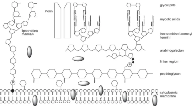
Mycobacteria produce an extremely uncommon cell wall structure; the peptidoglycan contains N -gly-colylmuramic acid instead of the usual N-acetylmuramic acid, found amongst most other bacteria. A far more dis-tinctive feature is that up to 60% of the mycobacterial cell wall is composed of lipids that consist basically of uncommonly long-chain fatty acids with 60 to 90 carbons, denominated mycolic acids (Brennan & Nikaido 1995). Mycolic acids are branched fatty acids that have a short and a long branch, with 22 to 24 and 40 to 64 carbons, respectively (Jarlier & Nikaido 1994); they are covalently linked to the polysaccharide that composes the cell wall, the arabinogalactan, which in turn is attached to
pepti-doglycan by a phosphodiester link (Brennan & Nikaido 1995). Approximately 10% of the arabinose residues in the arabinogalactan are substituted by mycolic acids (McNeil & Brennan 1991). The cell wall also contains several other free lipid species, which are not covalently attached to this basal skeleton (the mycolylarabinoga-lactan-peptidoglycan complex). These lipids can act as antigens in the host (Brennan & Nikaido 1995).
In 1982, Minnikin proposed a new cell wall model where the mycolic acid chains are packed side by side perpendicular to the cell surface, and this inner leaflet of long-chain fatty acids is covered by an outer leaflet com-posed of extractable lipids, thereby reproducing an asym-metric lipid bilayer. Recently, this model was updated by mycobacterial cell wall X-ray diffraction studies. In the ara-binogalactan polysaccharide, both galactan main chain and arabinan side branches are designed in a manner that would ensure maximum mobility between sugar residues. The mycolic acid residues are esterified to approximately two-thirds of the non-reducing termini of this highly branched polysaccharide (McNeil & Brennan 1991), as shown in Fig. 1.
Another distinguishing property shared among myco-bacteria is the fact that their cell wall retain carbol fuch-sin dye even in the presence of acidic alcohol, for this reason the rod-shaped mycobacteria are also known as acid fast bacilli (Glickman & Jacobs 2001). M. tubercu-losis produces a considerably diverse array of lipophilic molecules, which range from simple fatty acids, such as palmitate and tuberculostearate, to long-chain complex molecules, such as mycolic acids and phenolphthiocerol alcohols (mycoside attachment) (Cole et al. 1998). Al-though mycobacteria have various cell wall lipid types, some are limited to specific species, such as sulfolipids, solely present in M. tuberculosis and which are involved in its pathogenicity (Brennan & Nikaido 1995). Further-more, the mycobacterial cell wall fluidity gradient appears to have an opposite orientation to all Gram-negative bac-teria, as the more external regions are more fluid than the internal ones (Brennan & Nikaido 1995).
702 702 702 702
702 The resumption of consumption • Rodrigo Gay Ducati et al.
suggesting contribution of a lipophilic pathway to lipo-philic solute transport (Jarlier & Nikaido 1994).
Since mycobacteria are relatively resistant to drying, alkali, and many other chemical disinfectants, it is thus very difficult to prevent M. tuberculosis transmission in urban institution environments. This resistance, and the resistance to therapeutic agents, are both basically con-ferred by the extremely uncommon mycobacterial cell wall structure (Brennan & Nikaido 1995). The unusual cell wall also permits the microorganism to survive inside the macrophage, which would usually destroy phagocytosed pathogens (NSB Editorial Comment 2000).
Although the cell wall acts as an exceptional perme-able barrier, resistance to drugs in mycobacteria normally requires the participation of additional mechanisms, such as the removal of incorporated antibiotic molecules through chemical modification by β-lactamases that, syn-ergistically, confer significant resistance levels (Jarlier & Nikaido 1994). Thereby, since M. tuberculosis has a cell wall with a relatively high permeability, the inactivation of the second factor in synergism may allow an effective chemotherapy. M. tuberculosis cell wall has become a tar-get of the more recent researches towards the elucidation of the mechanism of action of many old drugs and the search of targets for the design of new ones (Brennan & Nikaido 1995). New data on genes specifically involved in its synthesis may represent potential drug targets (Young 1998).
Epidemiology and disease properties
Based on tuberculin skin test reactivity, epidemiolo-gists estimate that around one third of the world popula-tion (1.7 billion people) is infected with M. tuberculosis, and at risk of developing active TB. Statistical data indi-cate the occurrence of 8 to 10 million new TB cases and 3
million deaths annually, afflicting mostly the young and productive adults. Under the current conditions, it is ex-pected for this decade that 90 million people will develop the disease and 30 million will die from TB (Enarson & Murray 1996).
TB seems, to a certain point, under control in devel-oped countries such as Japan and United States, but de-tains a violent manifestation in other places like south-eastern Asia, Africa, and some regions of the Pacific, mostly due to complications of HIV infection and drug resistance (Brennan 1997). Approximately 95% of TB cases occur in developing nations, where 98% of the world TB death cases happen. According to the WHO databank, in 1998 Brazil occupied the 13th position among 22 coun-tries where TB was well disseminated. In 1999, an inter-esting study of the distribution of TB notified cases among Brazilian states revealed a decreasing ordered incidence among São Paulo, Rio de Janeiro, Bahia, Minas Gerais, and Rio Grande do Sul (Ruffino-Netto 2002).
Currently, multidrug-resistant (MDR) TB presents a high incidence, in an increasing order, in Latvia, India, Estonia, Dominican Republic, and Argentina, and low in-cidence in most occidental European and African coun-tries and United States (Fätkenheuer et al. 1999). The pres-ence of resistant strains has a direct relation to drug avail-ability and an inverse relation to treatment efficacy. A WHO and International Union Against Tuberculosis and Lung Disease (IUATLD) anti-TB drug-resistance global surveillance project made among 35 nations of 5 continents with standardized methods showed that during the 1994-1997 period, all the countries and regions analyzed presented M. tuberculosis strains resistant to at least one drug, usually isoniazid or streptomycin, suggesting that the disease represents a global problem (Pablos-Mendez et al. 1998).
703 703703 703703 Mem Inst Oswaldo Cruz, Rio de Janeiro, Vol. 101(7), November 2006
Human TB is an infectious disease caused by some mycobacteria of the “M. tuberculosis complex”, includ-ing M. bovis, M. africanum, and prevalently M. tubercu-losis. According to the WHO, TB kills more people than malaria and AIDS together. Annually, TB is responsible for the death of 100,000 children worldwide, and 161,800 new cases occur only in Brazil. From now until 2020, it is estimated that 1 billion more people will be infected, 200 million will develop the disease, and 70 million will die in case surveillance and control strategies continue as they are (Pasqualoto & Ferreira 2001).
The principal means of transmission occurs by infec-tive particles. Acinfec-tive TB patients will usually cough, as a result of typical chronic pulmonary inflammation, which constitutes the main dissemination mechanism for the pathogen to new hosts (Glickman & Jacobs 2001). The released particles from an ill patient are exhaled from the infected lungs into the air, being able to remain in sus-pension for hours, representing a highly contagious disease (NSB Editorial Comment 2000). Infection usually occurs from person to person through the inhalation of the infective particles (Pasqualoto & Ferreira 2001). Ex-periments with animal models demonstrate that particles in suspension containing 1 to 10 bacilli are enough to cause an infection. The main determinants of risk of in-fection are the concentration of bacilli in an exhaled particle from a source, its aerodynamic features, the ventilation rate, and the exposure period (Bloom & Murray 1992).
Usually, upon infection, inhaled bacilli are ingested by phagocytic alveolar macrophages, and can either be immediately eliminated or grow in the intracellular envi-ronment in localized lesions called tubercles. Two to six weeks past infection are usually followed by the estab-lishment of cellular immunity, and subsequent lympho-cyte and activated macrophage infiltration into the lesion, which leads to the elimination of most portion of the ba-cilli and the end of the primary infection, commonly with-out symptom presentation. The sole evidence of previous infection in these cases can be identified by the tubercu-lin skin test reactivity, or, in some cases, evidences of calcified lesions by X-ray.
In most cases, however, the bacilli can coexist pacifi-cally within its human host as a quiescent or dormant form of infection, establishing a large bacterial reservoir among infected individuals. People harboring latent infection have an active TB developing risk of approximately 5% after the first year and 10% during their life-time.
Although much of the bacterial load is usually elimi-nated, a great proportion of the infiltrating phagocytes and lung parenchymal cells are also killed, which pro-duces a characteristic solid caseous necrosis (granuloma or Gohn complex) where some bacilli have the opportunity to hide. In case host immune response predominates, the lesion is contained, causing simply residual damage to the lungs. However, in case the necrosis reaction expands, breaking into a bronchus, a lung cavity can be formed, which may allow a massive bacterial dissemination into the air through coughing. There can be even worse cases, such as when inflammatory cells liquefy the solid necrosis, creating a rich environment for bacillary proliferation
(Bloom & Murray 1992, Young 1998).
Approximately 15% of the patients with the active dis-ease present extra-pulmonary TB, which is caused by granuloma evolution due to excessive bacterial growth, invading the blood stream and disseminating the bacilli to various parts of the body. Also called miliary TB, it frequently occurs in the pleura, lymph nodes, liver, spleen, bones and joints, heart, brain, genital-urinary system, meningis, peritoneum, and skin.
The pathological and inflammatory processes produce typical TB symptoms such as weakness, fever, weight loss, night sweat, chest pain, respiratory insufficiency, and cough; advanced pathology may also cause blood vessel disruption, which leads to hemoptisis (Bloom & Murray 1992). For this reason, TB was also known as consumption, since the disease is developed at a leisurely pace and with multiple symptoms which lead to gradual debilitation and physical exhaustion.
Immune system in tuberculosis
Following intravenous mycobacterial infection in mice, the bacilli present (initially) a very short replication time in vivo, when macrophage activation begins by mac-rophage-derived pre-inflammatory cytokines, such as inter-leukin 6 (IL-6), IL-12, and tumor necrosis factor (TNF), besides the involvement of gamma interferon (INF-γ), initially derived from natural killer (NK) cells, in order to contain or inhibit bacterial growth. Approximately 2 weeks after initial infection, there is a considerable reduction of bacterial growth (representing a plateau in a growth plot) due to the activation and differentiation of specific lymphocytes, which are able to supply the lack of INF-γ required to increase the initial innate response, activating macrophages that induce the nitric oxide synthase 2 (iNOS) to produce nitric oxide, one of the main myco-bacteriostatic mediators or effector molecules in mice. Following infection with M. tuberculosis, there is a significant reduction of bacterial load in the liver, and also in the spleen (to a lesser extent). The remaining bacilli enter into a state of non-replicating persistence, although these still are fully viable (Ehlers 1999). Although the bacillus charge in this phase of the infection in mice does not mimic the latent state in the human host, it represents an equilibrium between the pathogen’s persistence and the host’s immune response (Glickman & Jacobs 2001). This dormant but yet viable bacillary form can re-establish its replication and develop the active disease in certain conditions of immune suppression, such as aging, corticosteroid therapy, CD4 cell charge reduction or treatments with iNOS inhibitors. Unfortunately, total bacillary elimination is improbable solely through the immune system, and considerably difficult to be reached by chemotherapy (Ehlers 1999).
704 704 704 704
704 The resumption of consumption • Rodrigo Gay Ducati et al.
infected area (NSB Editorial Comment 2000). Granuloma formation might sometimes occur at the moment where the above-mentioned plateau is reached, causing destabi-lization and destruction of adjacent tissues, and possibly necrosis, followed by cavity formation (Jagirdar & Zagzag 1996). Granulomas are a result of CD4-mediated delayed-type hypersensitivity reaction within parenchymal tissues. Accordingly, the same system that is responsible for bacterial growth decrease (host defense) is also intrinsically associated with tissue damage through granu-loma formation and necrosis (Ehlers 1999). Many TB symptoms, including tissue destruction which eventually liquefies lung infected portions, are preferentially mediated by the host’s immune response against the bacillus instead of the bacterial virulence itself (Glickman & Jacobs 2001).
Depending on the bacterial charge that persists in the primary lesions, there can be granuloma development and differentiation. Owing to an efficient systemic antibacte-rial response, liver-localized granulomas will frequently suffer size reduction and can sometimes disappear. In the lungs, where bacterial charge is constantly high, it can be observed evident chronic-progressive pathology. Progres-sive interstitial fibrosis can eventually occur, gradually replacing most of the lung airspaces with dense fibrotic tissue separating groups of alveolar sacs, giving the entire lung a honeycomb appearance (Ehlers 1999).
M. tuberculosis latency and reactivation
Latent TB is a clinical syndrome caused by exposure to M. tuberculosis, followed by establishment of infec-tion and host’s immune response to control bacillary growth, forcing it into a quiescent state in the infected tissue. It is characterized by a reduction of bacterial me-tabolism, as a consequence of the action of cellular im-mune response, and which can, to a certain point, con-tain, but not eradicate, the infection. Contrary to active TB, latent TB is not an infectious disease and, therefore, does not represent a public health threat. Since latent TB is not presented as a clinical illness, the sole form to be diagnosed is by tuberculin skin tests. A positive result indicates that the patient has already had contact with the microbe. The method relies on the intradermal inocula-tion of PPD. In certain cases, the infecinocula-tion can be identi-fied through a chest X-ray radiography that demonstrates scars of an old infection.
The bacterial intracellular survival is based on its ca-pacity to deal with the phagosome acidification in infected macrophages and prevent the phagosome-lysosome fu-sion. In most immunocompetent infected patients, there is the occurrence of T-cell and macrophage recruitment, and the establishment of secondary immune response, resulting in infection control. As the immune system be-gins to fail, latent infection can be reactivated, leading to the development of active TB, frequently several decades after initial infection. Reactivation can be induced by vari-ous factors, all of which compromise the immune system’s efficacy, such as HIV co-infection, malnutrition, aging, drug use, cancer, diabetes, chronic renal insufficiency and immunosuppressive drug therapy (Parrish et al. 1998).
It is well known that the conventional treatment can
reduce active TB risk in patients recently infected by the bacillus. Since 90% of the infected people develop im-mune response against the microorganism, it becomes interesting to establish an effective protection mechanism that complements the immune system action, curbing dis-ease development through drugs or recombinant strain vaccines. Antigenic expression induction during latent infection can optimize immune system activation against microorganisms that insist in persisting (Young 2001).
Tuberculosis and AIDS
The connection between TB and the human HIV was well documented for the first time in New York, where it was estimated that the risk of developing active TB among HIV and M. tuberculosis co-infected people was approxi-mately 8% per year, compared to 10% risk throughout life-time for people infected solely with the bacillus (Bloom & Murray 1992). As the immune system withers due to HIV infection, the probability to develop the disease increases up to 30 times (Pasqualoto & Ferreira 2001). An increase in TB susceptibility is associated to the first HIV infection stages, accelerating its progression to acquired immune deficiency syndrome (AIDS) (Young 1998). The incidence of double infection cases occurs mainly among workers in high productivity age, such as 15 to 59 years old (Narain et al. 1992). Among patients with AIDS (immune suppressed) there can be opportunistic infections, caused by the so called “atypi-cal mycobacteria”, which include the M. avium complex,
M. kansasii, M. fortuitum, and M. chelonae, although these species are essentially saprophytic (Brennan & Nikaido 1995).
705 705705 705705 Mem Inst Oswaldo Cruz, Rio de Janeiro, Vol. 101(7), November 2006
HIV infection has drastically changed TB’s epidemi-ology and “natural” history, causing an increase in its transmission dynamic, morbidity and mortality. TB diag-nosis among HIV-positive patients became more difficult to be performed due to various factors, such as: false negative tuberculin skin tests; pulmonary TB with atypi-cal chest X-ray findings or with sputum smears negative for acid fast bacilli; and extra-pulmonary TB (Fätkenheuer et al. 1999). It is evident that HIV epidemic favors the emergence of drug-resistant strains of the TB bacillus in co-infected patients since, in these cases, there is a higher treatment abandon rate (Brennan 1997). Mortality rates among HIV-positive patients infected with MDR-TB fre-quently exceed 80%, and the period between the diagno-sis and death usually ranges from 4 to 16 weeks (Riley 1993). For these reasons, MDR-TB is currently known as “the most malignant opportunistic infection yet asso-ciated with HIV infection” (Nolan 1997).
Diagnosis
The tuberculin skin test is the epidemiological sur-veillance method currently disseminated throughout the world, and can be used to detect infections from many years past or even very recent ones (Bloom & Murray 1992), and it is the only way to detect a latent infection by the Koch’s bacillus, through delayed type hypersensi-tivity against mycobacterial antigens (Glickman & Jacobs 2001). However, BCG vaccination also produces reac-tivity to PPD, making the use and trustworthiness of this method gradually lower as child BCG vaccination in-creases (Bloom & Murray 1992). It was demonstrated by an animal model experiment that there is a great discrep-ancy between immunological, bacteriological and micro-scopic methods for the detection of latent TB in infected animals, which suggests that none of these techniques is 100% sensitive. Based on this fact, it can be inferred that the tuberculin skin test used to detect latent TB in hu-mans gives distrustful results (Ehlers 1999).
Diagnosis and treatment of pediatric TB have several difficulties due to various factors, since young children rarely expectorate, tuberculin skin tests are not always easily interpreted, as false positive results can occur due to BCG vaccination, which is routinely given at birth; and false negative results can frequently occur in immune suppressed patients (Fätkenheuer et al. 1999). In order to diagnose MDR-TB, it is necessary to perform bacillary sensibility tests for anti-TB drugs (Bactec system), using established drug concentrations, and a control without drugs (as a reference). Another technique is the propor-tion method, in which it is defined which drugs and at what minimal concentrations occurs inhibition of at least 99% of bacterial growth (Petrini & Hoffner 1999). The progress of molecular techniques allowed the develop-ment of more sensitive and rapid methods for the detec-tion and identificadetec-tion of mycobacteria. Many of these methods are commercially available, with sensitivity and specificity usually superior to 90%. However, one of the main problems here relies on the cost of these methods, restricting their use to developed nations. Molecular de-tection methods usually initiate with genomic sequence amplification, generally through polymerase chain
reac-tion (PCR), which uses specific repetitive or DNA single copy sequences, and can give a specific and sensitive di-agnosis in a few hours (Bloom & Murray 1992, Caws & Drobniewski 2001). In vitro amplification of specific se-quences in the pathogen’s genome allows a rapid diagno-sis with a greater sensitivity and specificity degree than standard traditional methods that have been established along the past years. In a few hours, it is possible to identify relevant pathogenic clinical features, either di-rectly in samples or in precocious cultures, and detect antimicrobial resistance markers directly on samples. Thereby, TB diagnosis can now be confirmed in a single day instead of a 1 to 2 month period as in the past.
There are many mycobacterial genes that confer re-sistance to drugs caused by specific mutations. After the sequencing of these genes and the identification of their mutations, one can use several molecular detection meth-ods for drug resistance. Although the ideal one would be DNA sequencing, it is sometimes impracticable, forcing the option for alternative techniques, such as PCR single-strand conformation polymorphism analysis, heteroduplex analysis, mutation-specific priming, restriction enzyme analysis, and solid-phase hybridization methods. Other rapid detection systems have recently been developed as an alternative based on phenotypic methods, which can be adapted for use in susceptibility tests. One of these techniques uses mycobacteriophages with the lux gene inserted in the genome, for example; other methods include flow cytometry and reverse transcriptase PCR (Caws & Drobniewski 2001).
Molecular methods tend to have their use gradually increased in order to perform rapid diagnosis, patho-genical studies and epidemiological distribution of infec-tious diseases. The availability of genomic sequences from a great number of microbial pathogens will provide a bet-ter understanding of their evolutive genetics, virulence and interactions with its host (Gilbert 2002). Although molecular diagnosis has a modest prestige, human dis-ease diagnosis is clearly tending towards these innova-tive methods. According to Daniel Farkas, this molecular trend is due to the genomic Era, which turned available genomic sequences from important organisms, and also to the discovery of relevant targets to diagnose (Farkas 2002).
In order to have a better understanding of pathogenic-ity, it becomes necessary to know the classes or groups of proteins and their variants. Therefore, proteomic use and development becomes necessary in order to explain complex disease phenotypes. The identification of all pro-tein variants, their inter-relations, and the functional con-sequences generated by changes in their levels should be considered to understand the clinical presentation of complex diseases. There are some barriers to be overcome before there can be an implementation of molecular diag-nostic tests in clinical laboratories, such as: which test to be applied, the technology and equipment to be chosen, and factors like cost-effectiveness, precision, and staff training, among others (Fortina et al. 2002).
706 706 706 706
706 The resumption of consumption • Rodrigo Gay Ducati et al.
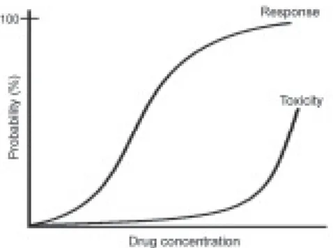
between monozygotic twins, it is believed that some of these differences in drug response are inherited. It is sup-posed that this can be attributed to polymorphisms in genes encoding drug-metabolizing enzymes, drug transporters and/or drug targets. However, it is recognized that many non-genetic factors influence the effects of medications in patients, including the nature and the severity of the disease that is being treated, the patient’s age, sex, and ethnicity, among others. The utility of pharmacogenetics in diagnosis will come from the ability to determine, based on genetic tests, the probability of a specific medication to produce the desired therapeutic effects (the efficacy) or the risk of an adverse response to the drug (the toxicity). It can divide a population of patients with the same diagnostic into sub-groups that have genetic differences in their metabolism and/or drug response susceptibility.
Chemotherapy efficacy rates to most diseases vary from 25 to 80%. Therefore, the ability to predict an effi-cient response based on genetic tests, performed before the beginning of therapy, has the potential to be of great clinical value. Since all drugs that produce an efficient response can, in specific conditions, induce adverse ef-fects, the availability of genetic tests that can identify the patients at risk of developing the rare, but still severe, adverse effect seems particularly attractive. These genetic determinants of drug effects remain stable during the patient’s lifetime, and, therefore, require a single mea-surement. At birth the child can have a blood sample col-lected in order to have the genome determined, which can be used throughout life to guide primary prevention strat-egies, make diagnoses on a molecular basis, and estab-lish particular drug therapy, that is, translate functional genomics into personalized medicine. It seems inevitable that, in the future, pharmacogenomics will come up to be an important part in the process of drug development (Johnson & Evans 2002).
Treatment
It happens then as it does to physicians in the treatment of Consumption, which in the commencement is easy to cure and difficult to understand; but when it has neither been discovered in due time nor treated upon a proper principle, it becomes easy to understand and difficult to cure. The same thing happens in state affairs; by foreseeing them at a distance, which is only done by men of talents, the evils which might arise from them are soon cured; but when, from want of foresight, they are suffered to increase to such a height that they are perceptible to everyone, there is no longer any remedy.
Nicollo Machiavelli, in The Prince, Luigi Ricci, Ed. (English edition, Oxford University Press, Oxford, 1933), chap. 3, p. 153.
About half of the new TB cases would be inevitable, as a consequence of the disease’s natural history and HIV co-infection. However, many of the exceeding cases, which result from the increase in active transmission, could be prevented by effective treatment program imple-mentation (Bloom & Murray 1992). In the XIX century, common “treatments” for TB included lung collapse and thoracoplasty (mutilating surgery where a few ribs were removed from the patient with cavitary TB), besides the isolation of infected patients to institutions, where the air
was thought to be cleaner (NSB Editorial Comment 2000). Previously, in the classic period of the Roman Empire, Clarissimus Galenus, son of the great mathematician and architect Nikon, used to prescribe to his patients treat-ments based on fresh milk, pure air, maritime trips, horse-back rides, and much rest in dry environments of higher altitude. TB treatment has evolved throughout time, from magical potions to rational drug use (Daniel 1997).
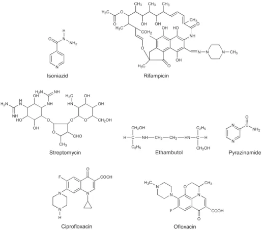
The principal objective of chemotherapy in TB pa-tients is the eradication of the whole bacillary load (Petrini & Hoffner 1999). The disease is caused by a well studied pathogen, against which there can be “magic bullets” – drugs that eliminate the bacteria without causing damage to man (Daniel 1997). Modern therapy relies on a combi-nation of potent bactericidal agents, such as isoniazid, rifampicin and pyrazinamide, in a treatment with six month duration. Sometimes during treatment, there can be an initial resistance of the bacillus to isoniazid, making it necessary to add other first-line drugs to the treatment, such as ethambutol and streptomycin. Whenever there is resistance to at least rifampicin and isoniazid, which char-acterizes MDR-TB (Telenti & Iseman 2000) it becomes necessary to extend the treatment period, and frequently rely on the use of second- or even third-line drugs, even though the increased toxicity stands as a negative factor. Very recently the Center for Diseases Control, CDC-USA, defined a new class of MDR, which was named exten-sively drug-resistant (XDR) TB, whose isolates were re-sistant to isoniazid and rifampicin and at least three of the six main classes of second line drugs (amino-glycosides, polypetides, fluoroquinolones, thioamides, cycloserine and para-aminosalicylic acid) (CDC 2006). In extreme cases, when an infection involves MDR or XDR strains (or when there is a portion of an organ considerably damaged), it becomes sometimes necessary to resort to surgical removal of granulomas, as an attempt to increase the likelihood of cure (Telenti & Iseman 2000). First-line drugs are mainly bactericidal, and combine a high degree of efficacy with a relative toxicity to the patient during treatment; these include isoniazid, rifampi-cin, streptomyrifampi-cin, ethambutol, pyrazinamide, and fluoro-quinolones (Fig. 2). Second-line drugs are mainly bacte-riostatic, which have a lower efficacy and are usually more toxic; these include para-aminosalicylic acid, ethionamide, and cycloserine (Fig. 3), among others (Goodman et al. 1996). Effective TB chemotherapy must include early bac-tericidal action against rapidly growing organisms and subsequent sterilization of dormant populations of bacilli. The first-line drugs exhibit early bactericidal activity against actively metabolizing bacilli and the bacteriostatic second-line drugs are reserved to strengthen the treatments with the presence of resistance (NSB Editorial Comment 2000). Among all first-line anti-TB agents, isoniazid has the greatest bactericidal activity against microorganisms growing actively in cavities, followed by rifampicin, streptomycin and quinolones. However, isoniazid can fre-quently cause fever, and is one of the drugs with the high-est degree of toxicity (Morehead 2000).
707 707707 707707 Mem Inst Oswaldo Cruz, Rio de Janeiro, Vol. 101(7), November 2006
chemotherapy is employed, since one of the greatest risks of mortality in TB is the delayed treatment. The treatment is normally consists of a medicinal association of regular use for a period long enough to avoid bacterial resistance and persistence. The treatment currently recommended by the WHO consists of the combined administration of isoniazid, rifampicin, pyrazinamide and streptomycin (or ethambutol) during the first 2 months, followed by the combination of isoniazid and rifampicin for at least 4 ad-ditional months. However, the long treatment period in-volves undesirable side effects to the administered drugs, leading patients to “give up” chemotherapy. The patient non-compliance led the WHO to invest in universal treat-ment adherence programs, through a process currently known as the directly observed treatment short-course (DOTS), where health care workers counsel patients, per-form progress surveillance, and make sure that each
medi-cation dose is correctly taken (NSB Editorial Comment 2000).
This revolutionary treatment aims initially at a bacte-riostatic action, inhibiting the synthesis of cell wall, nucleic acids and mycobacterial proteins, and, thereby, leading to a rapid elimination of most part of the infecting bacilli. The therapy also aims at a subsequent bactericidal action to consolidate the treatment through the elimina-tion of all remaining bacilli. DOTS combines five funda-mental elements: political commitment, microscopic ser-vices, drug supplies, surveillance systems and direct treat-ment observation. This strategy prevents the occurrence of new infections and, more importantly, makes MDR/ XDR-TB generation impractible (Pasqualoto & Ferreira 2001).
Patients with latent TB can also be treated on a chemotherapic basis; this treatment is based solely on isoniazid (monotherapy) and works only for patients with the latent infection that have not developed the active disease yet. According to the pediatrician Edith Lincon, inventor of this treatment in 1954, the therapy relies on the prophylactic administration of isoniazid for 6 to 9 months; this treatment makes disease development un-likely due to dormant bacilli elimination. The patients that most benefit from this kind of therapy are the ones more recently infected and not at advanced age. Although this chemotherapy has reached excellent results in the United States, where it is currently widely used, this type of pre-vention was not adopted by practically any other nation Fig. 2: first-line drugs are mainly bactericidal, combining a high efficacy with a relative low toxicity to patients undergoing treatment.
708 708 708 708
708 The resumption of consumption • Rodrigo Gay Ducati et al.
as part of the TB national control program (Daniel 1997). HIV-positive patients treated according to the WHO recommendations (DOTS) have sputum conversion and cure rates similar to HIV-negative treated patients. Ac-cording to the WHO, there are no treatments currently available that have yielded definitive cure results for ac-tive TB patients and MDR/XDR-TB control better than DOTS (Fätkenheuer et al. 1999). Even though the efficacy of the treatment for HIV-positive and negative patients is quite similar when dealing with drug-susceptible M. tu-berculosis infection, the choice for an appropriate TB treatment when drug resistance is suspicious can frequently be affected by the lack of rapid diagnosis tests. Drug-susceptibility tests require approximately 8 weeks to be concluded, a period usually greater than co-infected patient’s survival mean time (Riley 1993). Primary or initial resistance is defined by the identification of tolerance in individuals without previous medication; and acquired or secondary resistance is defined as the resistance resultant from previous inefficient treatments (Telenti & Iseman 2000). Usually, nations with a high primary resistance rates indicate the inefficiency of previous national TB control programs to curb the occurrence of transmission of resistant strains; secondary resistance to at least one drug is likely to reflect problems in programs in progress (Petrini & Hoffner 1999).
Curiously, the recognition of TB as a global threat and the interest in tackling this problem do not derive prima-rily from public health institutions, but rather from the World Bank and, most of all, by the IUATLD, which an-nually invests 4 million dollars in the establishment of control programs that detect approximately two-thirds of all cases, treat 65,000 cases, and provide cure rates rang-ing from 80 to 85% in some developrang-ing nations (Bloom & Murray 1992). However, this investment is still con-siderably below what is considered as necessary.
Since TB mortality is mainly attributed to the delayed detection of the disease, and is also associated to drug resistance and HIV co-infection, among others, it is ex-pected that patients without these factors will survive. However, the treatment of patients infected with drug-susceptible M. tuberculosis that have developed active disease with sub-therapeutic drug doses can easily lead to death, as the bacilli have a favorable condition to ex-pand infection and cause active disease (Morehead 2000).
Mechanism of drug action and MDR/XDR
M. tuberculosis is naturally resistant to many antibi-otics and chemotherapeutic agents, such as β-lactams, owing to the presence of hydrolytic and drug-modifying enzymes, such as periplasmic β-lactamases and amino-glycoside acetyltransferases, and drug efflux systems, besides the fact that they possess a highly hydrophobic cell wall that acts as a contention barrier that makes treat-ment more difficult. This bacterium is only susceptible to isoniazid, ethambutol, aminoglycosides (such as strepto-mycin) and rifamicins (such as rifampicin), among anti-biotics, and to fluoroquinolones (Fig. 2) among general chemotherapeutic agents (Brennan & Nikaido 1995). Within the same genus, obligatory parasite species such as M. tuberculosis have a relatively high permeability as
compared to soil mycobacterial species that have devel-oped a protection mechanism through the production of a cell wall with extremely low permeability, and, therefore, naturally more resistant (Jarlier & Nikaido 1994).
Many pathogenic bacteria possess resistance plasmids, which can effect a rapid MDR transition to drug-sus-ceptible wild-type strains, and might confer resistance to many antibacterial substances at once. This has never been observed in M. tuberculosis, but it is known that the resistant and multi-resistant phenotypes are caused by random chromosomal mutations in different genes of this organism, such as nucleotidic insertions, deletions, or substitutions (Petrini & Hoffner 1999).
The drug resistance in TB treatment is almost as old as the introduction of the first anti-TB drugs (Petrini & Hoffner 1999). After 4 to 6 weeks of treatment, the physi-cal debilitation symptoms begin to disappear, inducing many of the patients to interrupt the therapy. However, many of them end up developing the disease recurrently, making it necessary to initiate a new treatment, in case they are diagnosed, which creates favorable conditions for the selection of drug-resistant organisms (Bloom & Murray 1992). The first step towards the development of molecular detection methods of drug-resistance was the identification of genes and mutations involved in this process. Mycobacteria develop resistance to drugs spontaneously (“natural resistance”), and present different mutation rates for each drug. In the TB bacillus, these rates are equivalent to 1 in 105 to 106 for isoniazid,
1 in 108 for rifampicin (Riley 1993), 1 in 108 to 109 for
streptomycin, 1 in 107 for ethambutol, and 1 in 109 for
cycloserine (Gangadharam 1984). A cavitary lung lesion can shelter up to 109 organisms, and, thereby, it is probable
that there exists isoniazid or rifampicin resistant organisms. Mutation rates for both drugs is 1 in 1014, so
it is virtually impossible for M. tuberculosis to become spontaneously resistant to both drugs in patients correctly treated (Riley 1993). As monotherapy induces the selection of drug-resistant populations (“acquired resistance”), it becomes necessary to use a combined therapy, since the probability of a bacterial strain to develop resistance to two or more drugs at the same time is extremely low (Petrini & Hoffner 1999).
The first biochemical effect of isoniazid occurs in the first stages of mycolic acid synthesis. Isoniazid is a syn-thetic pro-drug that requires the product of the katG struc-tural gene for its activation (Telenti & Iseman 2000); this drug becomes an active compound once it is metabolized by the M. tuberculosis catalase-peroxidase enzyme, and inhibits the activity of the enoyl-ACP (CoA) reductase enzyme (encoded by the inhA gene) in the presence of NADH or NAD+ (reviewed in Basso & Blanchard 1998,
Schroeder et al. 2002, Basso & Santos 2005). Resistance to isoniazid is more complex, as it involves at least 4 genes:
katG, which mediates both susceptibility and resistance to isoniazid, and encodes the catalase-peroxidase enzyme;
inhA, which is involved in the elongation of fatty acids (Zhang et al. 1992); ahpC, which encodes the hydroper-oxide alquil reductase C; and oxyR, which is an important regulator of oxidative stress (Telenti & Iseman 2000).
709 709709 709709 Mem Inst Oswaldo Cruz, Rio de Janeiro, Vol. 101(7), November 2006
of the RNA polymerase enzyme from prokaryotic organ-isms to inhibit transcription, leading to bacterial death. Molecular detection of resistance is relatively easy to be analyzed, since about 96% of the rifampicin-resistance cases involve specific mutation in the rpoB gene, which codes for the enzyme’s beta chain, producing resistance to the drug through the decrease in binding affinity of rifampicin to the polymerase (Telenti et al. 1993). The mutation rate responsible for isoniazid resistance is 100 times greater than the one responsible for rifampicin resistance, and is usually the first modification in the susceptibility of wild-type M. tuberculosis (Petrini & Hoffner 1999). Since, in the United Kingdom, 90% of the rifampicin-resistant isolates are also isoniazid-resistant, a single positive resistance result to the former can be considered a strong indicator of MDR-TB (Caws & Drobniewski 2001).
Streptomycin acts as an inhibitor of prokaryotic pro-tein synthesis initiation. Two genes were identified as being involved with the resistance to this drug: rrs, which encodes the rRNA 16S; and rpsL, which encodes the ri-bosomal protein S12 (Telenti & Iseman 2000). Resistance to ethambutol is determined by mutations in the embA,
embB and embC genes, which encode enzymes involved in the synthesis of arabinan (Alcaide et al. 1997). Pyrazi-namide is a drug that functions only against M. tubercu-losis, and no other mycobacterial species. Pyrazinamide resistance appears to be conferred by mutations in the
pncA gene, which encodes pyrazinamidase, an enzyme that hydrolyses the drug to turn it active (Telenti & Iseman 2000). Current experimental evidence indicates that pyrazinamide enters M. tuberculosis by passive difusion, is converted to pyrazinoic acid by pyrazinamidase/nico-tinamidase enzyme activity, and is excreted by a weak efflux pump (Zhang & Mitchison 2003). Protonated pyrazinoic acid is then reabsorbed into the bacilli under acidic conditions and accumulates due to the inefficiency of the efflux pump, leading to cellular damage. Pyrazinoic acid and pyrazinamide could de-energize the membrane by collapsing the membrane potential and affect the mem-brane transport function at acidic pH. Unlike other anti-tubercular agents, PZA has no defined target of action. The mode of action of fluoroquinolones is on enzymes responsible for DNA topological conformation, topoisomerases, mainly DNA girases. Resistance to cipro-floxacine, one of the most active fluoroquinolones against
M. tuberculosis, is conferred by mutations to gyrA,gyrB
and lfrA genes, which encode the subunits A and B of DNA girase, and an efflux protein, respectively. Like most wild-type M. tuberculosis strains, many of the multi-resistant strains are susceptible to fluoroquinolones, since these bactericidal compounds increase the activity of isoniazid and rifampicin (Telenti & Iseman 2000). Some second line drugs, such as cycloserine, should be used in the lack of alternatives, as these present greater toxicity and, thereby, can cause dangerous psychic collateral effects (Petrin & Hoffner 1999).
The rate of MDR-TB mortality is estimated to range from 40 to 60%, which is similar to the mortality of pa-tients with untreated TB (Bloom & Murray 1992). Among some of the factors responsible for the increase in MDR/
XDR-TB incidence, there is the HIV/AIDS epidemic and the increase in TB incidence, especially in populations with easy access to anti-TB medication. The principal risk factors that contribute for MDR/XDR-TB emergence are patient noncompliance with the treatment and the inap-propriate administration of drugs by clinicians (Riley 1993). The Era of antibiotics has been constantly marked by cycles, which consist on the introduction of new anti-microbial agents and the subsequent emergence of resis-tance to these drugs (Swartz 2000).
Although MDR/XDR-TB patients manifest the dis-ease in a more aggressive form, there is no evidence that these patients, HIV co-infected or not, are more prone to transmit the infection than patients with drug-susceptible TB, nor that these drug-resistant strains are more infec-tious than drug-susceptible ones. In HIV-negative patients, MDR/XDR-TB frequently leads to considerable loss of weight, respiratory insufficiency, and the formation of lung cavitary lesions. In contrast, in HIV-positive patients, MDR/XDR-TB is more aggressively developed, leading to high mortality rates (Riley 1993). A study performed in Florida with HIV-positive patients revealed that, since diagnosis, the mean duration of survival is approximately 45 days for MDR-TB co-infected patients and 430 days for drug-susceptible TB co-infected ones (Fischl et al. 1992).
The WHO estimates that around 50 million people are infected with MDR-TB, which is more difficult and ex-pensive to be treated, and more likely to be fatal. In in-dustrialized nations, the complete treatment costs about US$ 2,000 per patient, much cheaper than the US$ 250,000 required to treat an MDR-TB patient (Pasqualoto & Ferreira 2001). M. tuberculosis resistant strains are usu-ally scarce in regions where there is little availability of drugs to fight the disease, since non-treated TB patients either die, get spontaneously cured, or become chronic bacilli disseminators, but, in any case, their infecting bac-teria will rarely develop any kind of drug-resistance. In contrast, easy drug access and inappropriate chemotherapy conditions will always inevitably lead to drug-resistance (Petrini & Hofnner 1999). Therefore, MDR/XDR-TB is less common in developing nations, although high TB rates are usually present, owing to the lack of medications. However, among developed nations, where there is a greater availability of anti-TB medications, the MDR/ XDR-TB rates tend to be much higher (Riley 1993). Inappropriate chemotherapy is defined as the use of a single drug, inappropriate combinations of drugs, short treatment periods resultant of patient noncompliance, and low absorption of the administered drugs. In these condi-tions, M. tuberculosis will be exposed to sub-lethal anti-bacterial concentrations, which impose a selection favor-able to the growth of resistant bacilli among an originally susceptible population. When dealing with the detection or suspicion of resistance, one should avoid the addition of a single drug into the treatment, even though it shows an initial activity, as this situation resembles a mono-therapy, which allows the development of resistance to a drug to a bacterial strain already resistant to a different agent (Petrini & Hoffner 1999).