D e cre ase d spe rm ato ge nic and
andro ge nic te sticular functio ns
in adult rats subm itte d to
im m o bilizatio n-induce d stre ss
fro m pre pube rty
1Departamento de Fisiologia, Faculdade de Medicina de Ribeirão Preto, and 2Departamento de Morfologia, Estomatologia e Fisiologia,
Faculdade de O dontologia de Ribeirão Preto, Universidade de São Paulo, Ribeirão Preto, SP, Brasil
S.A. Almeida1,
S.O . Petenusci2,
J.A. Anselmo-Franci2,
A.A.M. Rosa-e-Silva1 and
T.L. Lamano-Carvalho2
Abstract
We investigated whether chronic stress applied from prepuberty to full sexual maturity interferes with spermatogenic and androgenic testicu-lar functions. Male Wistar rats (40 days old) were immobilized 6 h a day for 60 days. Following immobilization, plasma concentrations of corticosterone and prolactin increased 135% and 48%, respectively, while plasma luteinizing hormone and testosterone presented a sig-nificant decrease of 29% and 37%, respectively. Plasma concentration of follicle-stimulating hormone was not altered in stressed rats. Chronic stress reduced the amount of mature spermatids in the testis by 16% and the spermatozoon concentration in the cauda epididymidis by 32%. A 17% reduction in weight and a 42% decrease in DNA content were observed in the seminal vesicle of immobilized rats but not in its fructose content. The growth and secretory activity of the ventral prostate were not altered by chronic stress.
Co rre spo nde nce
T.L. Lamano-Carvalho Faculdade de O dontologia de Ribeirão Preto, USP Av. Café, s/n
14040-904 Ribeirão Preto, SP Brasil
Fax: + 55-16-633-0999 E-mail: tllc@ forp.usp.br Research supported by FAPESP (No. 97/2498-1) and CNPq (No. 520128/96-6). S.A. Almeida is the recipient of a CNPq fellowship (No. 141513/95-0).
Received March 26, 1998 Accepted August 7, 1998
Ke y wo rds
•Immobilization
•Spermatogenesis
•LH
•FSH
•Testosterone
•Prolactin
Intro ductio n
Although short- or long-term immobili-zation is stressful to laboratory animals, the magnitude of the response depends on the intensity and duration of the stimulus (1-11). Experiments are in progress in our labora-tory using a model of aversive stimulation – 6 h of daily immobilization – to investigate the effects of stress on the onset of puberty in male rats. When applied from prepuberty (40 days of age) to early puberty (55 days of age) the stressful stimuli significantly
de-creased plasma luteinizing hormone (LH) concentration but caused a marked increase in plasma testosterone; a lower extent of testicular maturation was also found in pu-bertal stressed animals (Almeida SA, Petenusci SO, Anselmo-Franci JA, Rosa e Silva AAM and Lamano Carvalho TL, un-published results).
noiselessness throughout the experiment. Stressed and control rats were killed by decapitation in the morning after the last stressing session. Trunk blood was collected into heparinized tubes and plasma was
sepa-rated by centrifugation and stored at -20oC
for hormone determinations. Testosterone (T), prolactin (Prl), LH, and follicle-stimu-lating hormone (FSH) were measured by double-antibody radioimmunoassay as de-scribed by Zanato et al. (12). A commercial kit (Coat-A-Count Rat Corticosterone, DPC - Diagnostic Products Corporation, Los An-geles, CA) was used to measure plasma cor-ticosterone.
The seminal vesicle and ventral prostate
were removed, weighed and stored at -20oC
for colorimetric measurements of DNA and fructose (13). The sperm suspension was collected from the cauda epididymidis for evaluation of spermatozoon concentration, as described by Kempinas and Lamano Carvalho (14).
The testes were removed, weighed and immersed in Alfac fixing solution (85% etha-nol 80º, 10% formaldehyde and 5% glacial acetic acid) for 24 h. Seven-µm thick equato-rial paraffin sections were stained with he-matoxylin and eosin for histological and histometric analysis. Spermatid production was estimated by the amount of spermatids in the maturation phase (stages 15 to 18 of spermiogenesis, identified according to Ref. 15) counted in 10 seminiferous tubule cross-sections per animal.
The results were analyzed statistically by the non-parametric Mann-Whitney test.
Re sults
Sixty days of immobilization markedly increased plasma corticosterone (135%) and Prl (48%) concentrations. Plasma LH and T decreased by 29% and 37%, respectively, following chronic stress, while no signifi-cant change was observed in plasma FSH (Table 1).
spermatozoon concentration in the cauda epididymidis and on the accessory sex glands of adult rats.
Mate rial and Me tho ds
Male Wistar rats were submitted to im-mobilization from prepuberty (40 days of age) to full sexual maturity (100 days of age). The phases of sexual development were established according to Zanato et al. (12). The animals were housed four to a cage in plastic boxes (40 x 32 x 17 cm) under con-trolled environmental conditions (12-h light period starting at 8:00 h, temperature 23 ±
2oC). Laboratory chow and tap water were
given ad libitum.
The animals (N = 8) were immobilized inside plastic tubes dimensioned to produce stress without promoting pain (4.5 cm in diameter x 15.5 cm long up to 55 days of age; 5.0 cm in diameter x 25.5 cm long up to 100 days) for 6 h a day over a period of 60 days. Control rats (N = 8) were left undisturbed in their cages. Undesirable stress was avoided as much as possible by gentle handling and
Table 1 - Plasma hormone concentrations of chronically immobilized adult rats.
Rats w ere immobilized for 6 h/day from day 40 to 100 of life. Data w ere obtained by radioimmu-noassay and are reported as means ± SEM for 8 rats in each group. * P<0.05 compared to control (M ann-Whitney test).
Groups
Control Immobilized
Corticosterone 98.8 ± 19.4 231.9 ± 43.9*
(ng/ml)
Prolactin 19.0 ± 2.3 28.1 ± 4.0*
(ng/ml)
FSH 14.1 ± 0.8 12.4 ± 0.3 (ng/ml)
LH 5.19 ± 1.93 3.70 ± 0.40*
(ng/ml)
Testosterone 2.92 ± 0.28 1.83 ± 0.30*
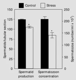
No change was observed in the testicular weight (1.77 ± 0.03 g in control and 1.69 ± 0.05 g in immobilized rats) or in the gross histological features. However, a significant decrease in spermatid production was de-monstrable by histometric evaluation, which revealed a 16% decline in the amount of maturing spermatids per seminiferous tu-bule cross-section. A 32% decrease in the concentration of spermatozoa stored in the cauda epididymidis was also observed in stressed rats (Figure 1).
Immobilization caused a significant re-duction in seminal vesicle weight (17%) and DNA content (42%) but not in in its quantity of fructose. Ventral prostate growth and se-cretory activity were not altered (Table 2).
D iscussio n
Stress caused by chronic immobilization was confirmed by a significant rise in plasma corticosterone. Literature data have shown that frequent stimulation with a low-inten-sity stressor often leads to habituation of the hypothalamic-pituitary-adrenal axis, while lower frequency and/or high-intensity stimuli can promote an exaggerated response (16). A recent review (17) summarizes the role of the hippocampus in the control of a variety of vegetative functions, such as ACTH se-cretion, and provides evidence that repeated restraint stress can promote hippocampal atrophy. Considering the inhibitory role of the hippocampus in glucocorticoid synthe-sis, stress-induced dendritic atrophy may have contributed to high corticosterone levels fol-lowing prolonged immobilization.
Plasma Prl is also expected to increase in response to stress (18,19), but the response is usually of short duration because of the suppressive effect of the concomitant in-crease in adrenal glucocorticoids (18). In the present study, however, Prl remained el-evated above control levels even 24 h after the last episode of repeated exposures to the stressor.
Our results support literature data show-ing no significant change in plasma FSH (7) and a decrease in plasma LH (7,9,10) after chronic immobilization of male rats. The stress-induced decrease in LH has been at-tributed to inhibition of gonadotropin releas-ing hormone (GnRH) secretion (10).
In the present study, immobilization of male rats from prepuberty to sexual maturity caused a significant decrease in T secretion. The inhibitory effect of chronic immobiliza-tion on plasma T has been a common finding in adult rats (5-7,10,11) attributed to re-duced LH concentration (7,20). The precise mechanism of the inhibitory effect of pro-longed stress on plasma T, however, is not
Table 2 - Effect of immobilization on rat seminal vesicle and ventral prostate w eight, and DNA and fructose content.
Rats w ere immobilized 6 h/day from day 40 to 100 of life. Data are reported as means ± SEM for 8 rats in each group. * P<0.05 compared to control (M ann-Whitney test).
Group Weight DNA Fructose
(g) (mg) (mg)
Seminal vesicle
Control 0.63 ± 0.03 0.33 ± 0.03 0.12 ± 0.01 Immobilized 0.52 ± 0.04* 0.19 ± 0.01* 0.11 ± 0.01
Ventral prostate
Control 0.35 ± 0.03 0.14 ± 0.01 0.26 ± 0.03 Immobilized 0.34 ± 0.02 0.16 ± 0.02 0.27 ± 0.03
Control Stress 250 200 150 100 50 0 150 125 100 75 50 25 0 S p e rm a to zo a ( n u m b e r/ m l x 1 0 7) S p e rm a ti d s /t u b u la r s e c ti o n Spermatid production Spermatozoon concentration * *
fully understood since the changes in andro-genic response are not always associated with altered LH levels (8,11,21). It was sug-gested that restraint stress may induce Leydig cell hyposensitivity to gonadotropin leading to a blockade of T biosynthesis at normal LH levels (21). More recent data have shown that increased glucocorticoids may act via glucocorticoid receptors on Leydig cells, thus suppressing the testicular response to go-nadotropins (11). The proopiomelanocortin (POMC)-derived peptides may also play an autocrine/paracrine role in mediating the stress-induced decline in testicular steroido-genesis (8).
In contrast to the considerations discussed above, studies under way in our laboratory have shown an increase in T concentration in early pubertal rats submitted to chronic immobilization from prepuberty (Almeida SA, Petenusci SO, Anselmo-Franci JA, Rosa e Silva AAM and Lamano Carvalho TL, unpublished results), indicating that the stressful stimulus probably acts in a different way on the gonadal axis during distinct phases of sexual development. Sympathetic inner-vation seems to modulate androgen biosyn-thesis, particularly at prepuberty and early puberty. Previous studies from our labora-tory dealing with the effects of chemical sympathectomy showed decreased steroido-genesis in prepubertal and early pubertal, but not in adult rats, at normal LH levels, and suggested that adrenergic stimulation plays a pivotal role in testicular steroidogenesis around the onset of puberty (22-24). If so, and since immobilization induces a signifi-cant rise in plasma epinephrine and norepi-nephrine levels (6), a sympathetic overstimu-lation might explain the increased T levels observed in pubertal stressed rats.
No change was observed in testicular weight or gross histological features after immobilization from prepuberty to sexual maturity; however, a significant decrease in both the production of maturing spermatids
and concentration of spermatozoa in the cauda epididymidis was observed. The de-creased androgenic status of stressed rats may be responsible, at least in part, for de-pressing spermatogenesis, since the stimula-tory action of both FSH and T is needed to initiate and maintain the process (15). In addition to the hormonal control, local regu-latory interactions occur between all testicu-lar cell types (interstitial, peritubutesticu-lar myoid, germ and Sertoli cells) and these cell-cell communications, involving growth factors and POMC peptides, mediate the cell growth and differentiation required for the initiation and maintenance of spermatogenesis (25). The stressful stimulus may have also inter-fered with elements of this elaborate para-crine control, impairing the spermatogenic process.
Although chronic immobilization did not disturb prostate growth or secretory activity, the seminal vesicle became atrophied. The atrophic seminal vesicle of stressed rats might be a consequence of decreased T secretion, in addition to high Prl levels, since the growth and secretory activity of male accessory glands are under the main control of testicu-lar androgens and Prl (26,27). There are, however, clear differences between the glan-dular response to androgens in distinct phases of sexual development: the prostate of peri-pubertal rats seems to be particularly respon-sive to testosterone when compared to younger or sexually mature animals, while the seminal vesicles showed decreasing re-sponsiveness with increasing ages (28). Prl acts both independently and in synergy with T by intensifying the growth-promoting in-fluence of androgens. However, while a nor-mal circulating Prl level is essential for nor-male reproductive functions, high levels produce adverse effects including glandular atrophy (26,29).
concentration in the cauda epididymidis of adult rats. In contrast to the findings in pubertal rats (Almeida SA, Petenusci SO, Anselmo-Franci JA, Rosa e Silva AAM and Lamano Carvalho TL, unpublished data), however, T secretion decreased in sexually mature stressed animals. Thus, besides the intensity and duration of the stressful stimulus, the phase of sexual de-velopment should also be taken into
ac-count in studies of the reproductive re-sponses to stress.
Ackno wle dgm e nts
The authors thank Dr. C.R. Franci for providing the facilities for hormone meas-urements, and Sonia A.Z. Baptista, Mauro F. Silva, Antonio de Campos and Edna A.S. Moraes for technical assistance.
Re fe re nce s
1. Zem janis R, Gondos B, Adey W R & Cockett ATK (1970). Testicular degenera-tion in M acaca nemestrina induced by immobilisation. Fertility and Sterility, 21: 335-340.
2. Cockett ATK, Elbadaw i A, Zemjanis R & Adey WR (1970). The effects of immobili-sation on spermatogenesis in subhuman primates. Fertility and Sterility, 21: 610-614.
3. Cockett ATK, Zemjanis R, Elbadaw i A & Adey WR (1971). M ale infertility: his-tochemical changes in the subhuman pri-mate testis after prolonged immobilisa-tion. Fertility and Sterility, 22: 565-572. 4. M eitner ER (1976). Einfluss der
Immobili-sat ion auf die Sperm iogenese. Act a
Anatomica, 95: 300-308.
5. Charpenet G, Taché Y, Forest M G, Haour F, Saez JM , Bernier M , Ducharme JR & Collu R (1981). Effects of chronic intermit-tent immobilisation stress on rat testicu-lar androgenic function. Endocrinology, 109: 1254-1258.
6. Collu R, Gibb W & Ducharme JR (1984). Role of catecholamines in the inhibitory effect of immobilisation stress on testos-terone secretion in rats. Biology of
Repro-duction, 30: 416-422.
7. Dem ura R, Suzuki T, Nakam ura S, Komatsu H, Odagiri E & Demura H (1989). Effect of immobilisation stress on testos-terone and inhibin in male rats. Journal of
Andrology, 10: 210-213.
8. M ann DR & Orr TE (1990). Effect of re-straint stress on gonadal proopiomelano-cortin peptides and the pituitary-testicular axis in rats. Life Sciences, 46: 1602-1609. 9. López-Calderón A, González-Quijano M I, Tresguerres JAF & Ariznavarreta C (1990). Role of LHRH in the gonadotropin re-sponse to restraint stress in intact male
rats. Journal of Endocrinology, 124: 241-246.
10. López-Calderón A, Ariznavarret a C, González-Quijano M I, Tresguerres JAF & Calderón M D (1991). St ress induced changes in testis function. Journal of
Ste-roid Biochemistry and M olecular Biology,
40: 473-479.
11. Orr TE & M ann DR (1992). Role of gluco-corticoids in the stress-induced suppres-sion of testicular steroidogenesis in adult male rats. Hormones and Behavior, 26: 350-363.
12. Zanato VF, M artins M P, Anselmo-Franci JAA, Petenusci SO & Lamano Carvalho TL (1994). Sexual development of male Wistar rats. Brazilian Journal of M edical
and Biological Research, 27: 1273-1280.
13. Kem pinas W G, Lam ano Carvalho TL, Petenusci SO, Favaretto ALV, Lopes R & Azoubel R (1988). Functional disturbance of rat sexual accessory glands in an early phase of lead intoxication. Gegenbaurs
M orphologisches Jahrbuch, 134: 791-798.
14. Kempinas WG & Lamano Carvalho TL (1988). A method for estimating the con-centration of spermatozoa in the rat cauda epididymidis. Laboratory Anim als, 22: 154-156.
15. Steinberger E & Steinberger A (1975). Spermatogenic function of the testis. In: Greep RO & Astw ood EB (Editors),
Hand-book of Physiology. Vol. 5. Williams &
Wilkins, Baltimore, 1-19.
16. Pitman DL, Ottenw eller JE & Natelson BH (1990). Effect of stressor intensity on habituation and sensitization of glucocor-ticoid response in rats. Behavioral
Neuro-science, 104: 28-36.
17. M cEw en BS & M agarinos AM (1997). Stress effects on morphology and func-tion of the hippocampus. Annals of the
New York Academy of Sciences, 821:
271-284.
18. Gala RR (1990). The physiology and m echanism s of t he st ress-induced changes in prolactin secretion in the rat.
Life Sciences, 46: 1407-1420.
19. Nonaka KO, M idlej M , Cobas C, Ramalho M J, Fregoneze JB, M achado A, Antunes-Rodrigues J & Castro e Silva E (1991). Effects of central epinephrine synthesis inhibition on stress-induced prolactin se-cretion in male rats. Brazilian Journal of
M edical and Biological Research, 24:
1071-1079.
20. Sapolsky RM (1985). Stress-induced sup-pression of testicular function in the w ild baboon: role of glucocorticoids.
Endocri-nology, 116: 2273-2278.
21. Charpenet G, Taché Y, Bernier M , Ducharme JR & Collu R (1982). Stress-induced testicular hyposensitivity to go-nadotropins in rats. Role of the pituitary gland. Biology of Reproduction, 27: 616-623.
22. Lamano Carvalho TL, Guimarães M A, Kempinas WG, Petenusci SO & Rosa e Silva AAM (1996). Effects of guanethi-dine-induced sympathectomy on the sper-matogenic and steroidogenic testicular functions of prepubertal to mature rats.
Andrologia, 28: 117-122.
23. Kempinas WG, Petenusci SO, Rosa e Silva AAM , Favaretto ALV & Lamano Carvalho TL (1995). The hypophyseal-tes-ticular axis and sex accessory glands fol-low ing chemical sympathectomy w ith guanethidine of pre-pubertal to mature rats. Andrologia, 27: 121-125.
Brazilian Journal of M edical and Biological
Research, 28: 1109-1112.
25. Skinner M K, Norton JN, M ullaney BP, Rosselli M , Whaley PD & Anthony CT (1991). Cell-cell interactions and the regu-lation of testis function. In: Robaire B (Edi-tor), The M ale Germ Cell:
Spermatogo-nium to Fertilization. Annals of the New
York Academy of Sciences, 637: 354-363.
26. M ann T & Lutw ak-M ann C (1981).
Secre-tory function of the prostate, seminal vesicle, Cow per’s gland and other acces-sory organs of reproduction. In: M ann T & Lutw ak-M ann C (Editors), M ale
Reproduc-tive Function and Semen. Springer-Verlag,
New York, 171-193.
27. Coffey DS (1988). Androgen action and the sex accessory tissues. In: Knobil E & Neill LJ (Editors), The Physiology of
Re-production. Raven Press, New York,
1081-1119.
28. Odell WD (1990). Sexual maturation in the rat. In: Gumbach M M , Sizonenko PC & Aubert M L (Editors), Control of the
On-set of Puberty. Williams & Wilkins,
Lon-don, 183-210.
29. Bartke A (1980). Role of prolactin in repro-duction in male mammals. Federation