Predicting the progress of colon cancer by DNA methylation markers
of the
p16
gene in feces - Evidence from an animal model
Wen-Chih Wu
1,2, Chih-Hsiung Hsu
2, Jen-Chun Kuan
3, Jih-Fu Hsieh
2, Chien-An Sun
2,4, Tsan Yang
5,
Chang-Chieh Wu
6and Yu-Ching Chou
21
Department of Surgery, Zuoying Branch of Kaohsiung Armed Forces General Hospital,
Kaohsiung, Taiwan.
2
School of Public Health, National Defense Medical Center, Taipei, Taiwan.
3Graduate Institute of Life Sciences, National Defense Medical Center, Taipei, Taiwan.
4Department of Public Health, College of Medicine, Fu-Jen Catholic University, New Taipei City, Taiwan.
5Department of Health Business Administration, Meiho University, Pingtung, Taiwan.
6
Department of Surgery, Tri-Service General Hospital, Taipei, Taiwan.
Abstract
A new noninvasive screening tool for colorectal neoplasia detects epigenetic alterations exhibited by gastrointestinal tumor cells shed into stool. There is insufficient existing data to determine temporal associations between colorectal cancer (CRC) progression and aberrant DNA methylation. To evaluate the feasibility of using fecal DNA methylation status to determine CRC progression, we collected stool samples from 14 male SD rats aged six weeks, and admin-istered subcutaneous injections of either 1,2-dimethylhydrazine or saline weekly.p16 DNA methylation statuses in tumorous and normal colon tissue, and from stool samples were determined using methylation-specific PCR. Addi-tionally,p16 methylation was detected in stool DNA from 85.7% of the CRC rats. The earliest change in p16 methylation status in the DMH-treated group stool samples occurred during week nine; repeatabilities were 57.1% in week 19 (p = 0.070) and 85.7% in week 34 (p = 0.005). A temporal correlation was evidenced between progression of CRC andp16 methylation status, as evidenced by DMH-induced rat feces. Using fecal DNA methylation status to de-termine colorectal tissue methylation status can reveal CRC progression. Our data suggests thatp16 promoter methylation is a feasible epigenetic marker for the detection and may be useful for CRC screening.
Keywords: colorectal cancer, DNA methylation, stool test for colorectal cancer. Received: April 5, 2012; Accepted: May 17, 2013.
Introduction
Colorectal cancer (CRC) accounts for more than 600,000 deaths each year worldwide (Jemalet al., 2011). In Taiwan in recent years, CRC has become the most common form of cancer occurrence (Promotion, 2010). Effective treatment is possible during the early stages of CRC, but the disease is generally asymptomatic. Unfortunately, the ther-apy is rarely successful in stage IV after the tumor cells have spread to lymph nodes and other organs (Wonget al., 2004). Thus, an effective screening test and early detection would provide substantial clinical benefits. The present technology CRC screening tools for an average-risk popu-lation fall into two categories: (1) physical examination, in-cluding colonoscopy, virtual colonoscopy, sigmoidoscopy,
double contrast barium enema (DCBE), and digital rectal exam (DRE) (Bretthauer, 2011), and (2) stool tests, includ-ing detection of occult blood and exfoliated mutant or epigenetic changes of DNA (Harrison and Benziger, 2011). Colonoscopy and sigmoidoscopy are highly specific and sensitive screening tools; however, these are less accept-able to the public because of invasive medical procedures. Fecal occult blood testing (FOBT), the most efficient noninvasive screening test for colorectal cancer, has a lim-ited impact on survival rate because of its low sensitivity to early stage CRC (Baeket al., 2009).
To provide early detection, it is important to develop a noninvasive and screening tool that is sensitive to CRC characteristics (Krishnan and Wolf, 2011; Labianca and Merelli, 2010). At the molecular level, CRC progression is accompanied by specific genetic and epigenetic changes (Schmid, 2010). Aberrant DNA hypermethylation of CpG islands within promoters is associated with the develop-ment and progression of colorectal cancer, which in turn
www.sbg.org.br
Send correspondence to Yu-Ching Chou. School of Public Health, National Defense Medical Center, No.161, Sec. 6, Minquan E. Rd., Neihu Dist., Taipei City, Taiwan. E-mail: [email protected].
leads to silencing of tumor suppressor genes such as
p16INK4a,MGMTandMLH1,SFRP2, andvimentingenes (Chenet al., 2005; Wang and Tang, 2008; Psofaki et al., 2010; Shimaet al., 2010).
A novel and non-invasive screening tool for detection of colorectal neoplasia is to assay the methylation alter-ations present in gastrointestinal tumor cells shed into stool. (Lenhardet al., 2005; Azuaraet al., 2010). However, exist-ing data was insufficient to determine temporal correlation between the progression of CRC and aberrant DNA methylation. To address this knowledge gap, we assayed
p16gene methylation changes in the feces of a rat model with chemically induced colorectal cancer. The assay could provide evidence that feces contain a detectable biomarker to diagnose early stage colorectal cancer. The aim of this study was to evaluate the feasibility of using fecal DNA methylation status to predict CRC progression.
Materials and Methods
Animals
All 14 male Sprague-Dawley rats used in the experi-ment were obtained from BioLASCO Taiwan Co. Ltd. (Taipei, Republic of China), six weeks of age and weighing 250-300 g. They were individually housed in ventilated stainless-steel cages with soft shavings air-conditioned room (temperature 22± 1 °C, humidity 50-60% with 12-hour light-dark cycles, lights on at 0700), and were pro-vided free access to food and water throughout the experi-ment. All procedures were approved by the Institutional Animal Care and Use Committee of National Defense Medical Center (certificate number IACUC-09-047), and were performed in accordance with National Institute of Health guidelines for the treatment of animals.
Generation of DMH induced tumors
Beginning at seven weeks of age, the rats received subcutaneous (s.c.) injections of either 1,2-dimethyl-hydrazine (DMH) (Sigma Chemical Co) at a dosage of 20 mg/kg body weight (n = 7), or the same volume of saline (n = 7) weekly for 30 weeks. Rats were sacrificed at age 34 weeks through inhaled overdose of CO2.
Collection of samples
Fecal pellets were collected weekly and placed in 2 mL microcentrifuge tubes for DNA extraction. Tissue samples were harvested after the rats were sacrificed. Sam-ples were resected with scissors and inspected for tumors. Tumor, normal tissue close to a tumor, and normal tissue samples were dissected with a razor blade and placed in 2 mL microcentrifuge tubers for DNA extraction. Each tis-sue sample was sectioned for standard histological prepara-tion and hematoxylin-eosin staining. Tissue and fecal samples were stored at -80 °C.
DNA extraction and sodium bisulfite treatment
Genomic DNA was extracted either from frozen tis-sue samples using a DNeasy Blood & Tistis-sue kit (QIA-GENE, Germany), or from fecal samples using a QIAamp DNA stool Mini kit (QIAGENE, Germany). The resulting DNA was modified with sodium bisulfite using an EZ DNA Methylation kit (Zymo Research, Orange, County, CA), and a methylated DNA positive control for MSP as-says was generated using SssI methylase (Zymo Research, Orange County, CA).
Tissue and fecal DNA methylation studies
The methylation status of tissue samples was deter-mined by methylation-specific PCR (MSP) of bisulfite treated DNA. MSP was carried out in a volume of 15mL with 7.5 mL of HotStart Taq Premlx (RBC Bioscience), 0.6mL of each primer, and 0.6 mL of bisulphite treated DNA. PCR conditions included denaturation at 95 °C for 10 min followed by 35 cycles at 95 °C for 30 s, annealing temperature at 55 °C for 35 s, 72 °C for 30 s, and a exten-sion at 72 °C for 4 min.
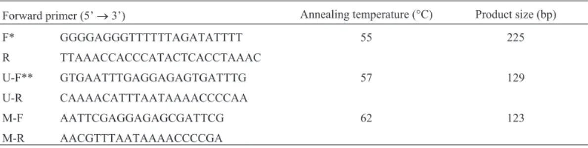
As only limited quantities of DNA are obtained from fecal samples (Zouet al., 2009) we adopted a nested strat-egy (Glockneret al., 2009). During the first stage, we car-ried out amplification by bisulfite-sequencing PCR (BSP) for 35 cycles using primers located in the flanking region of the CDKN2A promoter. In the following stage, MSP am-plification was carried out for 30 cycles using 1:100 dilu-tions of the first-stage products. Table 1 lists the MSP and BSP primer sets, and annealing temperatures.
Table 1- MSP and BSP primer sets and annealing temperature.
Forward primer (5’®3’) Annealing temperature (°C) Product size (bp)
F* GGGGAGGGTTTTTTAGATATTTT 55 225
R TTAAACCACCCATACTCACCTAAAC
U-F** GTGAATTTGAGGAGAGTGATTTG 57 129
U-R CAAAACATTTAATAAAACCCCAA
M-F AATTCGAGGAGAGCGATTCG 62 123
M-R AACGTTTAATAAAACCCCGA
Statistical analyses
All statistical analyses were performed using the SPSS 21.0 software package for Windows (SPSS Taiwan Corp.). We computed the sensitivity and specificity of the fecal DNA methylation assay by using the McNemar test to compare methylation statuses between fecal and tissue samples for rats treated with DMH (n = 7) and those in-jected with saline (n = 7); the Fisher’s exact test was used to evaluate the differences ofp16hypermethylation between the two groups in stool samples. A p value£0.05 was con-sidered statistically significant.
Results
There were no significant differences in body weight between experimental and control rats at euthanasia, and both groups exhibited a healthy appearance. All of the DMH-treated, but none of the control group rates devel-oped colorectal adenomatous cells (Figure 1).
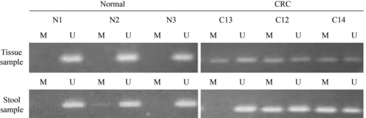
We analyzed the methylation status of thep16gene in DNA from tissue samples. A methylatedp16gene was de-tected in all intestinal adenoma tissue samples from the
DMH-treated group. By contrast, there was no hypermethyl-ation of thep16gene in the control group tissue samples (Figure 2). We then assessed thep16gene methylation status of tissue and stool samples from corresponding donor rats to ascertain the consistency ofp16hypermethylation between the two sample types. After euthanasia in week 34, p16
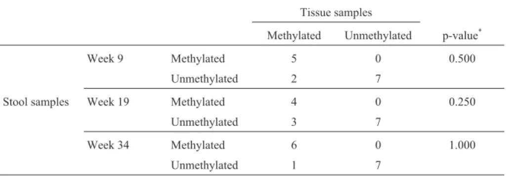
hypermethylation was found in the stool of 85.7% (6/7) of the DMH-treated rats but in none (0/7) of the control group. The McNemar testp-value was 1.000 (Table 2).
When retrospectively assaying stool samples to as-sess the temporal association between CRC progression and aberrant DNA methylation we detected the earliest in-stance of p16 hypermethylation in week nine. Samples from the DMH-treated group showed ap16 hypermethyl-ation ratio of 71.4% (5/7) against 0% (0/7) from the control group (McNemar test p = 0.500, Table 2); the repeatability of using fecal DNA methylation status for identifying CRC in our rat samples was 57.1% (4/7) during week 19 and 85.7% (6/7) in week 34 (McNemar test p = 0.250 and 1.000, respectively) (Table 2).
From the two groups of this study (DMH-treat and sa-line control), the methylation status in stool samples of
pe-Table 2- Consistency ofp16hypermethylation between tissue and stool samples.
Tissue samples
Methylated Unmethylated p-value*
Week 9 Methylated 5 0 0.500
Unmethylated 2 7
Stool samples Week 19 Methylated 4 0 0.250
Unmethylated 3 7
Week 34 Methylated 6 0 1.000
Unmethylated 1 7
*McNemar test.
riods of time was significantly different in week nine and 34 (Fisher’s exact testp-value were 0.021 and 0.005, re-spectively) and it was borderline significant in week 19 (p = 0.070) (Table 3). The repeatability of using fecal DNA methylation status was used as an indicator for CRC pro-gression (Figure 3). Samples were taken at the indicated rat age.
Discussion
The development and progression of colorectal can-cer follows the “adenoma-carcinoma” sequence, in which colorectal tumor cells develop via a worsening dysplasia of normal colonic mucosa (Fearon and Vogelstein, 1990). An uncomplicated surgical procedure can provide an effective
treatment if diagnosis is made during the early stages be-fore metastasis occurs (Kimet al., 2010). Thus, it is desir-able to find biomarkers that have high sensitivity and specificity towards CRC.
There is abundant evidence supporting the efficacy of FOBT in decreasing colorectal cancer mortality. However, the impact of FOBT on CRC incidence is lessened by the method’s low sensitivity (Heitmanet al., 2010; Leviet al., 2007). Mandelet al.(2000) reported that FOBT decreases CRC incidence by 17-20%. Conversely, Heresbachet al.
(2006) provided evidence that FOBT has an insignificant effect on CRC incidence. The incidence of CRC and its mortality rates are expected to increase due to our aging population, particularly if there are no new screening tools developed to replace FOBT.(Mariottoet al., 2006)
In a normal intestinal tract, the epithelium is con-stantly and rapidly renewed by the turnover of 5 x 10¹⁰ epi-thelial cells per day (Mehl, 1991). The shedding rate of carcinoma colonocytes is faster than that of normal cells, and we can use this characteristic to identify tumor cells and assay the status of genetic or epigenetic abnormalities. Promoter hypermethylation analysis of stool DNA is a promising noninvasive test for early diagnosis of CRC, and this area has received much research interest. An increasing number of genes is found to undergo promoter region hypermethylation in the tissue and stool of CRC patients, and Glockner et al. (2009) found a higher sensitivity to
TFPI2methylation in patients with CRC (73-89%), in pa-tients with adenomas (21-43%), and a high specificity to-ward TFPI2 methylation by patients with either CRC or adenomas (93-100%). Other reports observed high sensi-tivity (> 68%) and specificity (> 84%) towardsSFRP2and
GATA4methylation (Oberwalderet al., 2008; Wang and Tang, 2008; Hellebrekerset al., 2009)
There is substantial evidence supporting the value of using methylation analysis of stool DNA as a screening tool, although we cannot exclude the possibility of bias due to the nature of these cross-sectional studies (Thomaset al., 2005); the literature contains no reports of a sequential as-sociation between CRC and changes in gene methylation
Figure 3- The repeatability of using fecal DNA methylation status to identify CRC progression. Samples were taken at the indicated rat age.
Table 3- Differences inp16hypermethylation between DMH-treated and saline-control groups in stool samples.
Week DMH-treated Saline-control p-value*
9 Methylated 5(71.4) 0(0) 0.021
Unmethylated 2(28.6) 7(100)
19 Methylated 4(57.1) 0(0) 0.070
Unmethylated 3(42.9) 7(100)
34 Methylated 6(85.7) 0(0) 0.005
Unmethylated 1(14.3) 7(100)
*Fisher’s exact test.
The sensitivities in weeks 9, 19 and 34 were 71.4% (5/7), 57.1% (4/7) and 85.7% (6/7), respectively, in the DMH-treated group stool samples; the specificity of each week was 100% (7/7) in the normal saline group.
status in stool. Our findings answer questions about the consistency and temporal association between the progres-sion of CRC and aberrant DNA methylation.
There are currently two animal models commonly employed to investigate the rules of DNA methylation in progression of CRC: (1) using genetic manipulation of ani-mals to overexpress or down-regulate specific genes that are direct regulators of DNA methylation and methyl-ation-related gene expression; and (2) carcinogen-treated animal models with assessment of somatic epigenetic alter-ations that arise in the resulting tumors (Conerly and Grady, 2010). Our study design adopted the latter category; we used DMH to change the genome methylation status and in-duce CRC. DMH-inin-duced colon apoptosis in the rat model exhibits a characteristic aberrant crypt foci-adenoma-carcinoma sequence and associated increase in cellular pro-liferation of colonic epithelial cells similar to that observed in humans (Robertiset al., 2011).
Borinsteinet al.(2010) used an AOM-induced mice model to investigate the relationship between methylation of candidate genes, CRC, and normal tissues, and these au-thors reported 25% p16 methylation in CRC tissue, whereas we found 100% methylation to occur. We attribute the difference between Borinstein’s and our own findings to the use of different carcinogens and rat species. In human studies, the extent of p16 hypermethylation could be as high as 70% (Psofakiet al., 2010), which is in accordance with our results, indicating thatp16methylation is a high frequency CRC marker.
Although this study reveals a temporal correlation be-tween progression of colorectal cancer andp16methylation status in rat feces, the observed high frequency of p16
methylation in tumor samples may be due to the DMH car-cinogenic mechanism (Robertis et al., 2011). Thus, our findings may not represent the true temporal causal relation between the natural history of colorectal cancer andp16
methylation status in human feces.
Our study demonstrates thatp16hypermethylation in stool occurs before CRC develops and that changes in the methylation status of thep16gene in stool are statistically significant or borderline significant. The consistency of hypermethylatedp16between stool sample and tumor tissue demonstrates that detecting the hypermethylatedp16gene in stool to identify colorectal neoplasia provides a sensitive early detection, consistent and noninvasive screening tool.
Acknowledgments
This study was funded by a grant from Zuoying Branch of Kaohsiung Armed Forces General Hospital, Tai-wan, Republic of China. Grant number ZAFGH9901.
References
Azuara D, Rodriguez-Moranta F, de Oca J, Soriano-Izquierdo A, Mora J, Guardiola J, Biondo S, Blanco I, Peinado MA,
Moreno V, et al.(2010) Novel methylation panel for the early detection of colorectal tumors in stool DNA. Clin Colorectal Cancer 9:168-176.
Baek YH, Chang E, Kim YJ, Kim BK, Sohn JH and Park DI (2009) Stool methylation-specific polymerase chain reac-tion assay for the detecreac-tion of colorectal neoplasia in Korean patients. Dis Colon Rectum 52:1452-1459; discussion 1459-1463.
Borinstein SC, Conerly M, Dzieciatkowski S, Biswas S, Wash-ington MK, Trobridge P, Henikoff S and Grady WM (2010) Aberrant DNA methylation occurs in colon neoplasms aris-ing in the azoxymethane colon cancer model. Mol Carcinog 49:94-103.
Bretthauer M (2011) Colorectal cancer screening. J Intern Med 270:87-98.
Chen WD, Han ZJ, Skoletsky J, Olson J, Sah J, Myeroff L, Platzer P, Lu S, Dawson D, Willis J,et al.(2005) Detection in fecal DNA of colon cancer-specific methylation of the nonexpres-sed vimentin gene. J Natl Cancer Inst 97:1124-1132. Conerly M and Grady WM (2010) Insights into the role of DNA
methylation in disease through the use of mouse models. Dis Model Mech 3:290-297.
Fearon ER and Vogelstein B (1990) A genetic model for colo-rectal tumorigenesis. Cell 61:759-767.
Glockner SC, Dhir M, Yi JM, McGarvey KE, Van Neste L, Louwagie J, Chan TA, Kleeberger W, de Bruine AP, Smits KM,et al.(2009) Methylation of TFPI2 in stool DNA: A po-tential novel biomarker for the detection of colorectal can-cer. Cancer Res 69:4691-4699.
Harrison S and Benziger H (2011) The molecular biology of colorectal carcinoma and its implications: A review. Sur-geon 9:200-210.
Heitman SJ, Hilsden RJ, Au F, Dowden S and Manns BJ (2010) Colorectal cancer screening for average-risk North Ameri-cans: An economic evaluation. PLoS Medicine 7:e1000370. Hellebrekers DM, Lentjes MH, van den Bosch SM, Melotte V,
Wouters KA, Daenen KL, Smits KM, Akiyama Y, Yuasa Y, Sanduleanu S,et al.(2009) GATA4 and GATA5 are poten-tial tumor suppressors and biomarkers in colorectal cancer. Clin Cancer Res 15:3990-3997.
Heresbach D, Manfredi S, D’Halluin PN, Bretagne JF and Bran-ger B (2006) Review in depth and meta-analysis of con-trolled trials on colorectal cancer screening by faecal occult blood test. Eur J Gastroenterol Hepatol 18:427-433. Jemal A, Bray F, Center MM, Ferlay J, Ward E and Forman D
(2011) Global cancer statistics. CA Cancer J Clin 61:69-90. Kim MS, Lee J and Sidransky D (2010) DNA methylation
mark-ers in colorectal cancer. Cancer Metastasis Rev 29:181-206. Krishnan S and Wolf JL (2011) Colorectal cancer screening and
prevention in women. Womens Health (Lond Engl) 7:213-226.
Labianca R and Merelli B (2010) Screening and diagnosis for colorectal cancer: Present and future. Tumori 96:889-901. Lenhard K, Bommer GT, Asutay S, Schauer R, Brabletz T, Goke
B, Lamerz R and Kolligs FT (2005) Analysis of promoter methylation in stool: A novel method for the detection of colorectal cancer. Clin Gastroenterol Hepatol 3:142-149. Levi Z, Rozen P, Hazazi R, Vilkin A, Waked A, Maoz E,
Mandel JS, Church TR, Bond JH, Ederer F, Geisser MS, Mongin SJ, Snover DC and Schuman LM (2000) The effect of fecal occult-blood screening on the incidence of colorectal can-cer. N Engl J Med 343:1603-1607.
Mariotto AB, Yabroff KR, Feuer EJ, De Angelis R and Brown M (2006) Projecting the number of patients with colorectal car-cinoma by phases of care in the US: 2000-2020. Cancer Causes Control 17:1215-1226.
Mehl LE (1991) A mathematical computer simulation model for the development of colonic polyps and colon cancer. J Surg Oncol 47:243-252.
Oberwalder M, Zitt M, Wontner C, Fiegl H, Goebel G, Kohle O, Muhlmann G, Ofner D, Margreiter R and Muller HM (2008) SFRP2 methylation in fecal DNA - A marker for colorectal polyps. Int J Colorectal Dis 23:15-19.
Promotion BOH (2010) Cancer Registry Annual Report, 2007. Department of Health, Taiwan.
Psofaki V, Kalogera C, Tzambouras N, Stephanou D, Tsianos E, Seferiadis K and Kolios G (2010) Promoter methylation sta-tus of hMLH1, MGMT, and CDKN2A/p16 in colorectal adenomas. World J Gastroenterol 16:3553-3560.
Robertis M, Massi E, Poeta M, Carotti S, Morini S, Cecchetelli L, Signori E and Fazio V (2011) The AOM/DSS murine model for the study of colon carcinogenesis: From pathways to di-agnosis and therapy studies. J Carcinog 10:e9.
Schmid G (2010) The use of molecular markers in the diagnosis of colorectal cancer screening. Dig Dis 28:625-628.
Shima K, Nosho K, Baba Y, Cantor M, Meyerhardt JA, Giovan-nucci EL, Fuchs CS and Ogino S (2010) Prognostic signifi-cance of CDKN2A (p16) promoter methylation and loss of expression in 902 colorectal cancers: Cohort study and liter-ature review. Int J Cancer 128:1080-1094.
Thomas JR, Nelson JK and Silverman SJ (2005) Research meth-ods in physical activity. Human Kinetics, Champaign, 454 pp.
Wang DR and Tang D (2008) Hypermethylated SFRP2 gene in fe-cal DNA is a high potential biomarker for colorectal cancer noninvasive screening. World J Gastroenterol 14:524-531. Wong JM, Yen MF, Lai MS, Duffy SW, Smith RA and Chen TH
(2004) Progression rates of colorectal cancer by Dukes’ stage in a high-risk group: Analysis of selective colorectal cancer screening. Cancer J 10:160-169.
Zou H, Taylor WR, Harrington JJ, Hussain FT, Cao X, Loprinzi CL, Levine TR, Rex DK, Ahnen D, Knigge KL,et al.(2009) High detection rates of colorectal neoplasia by stool DNA testing with a novel digital melt curve assay. Gastroen-terology 136:459-470.
Editor: Carlos F.M. Menck