DNA methylation analysis of the tumor suppressor gene
CDKN2B
in Brazilian leukemia patients
Patrícia Santos Pereira Lima
1,2, Greice Andreoti Molffeta
2, Amélia Góes de Araujo
3,4,
Marco Antônio Zago
3,4and Wilson Araújo da Silva Jr.
2,41
Departamento de Ciências Naturais, Universidade Estadual do Sudoeste da Bahia,
Vitória da Conquista, BA, Brazil.
2
Laboratório de Genética Molecular e Bioinformática, Departamento de Genética,
Faculdade de Medicina de Ribeirão Preto, Universidade de São Paulo, Ribeirão Preto, SP, Brazil.
3Departamento de Clínica Médica, Faculdade de Medicina de Ribeirão Preto,
Universidade de São Paulo, Ribeirão Preto, SP, Brazil.
4
Centro de Terapia Celular and Centro Regional de Hemoterapia,
Faculdade de Medicina de Ribeirão Preto, Universidade de São Paulo, Ribeirão Preto, SP, Brazil.
Abstract
The aim of this work was to evaluate the methylation profile of thep15 (CDKN2B) gene in Brazilian patients with leu-kemia and to correlate theCDKN2B gene expression with the percentage of methylated CpG dinucleotides in its pro-moter region. Thirty-one samples from six patients with acute lymphocytic leukemia (ALL), four with chronic myeloid leukemia (CML), and 21 with acute myeloid leukemia (AML) were evaluated by MSP (Methylation-Specific PCR). TheCDKN2B gene was found to be methylated in four (67%) of the six ALL samples and in 16 (76%) of the 21 AML samples, but in none of the four CML samples analyzed. We observed a correlation between theCDKN2B mRNA ex-pression (RT-PCR) and the percentage of methylated CpG dinucleotides. Therefore, this study in Brazilian patients confirms that theCDKN2B gene is methylated in the majority of leukemia patients.
Key words:CpG island, DNA methylation,CDKN2B.
Received: August 23, 2007; Accepted: March 10, 2008.
Introduction
DNA methylation is a covalent modification and re-sults from the activity of a family of DNA methyltrans-ferase (DNMT) enzymes which catalyze the transfer of a methyl group (CH3) from S-adenosylmethionine (SAM) to the cytosine residues at CpG dinucleotides (Strathdee and Brown, 2002). The distribution of CpG dinucleotides in the human genome is not uniform, but there are small stretches (0.5 kb to several kb) of CpG-rich DNA regions termed CpG islands (Galmet al., 2006). It has been estimated that the human genome contains about 29,000 CpG islands (Nephew and Huang, 2003). These CpG islands are usually located in the vicinity of genes, are often found near the promoters of widely expressed genes, and typically extend into the first exon (Jones, 2003). In contrast to CpG dinu-cleotides, which are dispersed throughout the genome, the
cytosines within CpG islands, especially those associated with promoter regions, are normally unmethylated, allow-ing the expression of a gene. The exception to this unme-thylated state of CpG islands involves the imprinted genes and X-chromosome inactivation, and this indicates the tight association of promoter DNA methylation with transcrip-tional silencing during normal mammalian development (Galmet al., 2006).
The methylation pattern of normal cells is kept through successive cellular divisions in adult tissues and the heritage of this information is called epigenetic inheri-tance (Laird, 2003). Therefore, epigenetics is the study of modifications in gene expression that are not caused by al-terations of DNA sequence (Verma and Srivastava, 2002; Galmet al., 2006).
In tumorigenesis, the balance of the methylation state of normal cells is lost, and some of the possible alterations are: (I) transcriptional silencing of tumor suppressor genes by CpG island promoter hypermethylation; and (II) histone deacetylation and global genomic hypomethylation.
Hypo-Send correspondence to Patrícia Santos Pereira Lima. Laboratório de Genética Molecular e Bioinformática, Fundação Hemocentro de Ribeirão Preto, Rua Tenente Catão Roxo 2501, 14051-140 Ribei-rão Preto, SP, Brazil. E-mail: [email protected].
methylation contributes to carcinogenesis and is responsi-ble for the chromosomal instability, reactivation of transposable elements, and loss of imprinting (Esteller and Herman, 2002). Therefore, the profile of hypermethylation promoters differs according to each cancer type, because for each tumor type specific genes are methylated. In addi-tion to that, the epigenetic inactivaaddi-tion may affect all mo-lecular mechanisms involved in cell immortalization and transformation. Last but not least, it seems that epigenetic changes are among the several early steps in carcinogenesis (Mukai and Sekiguchi, 2002).
According to the estimates of cancer incidence for 2008 by INCA (Instituto Nacional do Câncer – Brazilian National Cancer Institute), leukemias will afflict 5220 men and 4320 women. Hematologic neoplasias can jeopardize the lymphoid or myeloid lineages. Lymphoid neoplasias start from lymphoid lineage cells in different stages of mat-uration. Myeloid neoplasias result from a pluripotential progenitor cell mutation which maintain the capacity, albeit in an imperfect manner, of differentiation and maturation for each one of the myeloid lineages (Zagoet al., 2001). Most acute leukemias appear to be the consequence of the collaboration between one class of mutations or gene rear-rangements that confer a proliferative and/or survival ad-vantage to hematopoietic progenitors and a second class of mutations that serve primarily to impair hematopoietic dif-ferentiation and subsequent apoptosis of cells (Kelly and Gilliland, 2002).
In hematological neoplasms, hypermethylation genes were identified in multiple fundamental pathways related to cancer, including cell cycle control (p15,p16,
Rb1,p27,p73), DNA repair (O6MGMT), apoptosis inhibi-tion (DAPK), tumoral metastasis (E-cadherin), and growth factors (ER,EphA3) (Frenchet al., 2002). One of the most frequent methylated genes in leukemia, mainly in ALM, isCDKN2B(Hermanet al., 1996; Issaet al., 1997; Wonget al., 2000; Toyotaet al., 2001; Claus and Lubbert, 2003).
In this work, we determined the methylation profile of theCDKN2Bgene in samples from Brazilian patients with acute myeloid leukemia (AML), acute lymphocytic leukemia (ALL), and chronic myeloid leukemia (CML). We also evaluated theCDKN2BmRNA expression and the percentage of methylated CpG dinucleotides by sequencing of sodium bisulfite-treated DNA.
Material and Methods
Samples
Genomic DNA was extracted from bone marrow cells of six ALL patients, four CML patients, 21 AML patients and one sample of normal bone marrow, using a Super Quik Gene Kit (AGTC). The AML samples were subdivided based on French-American-British (FAB) into: M0 (1), M1 (2), M2 (10), M4 (2), M5 (4) and M6 (2). All patients
signed a consent form approved by the Ethics Committee of the Institution.
Bisulfite DNA modification
Bisulfite DNA modification was accomplished in agreement with the technique described inCurrent Proto-cols In Human Genetics (Dracopoli et al., 1994). DNA (2µg) was denatured with NaOH (2 M) for 10 min at 37 °C. Ten millimoles of hydroquinone and 3 M of sodium bisulfite at pH 5, both freshly prepared, were added and mixed, and then samples were incubated at 50 °C for 16 h. The modified DNA was purified using a Wizard DNA clean-uppurification kit (Promega) according to the manu-facturer’s instructions and eluted into water. Modification was completed by treatment with NaOH 3 M for 5 min at room temperature, followed by ethanol precipitation. The DNA was resuspended in water and used immediately or stored at -80 °C.
Methylation-Specific PCR (MSP)
A modified DNA was used for two MSP reactions, both for theCDKN2Bgene: one reaction specific for meth-ylated DNA and other specific for unmethmeth-ylated DNA. The primer sequences used were:CDKN2B-F(M) 5’-CGTTCG TATTTTGCGGTT-3’;CDKN2B-R(M) 5’-CGTACAATA ACCGAACGACCGA-3’;CDKN2B-F(U) 5’ -TGTGATG TGTTTGTATTTTGTGGTT-3’; and CDKN2B-R(U) 5’-CCATACAATAACCAAACAACCAA-3’ (Hermanet al., 1996). For all reactions the amplification conditions were: bisulfite-modified DNA; 10X PCR buffer (Invitrogen); 50 mM MgCl2; 1.25 mM dNTP, and 300 ng/µL of each
primer. Reactions were hot-started at 95 °C for 5 min be-fore addition of 1.25 units ofTaqpolymerase (Invitrogen), followed by 35 cycles (30 s at 95 °C, 30 s at 61 °C for primer methylated or 60 °C for primer unmethylated, 30 s at 72 °C), and a final extension step of 4 min at 72 °C. Each PCR product was analyzed using 3% agarose gel, stained with ethidium bromide and directly visualized under UV il-lumination.
Sequencing of sodium bisulfite-treated DNA
chan-ged to 51 °C. The amplified products were sequenced di-rectly in a MegaBace 1000 sequencer.
RNA isolation and reverse transcriptase PCR (RT-PCR)
Total RNA was isolated using TRIZOL (Invitrogen) and the cDNA was synthesized with the use of a High Ca-pacity kit (Applied Biosystems). Amplification of
CDKN2B mRNA was performed with specific primers:
CDKN2B-RTF 5’-AACGGAGTCAACCGTTTCGG-3’ and CDKN2B-RTR 5’-TGTGCGCAGGTACCCTGCA A-3’ (Hoshinoet al., 2002). The PCR conditions were as follows: 10 X buffer (Biotools); 1.25 mM dNTP; 20 pmol of each primer, and 2 U ofTaq DNA polymerase (Bio-tools). PCR was initiated with one cycle at 95 °C for 1 min, followed by 30 cycles at 95 °C for 1 min, 55 °C for 1 min, and 72 °C for 1 min. The internal control was the glyceral-dehyde 3-phosphate dehydrogenase (GAPDH) gene. The amplified products were analyzed on 1.5% agarose gel, stained with ethidium bromide and directly visualized un-der UV illumination.
Results
Methylation analysis
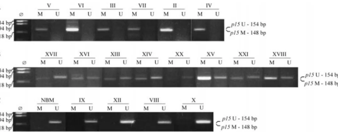
Different methylation patterns of theCDKN2Bgene were detected among leukemia types analyzed by MSP. The gene was methylated in four (67%) of the six ALL samples and in 16 (76%) of the 21 AML samples, but in none of the four CML samples analyzed. Regarding AML subtypes, the frequency distribution of CDKN2B gene methylation was: M0 1/1, M1 2/2, M2 9/10, M4 0/2, M5 3/4, and M6 1/2. In 12 AML samples, amplification was achieved with both primers, specific for methylated DNA and for unmethylated DNA, and therefore these samples were classified as hemimethylated (Figure 1). For fre-quency estimates, these hemimethylated samples were con-sidered as methylated.
Expression analysis
The expression ofCDKN2BmRNA was evaluated in 19 samples with available RNA (six methylated, five un-methylated, and eight hemimethylated) and in one normal bone marrow sample. In the methylated samples, no
CDKN2BmRNA expression was detected (Figure 2A). On the other hand,CDKN2Bexpression was detected in the unmethylated and in some hemimethylated samples (Fig-ures 2B and C). However, the level of expression was higher in the unmethylated than in the hemimethylated samples. The highest level of CDKN2Bgene expression was detected in the normal bone marrow sample.
Sequencing analysis
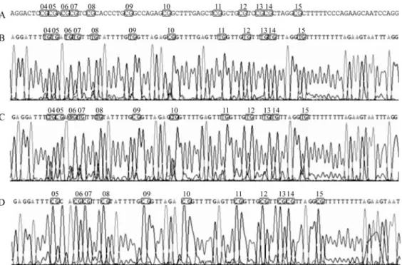
The region analyzed (498 bp) after bisulfite treatment of the DNA is located approximately -234 to +264 bp from the transcription start site, comprising two CpG islands ac-cording to the MethPrimer program (Li and Dahiya, 2002). However, the CpG dinucleotides analyzed by sequencing contained only the region located -15 to +208 bp (223 bp) from the transcription start site that shows 26 CpG di-nucleotides. The sequence amplified by MSP (147 bp), which shows 19 CpG dinucleotides, is within the se-quenced region (Figure 3).
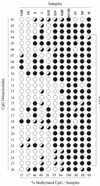
Thirteen out of the 31 samples analyzed by MSP were sequenced (one normal bone marrow, four methylated, four hemimethylated and four unmethylated). The methylation percentage for each sample was calculated as the number of methylated CpG dinucleotides in the total number of ana-lyzed CpGs (Figure 4). The normal sample did not show any methylated CpG (data not shown). Seven out of eight samples, which were amplified with the primer set specific for the methylated DNA, presented a methylated CpG fre-quency of 46% to 100%. The exception was sample XIX (Figure 4), considered to be hemimethylated by MSP, which showed only 26% of methylated CpG dinucleotides. In the unmethylated samples, the percentage of methylated CpGs ranged from 15% to 46%.
Discussion
TheCDKN2Bgene encodes a 15-kDa protein that is a cyclin-dependent kinase inhibitor. The role ofCDKN2Bas a tumor suppressor in acute myelogenous leukemia (AML) was established in several previous studies (Hermanet al., 1996; Issaet al., 1997; Wonget al., 2000; Toyotaet al., 2001; Claus and Lubbert, 2003). Hypermethylation of
CDKN2BCpG islands has been shown to occur in up to 80% of human AML, and this epigenetic state is associated with reduced expression (Markuset al., 2007).
In this study, analysis results of the methylated state of theCDKN2Bgene, performed by MSP, were confirmed
by direct sequencing and expression analysis. Our MSP re-sults showed thatCDKN2Bwas methylated in 76% of the AML samples. These results are in agreement with previ-ous studies that found theCDKN2Bgene to be methylated in more than 50% of the cases (Hermanet al., 1996; Issaet al., 1997; Toyotaet al., 2001; Claus and Lubbert, 2003).
Among patients with AML, Wonget al. (2000) re-portedCDKN2Bmethylation frequencies to be higher in subtypes M2, M3 or M4 than in M1, M5, M6 or M7. In nine of our 10 subtype M2 samples, the gene was methylated; for the other subtypes, the number of samples was too small to allow a correlation analysis.
In ALL, previous studies indicated different methyl-ation frequencies for theCDKN2Bgene, depending on the methodology used (Issa et al., 1997; Melki et al., 1999; Garcia-Maneroet al., 2002; Melki and Clark, 2002; Chim
et al., 2003). We found a higher frequency (67%) than that (40%) described by Chim and colleagues (2003), using the same methodology (MSP). This might be due to our smaller sample size (six samples) compared to theirs (25 samples). Even with a small number of LMC samples (four), our data showed the unmethylated state of the CDKN2B gene, which is in agreement with previous reports (Issa et al., 1997; Toyotaet al., 2001).
A sample of genomic DNA usually consists of a large pool of molecules that may display methylation heteroge-neity. This heterogeneity can be due to the fact that two al-leles of any given genomic locus in a cell may differ in their methylation patterns, and that the DNA sample was derived from multiple cells with potentially different methylation patterns (Siegmund and Laird, 2002). This could explain the simultaneous amplification by MSP using primer sets for both methylated and unmethylated DNA. This methyl-ation heterogeneity of samples can also be difficult to ana-lyze by direct sequencing of sodium bisulfite-treated DNA, once it does not allow the methylation state of individual al-leles to be determined. Hence, in some chromatograms the Figure 2 - CDKN2B mRNA expression evaluated by RT-PCR.
NBM = normal bone marrow. The samples are represented in roman num-bers. (A) methylated samples (B) hemimethylated samples. (C) unmethylated samples. (D) Comparative RT-PCR.GAPDHwas used to control the integrity of the RNA samples.
TCpG sequence was observed instead of TpG or CpG. We expected to find TpG in the case of an unmethylated di-nucleotide and CpG in the case of a methylated dinucleo-tide. The term hemimethylated was also used to designate those CpG nucleotides for which the methylation state could not be defined by sequencing, as it occurred in CpG 04, 06 and 10 (Figure 5C).
In general, the amount of methylated CpG sites within a locus, as well as the number of methylated loci, in-crease in more advanced stages of cancer (Nephew and Huang, 2003). The consequence of this dynamic epigenetic
gene silencing is that the degree of loss of protein produc-tion is not uniform throughout the tumor-cell populaproduc-tion, unlike the one that is produced by the genetic changes (Jones and Baylin, 2002). We analyzed the expression of
CDKN2BmRNA, which in general agreed both with the MSP results and with the percentage of methylated CpG dinucleotides, as determined by direct PCR sequencing. In the methylated samples that showed more than 80% of methylated CpG dinucleotides, no CDKN2B mRNA ex-pression was observed by RT-PCR (Figures 2A and 4). In the unmethylated samples, a relationship between the ex-pression level and the percentage of methylated CpG was observed, as in sample X (46% methylated CpG) and sam-ples VIII and IX (15% and 17% methylated CpG, respec-tively) (Figures 2C and 4). TheCDKN2Bgene expression was very low when the gene was found to be hemimethyl-ated by MSP (Figure 2B).CDKN2BmRNA expression dif-fered between the normal bone marrow and the unmethylated samples (Figure 2C). These unmethylated samples showed a low percentage of methylated CpG, which seems to correlate with the levels of mRNA tran-scription.
The percentage of methylated CpGs was similar (46%) in samples X and I (unmethylated and methylated, respectively). However, their level of gene expression and the number of hemimethylated CpGs (Figures 2D and 4) were different. There were 10 hemimethylated CpGs in sample I, while in sample X only 2 hemimethylated CpGs were identified. Hence, the methylation density of the re-gion analyzed was higher in sample I than in sample X, and this might have caused the apparent difference inCDKN2B
gene expression observed between these samples.
Another difference between the samples was the state of the methylated CpG 3 dinucleotide at the transcription factor Sp1 site. Sp1 is associated with the transcription of theCDKN2Bgene and is a regulator of cell cycle progres-sion (Pagliucaet al., 2000). Many factors are known to bind CpG-containing sequences, and some of them fail to bind when the CpG is methylated, therefore inhibiting transcrip-tion (Bird, 2002). CpG 3 and CpG 25 are part of two Sp1 sites (Figure 4), and in seven of the eight sequenced sam-ples which were amplified by the specific primer set for methylated DNA at least one of these dinucleotide CpGs was methylated (Figure 4).
Methylation of specific targets may explain the obser-vation that different hematopoietic malignancies harbor distinct methylation signatures (Rush and Plass, 2002). The cell type-specific pattern of hypermethylation suggests that the methylation of certain CpG islands may be used as dis-ease marker and is also useful in the detection of minimal residual disease after chemotherapy. This pattern may therefore influence any adjuvant treatments (Melki and Clark, 2002) or be used as a marker for disease progression (Laird, 2003). Agrawal et al. (2007) showed increased
CDKN2Bmethylation levels in the bone marrow of patients Figure 4- Scheme of direct bisulfite sequencing of region containing 26
with acute leukemias in clinical remission and, according to these authors, the presence of aberrant DNA methylation in remission is a powerful indicator of a high risk of leukemia relapse.
In this first report on the methylation status of the
CDKN2Bgene in Brazilian leukemia patients, CDKN2B
was found to be methylated, in agreement with data ob-tained in similar analyses performed in leukemia patients of other countries.
Acknowledgments
Support for this work was provided by the Brazilian agencies FAPESP, CNPq, Center for Cell-based Therapy and UESB. We also thank Marli H. Tavella, Cristiane F. Ayres, Adriana A. Marques and Anne Marie R. D. dos Santos for their excellent technical assistance.
References
Agrawal S, Unterberg M, Koschmieder S, Stadt UZ, Brunnberg U, Verbeek W, Buchner T, Berdel WE, Serve H and Mul-ler-Tidow C (2007) DNA methylation of tumor supressor genes in clinical remission predicts the relapse risk in acute myeloid leukemia. Cancer Res 67:1370-7.
Bird A (2002) DNA methylation patterns and epigenetic memory. Genes Dev 16:6-21.
Chim CS, Wong ASY and Kwong YL (2003) Epigenetic inactiva-tion of INK4/CDK/RB cell cycle pathway in acute leuke-mia. Ann Hematol 82:738-742.
Claus R and Lubbert M (2003) Epigenetic targets in hemato-poietic malignancies. Oncogene 22:6489-96.
Dracopoli NC, Haine JL, Moir DT, Morton CC, Seidman CE, Seideman JG and Smith DR (1994) Current Protocols in Hu-man Genetics. John Wiley & Sons, New York, 750 pp. Esteller M and Herman JG (2002) Cancer as an epigenetic
dis-ease: DNA methylation and chromatin alterations in human tumours. J Pathol 196:1-7.
French SW, Dawson DW, Miner MD, Doerr JR, Malone CS, Wall R and Teitell MA (2002) DNA methylation profiling: A new tool for evaluating hematologic malignancies. Clin Immunol 103:217-230.
Galm O, Herman JG and Baylin SB (2006) The fundamental role of epigenetics in hematopoietic malignancies. Blood Rev 20:1-13.
Garcia-Manero G, Daniel J, Smith TL, Kornblau SM, Lee M-S, Kantarjian HM and Issa J-PJ (2002). DNA methylation of multiple promoter-associated CpG islands in adult acute lymphocytic leukemia. Clin Cancer Res 8:2217-24. Herman JG, Graff JR, Myohanen S, Nelkin BD and Baylin SB
(1996) Methylation-specific PCR: A novel PCR assay for methylation status of CpG islands. Proc Natl Acad Sci USA 93:9821-6.
Hoshino K, Asou N, Okubo T, Suzushima H, Kiyokawa T, Kaw-ano F and Mitsuya H (2002) The absence of thep15INK4B gene alterations in adult patients with precursor B-cell acute lymphoblastic leukaemia is a favourable prognostic factor. Br J Haematol 117:531-40.
Issa J-PJ, Baylin SB and Herman JG (1997) DNA methylation changes in hematologic malignancies: Biologic and clinical implications. Leukemia 11:S7-S11.
Jones PA (2003) Epigenetics in carcinogenesis and cancer pre-vention. Ann NY Acad Sci 983:213-9.
Jones PA and Baylin SB (2002) The fundamental role of epige-netic events in cancer. Nat Rev Genet 3:415-28.
Kelly LM and Gilliland DG (2002) Genetics of myeloid leuke-mias. Annu Rev Genomics Hum Genet 3:79-98.
Laird PW (2003) The power and the promise of DNA methylation markers. Nat Rev Cancer 3:253-66.
Li L-C and Dahiya R (2002) MethPrimer: Designing primers for methylation PCRs. Bioinformatics 18:1427-31.
Markus J, Garin MT, Bies J, Galili N, Raza A, Thirman MJ, Beau MML, Rowley JD, Liu PP and Wolff L (2007) Methyl-ation-independent silencing of the tumor suppressorINK4b (p15) by CBFβ-SMMHC in acute myelogenous leukemia with inv (16). Cancer Res 67:992-1000.
Melki JR and Clark SJ (2002) DNA methylation changes in leu-kaemia. Semin Cancer Biol 12:347-57.
Melki JR, Vincent PC and Clark SJ (1999) Concurrent DNA hypermethylation of multiple genes in acute myeloid leuke-mia. Cancer Res 59:3730-40.
Mukai T and Sekiguchi M (2002) Gene silencing in phenomena related to DNA repair. Oncogene 21:9033-42.
Nephew KP and Huang TH-M (2003) Epigenetic gene silencing in cancer initiation and progression. Cancer Lett 190:25-33. Pagliuca A, Gallo P and Lania L (2000) Differential role for
Sp1/Sp3 transcriptional factors in the regulation of the pro-moter activity of multiple cyclin-dependent kinase inhibitor genes. J Cell Biochem 76:360-7.
Rush LJ and Plass C (2002) Alterations of DNA methylation in hematologic malignancies. Cancer Lett 185:1-12.
Siegmund KD and Laird PW (2002) Analysis of complex methyl-ation data. Methods 27:170-8.
Strathdee G and Brown R (2002) Aberrant DNA methylation in cancer: Potential clinical interventions. Exp Rev Mol Med 4:1-17.
Toyota M, Kopecky KJ, Toyota M-O, Jair K-W, Willman CL and Issa J-PJ (2001) Methylation profiling in acute myeloid leu-kemia. Blood 97:2823-9.
Verma M and Srivastava S (2002) Epigenetics in cancer: Implica-tions for early detection and prevention. Lancet Oncol 3:755-63.
Wong IHN, Ng MHL, Huang DP and Lee JCK (2000) Aberrant p15promoter methylation in adult and childhood acute leu-kemias of nearly all morphologic subtypes: Potential prog-nostic implications. Blood 95:1942-49.
Zago MA, Falcão RP and Pasquine R (2001) Hematologia: Fun-damentos e Prática. 1st ed. Ateneu, São Paulo, 1042 pp.
Internet Resources
INCA (Instituto Nacional do Câncer), http://www.inca.gov.br (March 5, 2008).
Associate Editor: Emmanuel Dias Neto