Parasitism of two zoonotic reservoirs
Dasyprocta leporina
and
D
.
fuliginosa
(Rodentia) from Amazonas, with Trichostrongylina
nematodes (Heligmonellidae): description of a new genus and
a new species
Alessandra Queiroga Gonçalves, Roberto Magalhães Pinto*/
+,
Marie-Claude Durette-Desset**
Laboratório de Doenças Parasitárias *Laboratório de Helmintos Parasitos de Vertebrados, Instituto Oswaldo Cruz -Fiocruz, Av. Brasil 4365, 21045-900 Rio de Janeiro, RJ, Brasil **Département de Systématique et Evolution, UMR 7138 associée au
C.N.R.S, Muséum National d’Histoire Naturelle, Paris, France
A new genus and a new species of Heligmonellidae nematodes are described parasiting the stomach of three agoutis (two Dasyprocta fuliginosa and one D. leporina) captured in the middle and high Negro river microregion, state of Amazonas, Brazil. The new genus, as well as its type-species, are closely related to the trichostrongylids included in Fuellebornema, particularly on what concerns the pattern of the caudal bursa, but differing from them by the characteristics of the synlophe, that presents a poorly developed carene, when compared to the referred number of body ridges in Freitastrongylus n. gen. and consequently in F. angelae n. sp.,in which the ridges are well developed and the carene at mid-body has a similar size when compared to the ridge situated in front of the right field (ridge no. 5). Caudal bursa is of the type 1-4, with rays 9 shorter than rays 10, with a very long genital cone.
Key words: Freitastrongylus angelae n. gen., n. sp. - Nematoda - Trichostrongylina - Heligmonellidae - Dasyprocta spp.
-rodents - Caviomorpha - Brazil
The description of new parasites harbored by hosts of this group of rodents is of great interest, since para-sites are also regarded as excellent biological markers. The report of nematodes occurring in hosts from the Ama-zon region can enlarge the knowledge of the distribu-tion of the Brazilian helminth fauna, mainly if one con-siders that, nowadays, rainforests are of great concern worldwide due to their endangered biodiversity.
To date, eight species of trichostrongylid worms (six belonging to the Heligmonellidae, two to the Viannai-idae) have been described on the basis of nematodes re-covered from specimens of Brazilian agoutis (Durette-Desset et al. 2006).
This paper is related to the description of a new ge-nus and a new species of trichostrongylid nematodes re-covered from specimens of Dasyprocta leporina (Linnaeus, 1758) and D. fuliginosa Wagler, 1832 from the state of Amazonas, Brazil.
MATERIALS AND METHODS
Capture and necropsy of the hosts specimens were authorized by the Ibama (Instituto Brasileiro de Meio Ambiente e Recursos Renováveis), Brazil, process no. 02001.002659/97-02, permits no. 056/2000 – Difas/ Direc (validity 01/04/00 to 01/04/01) and no. 012/2002-Coefa (validity 22/01/02 to 22/04/02). Nematodes were
recovered from stomach of two specimens of D. fuliginosa and one specimen of D. leporina in the middle and high Negro river microregion, state of Ama-zon, Brazil. Animals were captured between 2000 and 2002 in piassaba palm trees (Leopoldinia piassaba Wallace, 1853) plantations, maintained in the waterways of the Aracá river (left-side tributary of Negro river, Barcelos municipality) and of the Preto river (right-side tributary of the Padauiri river, Santa Isabel do Rio Negro municipal-ity). Agoutis were identified by taxidermists and depos-ited in the National Museum of Rio de Janeiro, Brazil.
Worms, briefly rinsed alive in a 0.85% NaCl solu-tion, were fixed in hot A.F.A (70º GL ethanol, 93%; form-aldehyde, 5%; glacial acetic acid, 2%) and stored as wet material in the same solution. Some nematodes were further dehydrated in an ethanol series, clarified in gla-cial acetic acid, phenol and preserved as whole mounts in beechwood creosote and Canada balsam solution 1:1. Type material and paratypes are deposited in the Coleção Helmintológica do Instituto Oswaldo Cruz (CHIOC) and in Muséum National d’Histoire Naturelle (MNHN), Paris. The nomenclature which is used above the family group follows Durette-Desset and Chabaud (1993). The synlophe was studied according to the Durette-Desset (1985), Durette-Desset and Digiani (2005) method. The cuticular ridges are numbered from left to right for the dorsal and for the ventral sides, the sides being delim-ited by the lateral fields. The nomenclature used for the study of the caudal bursa follows Durette-Desset and Chabaud (1981). Classification of the host is in accor-dance with Woods (1993). Figures were made with the aid of a drawing tube connected to a bright-field micro-scope. Measurements are in micrometers (µm) unless otherwise indicated.
+Corresponding author and research fellow CNPq. E-mail:
RESULTS
Definition of Freitastrongylus n. gen.: Heligmonel-lidae, Pudicinae. Synlophe with carene poorly developed in relation to other ridges, 4 dorsal, 6 ventral continous ridges at mid-body. Size of ridges decreasing from lateral to me-dian sides of the body. Caudal bursa of type 1-4. Dorsal ray well developed divided mid-way. Rays 9 shorter than rays 10, arising proximally to the middle region of dorsal ray. Female monodelphic. Parasites of Dasyproctidae.
Type and only species: Freitastrongylus angelae n. gen., n. sp.
Freitastrongylus angelae n. gen. n. sp. (Figs 1-26)
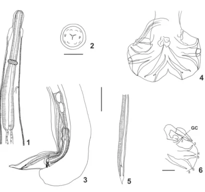
Small nematodes loosely coiled along ventral side. Ex-cretory pore within median third of oesophagus, deirids anterior to oesophagheal intestinal junction (Fig. 1).
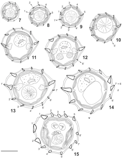
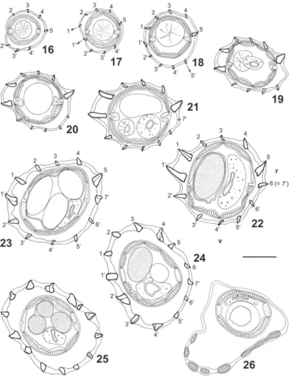
Synlophe - (studied in one male and one female paratypes). In both sexes, cuticle bearing longitudinally uninterrupted ridges with chitinoid struts. Ridges appear-ing at different levels between cephalic vesicle and nerve ring (Figs 7 to 10, 16 to 18) except ridge 6’ appearing just posterior to oesophageal intestinal junction (Figs 12, 20) and ridge 7’ appearing at end of anterior third of body in male (Fig. 13) and end of anterior fifth of body in female (Fig. 21). In posterior part of body, arising of new ridges only on ventral side, adjacent to right lateral field (Figs 15, 24, 25). Ridges disappearing anterior to caudal bursa in male and at level of vulva in female.
All along the body, except in ovejector region, pres-ence of carene made up of two left ridges, ventral being slightly more developed. Dorsal ridge appearing first, about 40 posterior to cephalic vesicle (Fig. 8) then ven-tral ridge (Figs 9, 17). In both sexes size of carene de-creasing in posterior quarter of body; in female, ridges of carene of equivalent size as of other ridges at level of distal uterus (Figs 23-25).
Number of ridges - In both sexes 6 (3 dorsal, 3 ven-tral) just posterior to cephalic vesicle (Fig. 7), 10 (carene, 4 dorsal, 4 ventral) at level of oesophageal in-testinal junction (Figs 11, 19), 12 (carene, 5 dorsal, 5 ventral) at mid-body (Figs 14, 22), 13 (carene, 4 dorsal, 7 ventral) in posterior quarter of body in male (Fig. 15). In female, 14 (7 dorsal, 7 ventral) at level of distal uterus (Fig. 25). At mid-body, right ventral ridge no. 7’ migrates towards dorsal side and becomes ridge no. 6 (Figs 14, 22). In female, presence of comaretes in posterior part of body (Figs 23 to 25). Just anterior to vulva, ridges are replaced by a chinoid thickening present only on ventral side (Fig. 26).
At mid-body in both sexes double gradient of size decreasing from lateral to median sides.
At mid-body, two axes of orientation directed from right-ventral to left dorsal side. Right axis inclined at 88° in male, 78° in female to sagittal axis. Left axis in-clined at 72° in male, 60° in female (Figs 14, 22). In female, at level of ovejector, orientation of ridges not clearly defined some ridges orientated perpendicularly to body surface (Figs 24, 25).
Holotype male - 7.1 mm long, 126 wide at mid-body; cephalic vesicle 79 long by 32 wide; nerve ring, excre-tory pore and deirids situated at 280, 338 and 523 from apex, respectively; oesophagus, 546 long.
Sub-symmetrical caudal bursa with dorsal lobe well developed. Pattern of type 1-4 (Fig. 4). Rays 3 originate first from common trunk to rays 3 to 6. Rays 4 most developed. Thick common trunk to rays 8 and dorsal ray. Rays 8 slightly shorter than dorsal ray. Dorsal ray deeply divided at mid- half into two branches, each branch giv-ing rise to 2 small branches, small rays 9 (external branches) and rays 10 (internal branches) (Fig. 4).
Filiform alate spicules 707 long with sharp distal extremities (Fig. 5). Spicule length/ body length ratio 10%. Complex genital cone with two parts: a proximal very elongated part 216 long and 29 wide at base and a bilobate distal part (Fig. 6). Papilla Zero and papillae 7 not observed. Gubernaculum absent.
Figs 7-15. Freitastrongylusangelae n. gen., n. sp. - Paratype male, 7.1 mm long, transverse sections of body; 7: just posterior to cephalic vesicle;
8-10: anterior to nerve ring; 8: 40 µm posterior to cephalic vesicle; 9: 80 µm posterior to cephalic vesicle; 8-10: 160 µm posterior to cephalic vesicle; 11: at oesophageal intestinal junction; 12: just posterior to oesophageal intestinal junction; 13: at third of body (2.7 mm from apex); 14: at mid-body (3.8 mm from apex); 15: at posterior quarter of body (500 µm anterior to caudal bursa); Bar = 50 µm; v: ventral side, r: right side. All ridges orientated as 14. Arrows indicate the origin of a ridge.
Main measurements (average and range) of ten paratypes. Body 7.7 (7.3-8.1) mm long and 130 (115-140) wide at mid-body, cuticular dilatation included; cephalic vesicle 82 (72-90) long and 40 (36-45) wide; nerve ring (n = 5), excretory pore (n = 7), and deirids (n = 8) situated at 271 (258-285), 351 (338-367), 539 (493-595) from apex, respectively; oesophagus 561 (518-613); spicules 723 (695-777) long, spicules/body length ratio 10% (9-11)%. Genital cone (n = 8) 218 (202-230) long, 30 (23-35) wide.
at morula stage 80 long by 41 wide on average. Uterus length/body length ratio 14.4%. Tail conical, 61 long, dorsally curved (Fig. 3).
Main measurements (average and range) of ten paratypes. Body, 10.8 (8.6-12.7) mm long and 150 (112-170) wide at mid-body, cuticular dilatation included; cephalic vesicle 81 (65-93) long by 44 (40-53) wide. Nerve ring (n = 7), excretory pore (n = 6), and deirids (n = 9) situated at 243 (225-278), 341 (297-390), and 551 (463-628) from apex, respectively; oesophagus, 578 (493-660) long; vulva (n = 7) situated at 179 (144-205) from caudal extremity; vagina vera (n = 6) 21 (15-25); ovejector with vestibule (n = 7) 158 (150-165) long; sphincter (n = 8) 38 (28-50) long by 35 (25-38) wide; infundibulum (n = 5) 198 (163-245) long; uterus (n = 8) 1980 (1260-2680) long with 111 (80-144) eggs at morula stage, 72 (58- 86) long by 40 (36-47) wide; tail (n = 6) 62 (54-68) long; uterus length/ body length ratio 18.2 % (12.4-21.0).
Figs 16-26. Freitastrongylus angelae n. gen., n. sp. - Paratype female, 9.5 mm long, transverse sections of body; 16-18: anterior to nerve ring; 16: 40 µm posterior to cephalic vesicle; 17: 80 µm posterior to cephalic vesicle; 18: 120 µm posterior to cephalic vesicle; 19: at oesophageal intestinal junction; 20: just posterior to oesophageal intestinal junction; 21: at fifth of body (1.7 mm from apex); 22: at mid-body (5.1 mm from apex); 23-25: at level of distal uterus 23: 700 µm anterior to vulva; 24: 600 µm anterior to vulva; 25: 500 µm anterior to vulva; 26: just anterior to vulva; Bar = 50 µm; v: ventral side; r: right side. All ridges orientated as 14. Arrows indicate the origin of a ridge.
Type material - Holotype male CHIOC no. 35044 a, allotype female CHIOC no. 35044 b.
Paratypes - CHIOC no. 35044 c-n (whole mounts) and no. 34844 (wet material). Other four male and four female paratypes were deposited in MNHN no. 319 MQ. Type host - Dasyprocta leporina (Linnaeus, 1758) – necropsy CHIOC no. 26463.
Other host - Dasyprocta fuliginosa Wagler, 1832 – necropsies CHIOC nos. 26464, 26465.
Voucher specimens - CHIOC no. 35045 a-b (whole mounts), CHIOC no. 34845, 34846 (wet material).
of the Malalarrá waterway, right margin of the Preto river, Santa Isabel do Rio Negro municipality, state of Amazonas and Três Barracas settlement, Jauari water-way, left margin of the Aracá river, Barcelos municipal-ity, state of Amazonas.
Site of infection - Stomach.
Etymology - The generic name is to honor the late Dr João Ferreira Teixeira de Freitas, one of the most outstanding Brazilian helminthologists and the specific name is after Dr Angela Cristina Verissimo Junqueira, for her efforts to improve the knowledge of the mam-malian zoonotic reservoirs in the investigated area.
Collector/dates - AQ Gonçalves/ April 27, 2000; February 26, 2001; February 06, 2002.
DISCUSSION
The heligmosomoid nematodes studied here, possess the main features of the Pudicinae (Heligmonellidae): axes of orientation oblique, carene present, dorsal ray deeply divided, female tail lacking spine (Durette-Desset 1985). They belong to the suprageneric group I (after Durette-Desset & Justine 1991) which includes Helig-mostrongylus Travassos, 1937, Fuellebornema Travas-sos and Darriba, 1929, Sciurodendrium Durette-Desset, 1971, and Pseudoheligmosomum Travassos, 1937. These genera share one synapomorphy i.e. rays 9 shorter than rays 10. Within these genera, the specimens, para-sitic in Dasyprocta spp. are closely related to Fuellebornema particularly by the same pattern of the caudal bursa. The synlophe also presents some similari-ties with that of Fuellebornema at mid-body: 5 dorsal and 6 ventral ridges, axes of orientation strongly inclined to the sagittal axis. Therefore it differs by the relative size of the dorsal and ventral ridges in relation to the carene. In Fuellebornema, the carene is strongly devel-oped and the other ridges small and of equivalent size (Durette-Desset 1970, Cassone & Durette-Desset 1991). In the Brazilian specimens studied here, all the ridges are well developed and the carene at mid-body has a similar size to that of the ridge situated in front of the right field (ridge no. 5). In addition there is a sole axis of orientation in the Fuellebornema species of which the synlophe is known.
The presence of a very long genital cone is not char-acteristic of the new genus Freitastrongylus. It is present in Fuellebornema almeidai Travassos, 1937 a parasite of D. leporina from Brazil (Travassos 1937) but also in other parasites all belonging to the Heligmonellidae such as Heligmonella moreli Gibbons, Durette-Desset & Daynes, 1977 (Heligmonellinae), a parasite of Dremomys lokriah (Hodgson, 1836) from Nepal (Gib-bons et al. 1977) or Brevistriata skrjabini Shulz & Lubimov, 1932 (Brevistriatinae), a parasite of chipmunks [Tamias sibiricus (Laxmann, 1766)] and red squirels (Sciurus vulgaris Linnaeus, 1758) from Soviet Far East (Shulz & Lubimov 1932), Otaru (Northern Japan) (Asakawa & Ohbayashi 1986), and France (Pisanu et al. 2007). Contrary to this, the orientation and the shape of the genital cone are original.
To date the genus Fuellebornema is made up of seven species, described from the Neotropical region (Brazil, Colombia, Paraguay) all parasitic in Dasyprocta spp. (Durette-Desset et al. 2006). Since Fuellebornema and Freitastrongylus n. gen. are morphologically closely related this suggests that they may have differentiated in the same host, Dasyprocta. According to the rules of the evolution of the synlophe in the Heligmosomoidea (Durette-Desset 1985), the synlophe of Freitastron-gylus n. gen. is more primitive than that of Fuellebor-nema: carene less developed, right ridge as developed as ridges of the carene, other cuticular ridges well de-veloped, ridges present all around the body (absent in the latero ventral quadrant in Fuellebornema), gradient of size of the ridges present, axis of orientation less in-clined to sagittal axis. In addition, the strong develop-ment of the right ridge and the presence of a gradient of size of the cuticular ridges are characteristic of the Heligmonellinae from which the Pudicinae originate. In view of this, the genus Freitastrongylus n. gen. may be considered as having an intermediary systematic posi-tion between the Heligmonellinae and the Pudicinae.
ACKNOWLEDGEMENTS
To José R Coura and Angela CV Junqueira, from the Labo-ratory of Parasitic Diseases, Oswaldo Cruz Institute, for the in-vitation to collect the samples for this study; to the National Health Foundation for logistic support; to Cibele R Bonvicino, National Cancer Institute, and Gilson E Iack-Ximenes, Museum of Zool-ogy, University of the State of São Paulo, São Paulo, Brazil, for the identification of the rodent hosts; to Jean-François Dejouanet, Muséum National d’Histoire Naturelle, Paris, for technical as-sistance with the drawings.
REFERENCES
Asakawa M, Ohbayashi M 1986. The first record of Brevistriata bergerardi Durette-Desset, 1970 from an Asiatic chipmunk
Tamias sibiricus lineatus Siebold in Hokkaido, Japan. Jpn J Vet Res 34: 291-294.
Cassone J, Durette-Desset MC 1991. Cinq espèces (donc trois nouvelles) de nématodes Trichostrongyloides coparasites de Dasyprocta azarae au Paraguay. Rev Suisse Zool 98:
229-242.
Durette-Desset MC 1970. Nématodes Héligmosomes d’Amé-rique du Sud. VI. Étude de cinq espèces, parasites de ron-geurs Dasyproctidés. Bull Mus Nat Hist Nat 42: 590-600. Durette-Desset MC 1985. Trichostrongyloid nematodes and their
vertebrate hosts: reconstruction of the phylogeny of a para-sitic group. Adv Parasitol24: 239-306.
Durette-Desset MC, Chabaud AG 1981. Nouvel essai de classi-fication des nématodes Trichostrongyloidea. Ann Par Hum Comp56: 297-312.
Durette-Desset MC, Chabaud AG 1993. Nomenclature des Strongylida au-dessus du groupe famille. Ann Par Hum Comp 68: 111-112.
Durette-Desset MC, Digiani MC 2005. The axis of orientation of the synlophe in the Heligmosomoidea (Nematoda, Tri-chostrongylina): a new approach. Parasite 12: 195-202.
Durette-Desset MC, Gonçalves AQ, Pinto RM 2006. Trichos-trongylina (Nematoda, Heligmosomoidea) coparasites in
Dasyprocta fuliginosa Wagler (Rodentia, Dasyproctidae) from Brazil, with the re-establishment of the genus Avellaria
Freitas & Lent and the description of two new species. Revta Bras Zool 23: 509-519.
Gibbons LM, Durette-Desset MC, Daynes P 1977. A review of the genus Impalaia Monnig, 1923 (Nematoda:
Trichostron-gyloidea). Ann Parasitol Hum Comp 52: 435-446.
Pisanu B, Jerusalem C, Huchery C, Marmet J, Chapuis JL 2007. Helminth fauna of the Siberian chipmunk, Tamias sibiricus
Laxmann (Rodentia, Sciuridae) introduced in suburban French forests. Parasitol Res 100: 1375-1379.
Schulz RE, Lubimov MP 1932. Longistriata skrjabini n. sp.
(Nematoda, Trichostrongylidae) from the Ussuri Squirrel.
Parasitology 24: 50-53.
Travassos L 1937. Revisão da família Trichostrongylidae Leiper, 1912, Instituto Oswaldo Cruz, Rio de Janeiro, 512 pp.