online | memorias.ioc.fiocruz.br
Potential of laticifer fluids for inhibiting
Aedes aegypti
larval
development: evidence for the involvement of proteolytic activity
Márcio V Ramos1/+, Danielle A Pereira1, Diego P Souza1, Eliane S Araújo1, Cléverson DT Freitas1, Mariana G Cavalheiro1, Mayara Patricia V Matos1, Ana FU Carvalho2/+
1Departamento de Bioquímica e Biologia Molecular 2Departamento de Biologia, Universidade Federal do Ceará, Campus do Pici, Caixa
Postal 6033, 60451-970 Fortaleza, CE, Brasil
It has been shown previously that the laticifer fluid of Calotropis procera (Ait.) R.Br. is highly toxic to the egg hatching and larval development of Aedes aegypti L. In the present study, the larvicidal potential of other laticifer fluids obtained from Cryptostegia grandiflora R.Br., Plumeria rubra L. and Euphorbia tirucalli L. was evaluated. We attempted to correlate larvicidal activity with the presence of endogenous proteolytic activity in the protein fraction of the fluids. After collection, the fluids were processed by centrifugation and dialysis to obtain the soluble laticifer protein (LP) fractions and eliminate water insoluble and low molecular mass molecules. LP did not visibly affect egg hatching at the doses assayed. LP from Cr. grandiflora exhibited the highest larval toxicity, while P. rubra was almost inactive. E. tirucalli was slightly active, but its activity could not be correlated to proteins since no protein was detected in the fluid. The larvicidal effects of LP from C. procera and Cr. grandiflora showed a significant rela-tionship with the proteolytic activity of cysteine proteinases, which are present in both materials. A purified cysteine proteinase (papain) from the latex of Carica papaya (obtained from Sigma)was similarly effective, whereas trypsin and chymotrypsin (both serine proteinases) were ineffective. The results provide evidence for the involvement of cysteine proteinase activity in the larvicidal action of some laticifer fluids. C. procera is an invasive species found in areas infested with Ae. aegypti and thus could prove useful for combating mosquito proliferation. This is the first report to present evidence for the use of proteolytic enzymes as chemical agents to destroy Ae. aegypti larvae.
Key words: Aedes aegypti - biological control - cysteine proteinases - latex - larvae - papain
Financial support: FUNCAP, CNPq, PADCT, RENORBIO, Interna-tional Foundation for Science (3070-3 to MVR)
+ Corresponding authors: [email protected] Received 11 November 2008
Accepted 21 July 2009
Dengue fever, recognised by the WHO as a “ma-jor international public health concern” (WHO 2009), peaked in Brazil in 1998 with 528,388 cases and again in 2002 with 784,000 cases (Siqueira Jr et al. 2005). Ac-cording to data published by the Brazilian government, 438,949 cases of classic dengue and 926 cases of yellow fever were registered in Brazil in 2007. Since January 2008, the whole country has experienced an explosion in the number of new cases that reached one per minute in Rio de Janeiro (PAHO/WHO 2008). However, these statistics may be underestimates due to unreported cases. Regardless, the problem has become more criti-cal each year, likely as a result of unsuccessful gov-ernmental actions. Strategies to combat the mosquito vector are still limited, but include the dissemination of chemicals and/or insecticidal bacteria in the envi-ronment. In fact, synthetic pesticides, chitin synthesis inhibitors (BPU’s), hormone analogues and some bac-terial species have been the only chemical and biologi-cal strategies developed to reduce insect proliferation (Braga & Valle 2007). However, such strategies have
proven to be relatively ineffective or undesirable due to the development of resistance in mosquito populations, negative environmental impacts and the fact that the bacteria show rapid sedimentation from the larval feed-ing zone, UV light sensitivity and a narrow host range that has hampered their development (Porter 1996). In light of these problems, the search continues for new sources of natural products that could be applied to combating the spread of dengue. Increasing insecticide resistance requires the development of strategies for prolonging the use of highly effective vector control compounds. The use of combinations of multiple in-secticides and phytochemicals is one such strategy that may be suitable for mosquito control. New sources of natural materials have been reported in the literature that can inhibit growth, reproduction and oviposition (Consoli & Oliveira 1994) or act as repellents (Carvalho et al. 2003). We previously described the effects of latex from Calotropis procera (Ait.) R.Br. (Apocyna- ceae) on egg hatching and larvae of Aedes aegypti
In the present study, the larvicidal effects of other la-ticifer fluids were investigated. In addition, we determined the contribution of endogenous proteolytic activity (com-monly present in latex fluids) to the larvicidal effect.
MATERIAL AND METHODS
Chemicals - N-α-benzoyl-DL-arginine β-naphthyl-
amide hydrochloride (BANA), trans-epoxysuccinyl-l-leucylamido (4-guanidio)-butane (E-64), ethylene-diaminetetraacetic acid (EDTA), ethylene glycol-bis(β-aminoethyl ether) N,N,N`,N`-tetra acetic acid, phenylmethylsulphonylfluoride, 4-(dimethyl-amino) cin- namaldehyde, pepstatin, papain (EC 3.4.22.2), trypsin type I from bovine pancreas, (EC 3.4.21.4), chymotrypsin (EC 3.4.21.1) and azocasein were purchased from Sigma Chem-ical Co. (St. Louis, MO, USA). Iodoacetamide (IAA), Coo-massie brilliant blue (R-350) and molecular mass markers were from Amersham Biosciences (Uppsala, Sweden). Bo-vine serum albumin (BSA) was from Inlab (lot 826.787). All other chemicals were of analytical grade.
Plant material - Healthy and non-cultivated plants of
C. procera (Ait.) R.Br., Cryptostegia grandiflora R.Br., Plumeria rubra L. (all Apocynaceae) and Euphorbia tirucalli L. (Euphorbiaceae)growing in the vicinity of Fortaleza-Brazil, were used as the source of fresh latex. Each plant sample was identified by the botanist Ed-son Paula Nunes and the vouchers 32663 (C. procera), 040409 (Cr. grandiflora), 15018 (P. rubra) and 38702 (E. tirucalli)were deposited at the Prisco Bezerra Her-barium of the Universidade Federal do Ceara, Fortale-za, Brazil. C. procera is an invasive species, occurring throughout Northeast Brazil. This plant produces gen-erous amounts of latex and thus its availability is not a limiting factor for use.
Latex processing - The latex of C. procera was col-lected from healthy plants by small incisions near the youngest leaves and the liquid was left to flow into dis-tilled water to produce a 1:1 (v/v) mixture. The other latices were collected from the ends of branches and processed following the same procedure detailed below. The mixtures (water and latex) were gently stirred dur-ing collection to overcome the tendency of the latices to coagulate. The samples were centrifuged at 5,000 g
for 10 min at 4°C. The rubber-like precipitates produced after centrifugation were discarded and the supernatants were exhaustively dialysed against distilled water us-ing membranes of 8,000 molecular mass cut-off. Each sample was maintained for 60 h at 8°C and the dialysis water was renewed three times daily. The retained ma-terials were centrifuged again using the conditions de-scribed above. The clean, rubber-free supernatants were lyophilised and used in all subsequent experiments. This procedure eliminates water-insoluble and low molecular mass molecules while retaining almost all proteins.
Eggs and larvae of Ae. aegypti - Eggs of Ae. ae-gypti were obtained from Núcleo de Endemias Trans-missíveis por Vetores (NUVET), Secretaria de Saúde, Ceará, Brazil. The eggs were deposited on paper strips, submerged in distilled water containing 1% ethanol (1 L) and allowed to develop at 25-28°C under a 12-h
light/dark cycle until reaching the third larval stage. About 20 mg of soybean seed flour was added to the water as a nutrient source.
Insecticidal bioassays - The bioassays were devel-oped according to WHO procedures (2005) but on a laboratory scale to allow the simultaneous evaluation of bioactivity and investigation of biochemical parameters. Larger-scale assays are currently being planned in ac-cordance with NUVET strategies.
To investigate the effect of LP on egg hatching, the lyophilised LP fractions were dissolved in distilled water containing 1% ethanol to achieve 1 mg/mL. The LP fraction at this concentration is able to inhibit egg hatching and is active against third instars (Ramos et al. 2006). BSA was used as a negative control. All solutions were prepared immediately before use. Undamaged eggs of Ae.aegypti adsorbed on dry paper strips (1 cm2)
were selected, counted and subsequently placed in test tubes containing 10 mL of each sample in triplicate. Egg hatching was monitored at 24 and 72 h. The assay was conducted at 25-28°C and under a dark/light cycle of 12 h. Ethanol (1%) in distilled water without protein was also used as control.
Assays to evaluate the toxicity of LPand of the puri-fied proteolytic enzymes EC 3.4.22.2, EC 3.4.21.1 and EC 3.4.21.4to third instars were carried out as follows: larvae were grown as described above. LP were dis-solved in distilled water containing 1% ethanol (as rec-ommended by WHO) at 1 mg/mL, excepting LP of C. procera and Cr. grandiflora, which were tested at dif-ferent concentrations and conditions [with or without di-thiothreitol (DTT) and IAA] to investigate correlations between toxicity and proteolytic activity. The purified enzymes listed above were dissolved in 50 mM PBS buf-fer (pH 6.0 for EC 3.4.22.2 or pH 7.0 for trypsin and EC 3.4.21.1) containing 1% ethanol. Ten millilitres of each LP sample were added to glass tubes (20 x 100 mm) in triplicate and 10 third instars were then added into each tube. Care was taken to use only undamaged individu-als. The assays were conducted under a 12-h light/dark cycle at 28°C for 48 h. Results were collected at 1, 3, 24 and 48 h after inoculation. Three independent ments were performed. Upon completion of the experi-ments, the larvae were disposed of following a 30 min boiling treatment.
Samples of C. procera and Cr. grandiflora were prepared as described above at different concentrations, mixed with 3 mM DTT and maintained at 25°C for 10 min before the addition of larvae. Similarly, samples were treated with DTT followed by E-64 (12 µM) and maintained at 25°C for additional 30 min prior to the assay. DTT acts as an activator of cysteine proteinase enzymes and cysteine proteinase activity is found in LP from C. procera (Freitas et al. 2007). E-64 and IAA are both potent and irreversible inhibitors of cysteine protein- ase proteolytic activity (Patel & Jagannadham 2003).
Electrophoresis - Electrophoresis of proteins was performed to visualise the proteins from LP samples and to develop zymograms. Polyacrylamide gel electropho-resis in the presence of sodium dodecyl sulphate (SDS) (PAGE-SDS) and 2-mercaptoethanol was performed as described by Laemmli (1970). Protein samples were dis-solved in sample buffer [0.0625 M Tris buffer (pH 6.8) containing 2% SDS]. Electrophoresis was performed at 25 mA per gel at 25°C for 4 h. Gels were stained with Coomassie brilliant blue (R-350) solution in water: ace-tic acid: methanol (8/1/3.5, v/v/v) and then de-stained with the same solution minus the dye.
Total proteolytic activity - Azocasein was used as a non-specific substrate to investigate total proteolytic ac-tivity in LP from Cr. grandiflora, P. rubra and E. tiru-calli. A detailed study about the proteolytic activity of latex from C. procera was recently published (Freitas et al. 2007) and in the present work, we only sought to char-acterise the toxicity to Ae. aegypti. The reaction mixture contained 50 or 100 µL (1 mg/mL) of the LP pre-incubated with 3 mM DTT for 10 min. Then, 200 µL of 1% azoca-sein in 50 mM PBS buffer (pH 6.0) was added. The reac-tions were performed at 37°C and stopped 60 min later by adding 300 µL of 20% trichloroacetic acid. The tubes, in triplicate, were centrifuged (5,000 g for 10 min at 25°C) and the supernatants were alkalinised with 400 µL of 2.0 N NaOH solution. The absorbance of the sample at 420 nm was then measured (Xavier-Filho et al. 1989). One unit of activity (AU) was defined as the amount of enzyme ca-pable of changing the absorbance at 420 nm by 0.01.
Assays for cysteine proteinases (EC 3.4.22) were conducted utilising BANA as substrate. Aliquots of 100 µL of the LP [1 mg/mL for C. procera, P. rubra, E. tirucalli and 0.5 mg/mL Cr. grandiflora in 50 mM PBS (pH 6.0)] were pre-incubated with 40 µL of an activa-tion soluactiva-tion in the presence of 3 mM DTT and 2 mM EDTA for 10 min. Then, 200 µL of BANA was added [1 mM in 50 mM PBS (pH 6.0)]. After 30 min, at 37°C, the reactions were stopped by adding 500 µL of 2% HCl in ethanol and 500 µL of 0.06% 4-(dimethyl-amino) cinnamaldehyde. Forty minutes later, the absorbance of the samples was measured at 540 nm (Abe et al. 1992). One AU was defined as the amount of enzyme that in-creases the absorbance at 540 nm by 0.01. All assays were performed in triplicate. The results of each series were expressed as the mean value ± SEM. EC 3.4.22.2, the cysteine proteinase purified from the latex of Carica papaya (obtained from SIGMA), was assayed for both proteolytic and larvicidal activities for use as a reference. As an additional attempt to verify whether other prote-olytic activity besides cysteine proteinase was associ-ated with larval death, EC 3.4.21.4 and EC 3.4.21.1, both serine proteinases (obtained from Sigma), were assayed at 0.05 and 0.1 mg/mL in 50 mM phosphate buffered sa-line pH 7.0 (PBS) with azocasein (1%) as the substrate and tested for larvicidal activity as described above.
Detection of proteases by zymography - Electropho-resis was carried out according to the method described by Schagger and Jagow (1987). LP fractions were dis-solved in 1 M Tris-HCl buffer (pH 6.8) containing 10%
SDS, 10% glycerol and in the presence or absence of 5% 2-mercaptoethanol. The sample was separated by elec-trophoresis through 12.5% polyacrylamide gels contain-ing 0.1% gelatin (as substrate for proteolysis) at 25°C, as described by Macedo et al. (2004). After electrophore-sis, the gels were immersed in water containing 2.5% of Triton X-100 (renaturing solution) and gently shaken for 30 min at 25°C to wash out the SDS and allow protein re-naturation. The gels were then incubated in 50 mM PBS (pH 6.0) containing 3 mM DTT and 2 mM EDTA for 3 h at 37°C. The incubated gels were stained with 0.2% Coo-massie brilliant blue R-350 in 40% methanol and 10% acetic acid followed by treatment using the same solu-tion without the dye. Enzymatic activity was detected as transparent bands by direct observation.
Statistical analysis - Results are reported as mean ± SEM for triplicates. Statistical significance (p < 0.05 or p < 0.001) was assessed by analysis of variance followed by Tukey’s test.
RESULTS
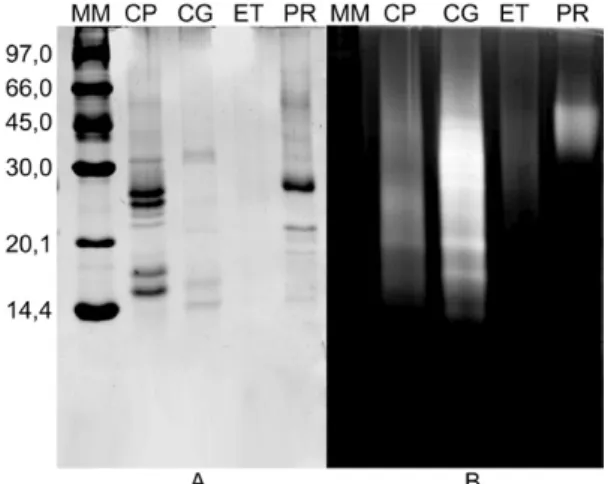
Protein content and larvicidal activity of latex flu-ids- The quantities of soluble protein in LP fractions were initially estimated by the principle of protein-dye binding (Bradford 1976). The latex of C. procera was the richest in protein, while Cr. grandiflora and P. ru-bra showed moderate levels. On the other hand, neither soluble protein nor proteolytic activity against azocasein or BANA was detected in E. tirucalli material (Table I). This result was further confirmed by electrophoresis (Fig. 1). Proteolytic activity was found in the LP of Cr. grandiflora and P. rubra by photocolourimetric assay using azocasein (Table I) and by zymography using gel-atin as the substrate (Fig. 1). As previously demonstrated (Freitas et al 2007, Oliveira et al. 2007), the LP of C. procera were active on both systems. It is noteworthy that Cr. grandiflora, when assayed under identical
con-Fig. 1: electrophoresis (A) and Zymogram (B) of laticifer proteins (LP)
of Calotropis procera (CP), Cryptostegia grandiflora (CG), Euphorbia
tirucalli (ET) and Plumeria rubra (PR). A total of 5 µg of LP was applied
ditions using BANA as the substrate, exhibited a pro-teolytic activity 32-fold higher than that of C. procera
and 57-fold higher than that of P. rubra. Since BANA is a specific substrate for EC 3.4.22 (Barrett 1972), this activity likely predominates in the studied protein ma-terials. In fact, cysteine proteinase activity is the only proteolytic activity found in LP from Cr. grandiflora
(CgLP), whereas LP from P. rubra (PrLP) exhibit poor cysteine and serine proteinase activity and completely lack aspartic and metallo-proteinase activity (data not shown). A detailed study of the protein profile and en-zymatic antioxidative and proteolytic activities of CgLP, PrLP and laticifer proteins of E. tirucalli (EtLP) will ap-pear elsewhere. The abundance of proteolytic activity in both Cr. grandiflora and C. procera LP prompted us to further analyse these samples in an attempt to correlate larvicidal effects and proteolytic activity.
LP did not prevent egg hatching when assayed at 1 mg/mL (Table II). However, the effect on larval growth was markedly different among the LP samples. Both,
C. procera and Cr. grandiflora exhibited strong toxic-ity to larvae, while E. tirucalli and P. rubra displayed fair activity, being similar to the controls. These data
were recorded at the end of the observation period (72 h) and thus it appears that LP act on larvae rather than eggs. There was a correlation between the toxicity of LP sample and the amount of proteolytic activity (Table I). Samples exhibiting stronger proteolytic activity also showed higher toxicity to larvae (Table III).
Correlations between larvicidal effects and cysteine proteinase activity-LP of Cr. grandiflora were very ef-fective in eliminating third instars of Ae. aegypti (Table III). E. tirucalli had moderate activity, while P. rubra
was inactive. LP of C. procera and Cr. grandiflora were tested at different concentrations in the presence and ab-sence of DTT (an efficient activator of EC 3.4.22) and E-64 (a specific and potent inhibitor of this activity). We observed that increasing concentrations of C. procera
LP elevated the mortality of third instars. This effect was substantially augmented after treating the LP with DTT, suggesting the involvement of cysteine proteinase activ-ity in the deleterious effects upon larvae. Furthermore, the larvicidal activities of C. procera (CpLP) and CgLP were drastically reduced when the samples were pre-treated with IAA, an irreversible inhibitor of EC 3.4.22,
TABLE I
Protein content and proteolytic activity of studied laticifer fluids
Protein samplesa
(laticifer proteins)
Protein contentb
(μg/100 μL)
Proteolytic activity upon azocasein (AU/μg protein)
Proteolytic activity upon BANA (AU/μg protein)
Calotropis procerac 132.0 ± 3.9 0.30 ± 0.03 0.42 ± 0.00
Cryptostegia grandiflora 22.8 ± 9.1 3.27 ± 0.13 15.0 ± 0.73
Plumeria rubra 41.8 ± 1.7 0.74 ± 0.07 0.26 ± 0.02
Euphorbia tirucalli nd nd nd
a: samples were pre-treated with dithiothreitol (DTT) to activate cysteine proteinases;AU: activity unit; b: estimated according
to Bradford method (1976); BANA: N-α-benzoyl-DL-arginine β-naphthylamide hydrochloride; c: C. procera was used as refer-ence; nd: not detected. Results are expressed as mean ± SD of six independent measurements.
TABLE II
Effects of laticifer proteins upon egg hatching of Aedes aegypti
Samples (1 mg/mL)
Eggs observed n
Hatching after 24 h %
Larvae (alive) after 72 h n (%)
Larvae(dead)after 72 h n
Mortality (%)
Ethanol (1%) 103. 67 ± 2.73 88.26 ± 7.42a 76.00 ± 6.66 (73.3) 15.67 ± 2.72 17.00 ± 1.49a
BSA 132.50 ± 11.03 48.44 ± 9.77b 52.50 ± 10.21 (81.7) 13.00 ± 5.72 18.5 ± 4.49a
CpLP 130.00 ± 13.06 65.65 ± 2.56a 13.66 ± 4.91 (10) 72.33 ± 15.95 82.00 ± 8.89b
CgLP 106.33 ± 3.76 86.75 ± 8.43a 1.33 ± 0.33 (1.2) 90.33 ± 6.77 98.33 ± 0.66b
EtLP 104.66 ± 3.28 93.95 ± 5.11a 72.00 ± 5.69 (68.7) 26.66 ± 5.37 27.33 ± 5.24a
PrLP 116.33 ± 13.02 91.93 ± 4.05a 84.66 ± 8.96 (72.9) 22.00 ± 1.53 20.66 ± 2.18a
Values are means ± SEM of triplicates. Different letters in the same column differ significantly (p < 0.05 for hatching and p < 0.001 for mortality data) from the control (ethanol 1%). Statistical significance was assessed by analysis of variance followed by Tukey’s test. BSA: bovine serum albumin; CgLP: laticifer proteins of Cryptostegia grandiflora; CpLP: laticifer proteins of
compared to corresponding samples that had been acti-vated with DTT to get maximum proteolysis (Table III). DTT, E-64 and IAA did not exhibit toxicity at the con-centrations at which they were assayed (data not shown). The fact that Cr. grandiflora and C. procera were highly active while E. tirucalli and P. rubra showed negligible effects on larvae also supports the hypothesis that prote-olytic activity is involved in the deleterious effects. Cr. grandiflora, whichexhibited stronger proteolytic activ-ity (Table I), was also more active than the C. procera
samples (Table III).
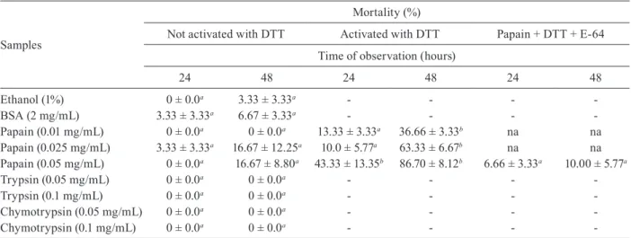
To emphasise the importance of cysteine proteinase activity for larval toxicity, EC 3.4.22.2 (obtained from Sigma), a cysteine proteinase purified from the latex of C. papaya (Caricaceae), was assayed (Nitsawang et al. 2006). We found that the proteolytic activity of EC 3.4.22.2 was inhibited by E-64 (Fig. 2). Table IV sum-marises the larvicidal effects of EC 3.4.22.2, which ex-hibited toxic effects upon third instars, mainly after acti-vation by DTT. This effect was drastically reduced when the enzyme was inhibited by E-64. According to the re-sults, there seems to be a direct correlation between the proteolytic activity of this cysteine proteinase of laticifer origin and toxicity to Ae. aegypti larvae. EC 3.4.21.4 and EC 3.4.21.1, both serine proteinases, were assayed for
larvicidal activity at 0.05 and 0.1 mg/mL (Kraut 1977). The proteolytic activity of these enzymes (0.1 mg/mL) against azocasein was estimated to be 4.7 and 2.0 AU/ µg, respectively. These values are similar to that of EC 3.4.22.2 (3.2 AU/µg) (Fig. 2). However, the serine protei-nases did not exhibit any larvicidal effects (Table IV). Similarly, no larval mortality was documented when lar-vae were exposed to PBS alone.
DISCUSSION
Through the years, the dengue mosquito has be-come progressively adapted to human civilisation. This trend, which has been confirmed by health agents in-volved in governmental programs for dengue combat, is evidenced by the number of cases of the disease re-ported by the World Health Organisation (PAHO/WHO 2008), which has increased drastically mainly in urban areas. Consequently, the use of traditional chemical or-ganophosphorates is not necessarily the best strategy to combat the mosquito. The dissemination of these in-secticides in the air does not effectively kill mosquitoes in dwellings and cannot be used indoors due to side effects and general toxicity. An alternative or comple-mentary strategy should include the use of novel bioac-tive compounds with lower toxicities that are able to
TABLE III
Larvicidal effect of laticifer proteins upon third instars of Aedes aegypti
Samples
Mortality (%)
3 h 24 h 48 h
Ethanol (1%) 0 ± 0.0a 0 ± 0.0a 0 ± 0.0a
BSA (1 mg/mL) 0 ± 0.0a 0 ± 0.0a 0 ± 0.0a
CpLP(0.1 mg/mL) 0 ± 0.0a 3.33 ± 3.33a 6.66 ± 3.33a
CpLP + DTT (0.1 mg/mL) 0 ± 0.0a 10.00 ± 5.77a 36.66 ± 6.67b
CpLP(0.25 mg/mL) 0 ± 0.0a 3.33 ± 3.33a 13.33 ± 3.33a
CpLP + DTT (0.25 mg/mL) 0 ± 0.0a 16.67 ± 3.33a 40.0 ± 5.78b
CpLP(1 mg/mL) 0 ± 0.0a 30.0 ± 10.00a 56.66 ± 3.33b
CpLP + DTT (1 mg/mL) 0 ± 0.0a 80.0 ± 11.56b 100.0 ± 0.0b
CpLP(2 mg/mL) 0 ± 0.0a 56.66 ± 16.67b 70.0 ± 10.00b
CpLP+ DTT (2 mg/mL) 0 ± 0.0a 90 ± 5.78b 100 ± 0.00b
CPLP + DTT + E-64 (2 mg/mL) 0 ± 0.0a 60.0 ± 10.0b 87.0 ± 8.83b
CpLP (2 mg/mL + IAA) 0 ±0.0a 13.00 ± 8.82b 60 ± 0.00b
CgLP(1 mg/mL) 0 ± 0.0a 83.33 ± 3.33b 96.66 ± 3.33b
CgLP + DTT + E-64 (1 mg/mL) 0 ± 0.0a 0 ± 0.0a 36.66 ± 6.67b
CgLP (0.25 mg/mL) 10.00 ± 1.44b 90.00 ± 5.78b 93.33 ± 3.33b
CgLP(0.25 mg/mL + DTT) 0 ± 0.0a 100 ± 0.0b 100 ± 0.00b
CgLP(1mg/mL + IAA) 0 ± 0.0a 0 ± 0.0a 21.42 ± 15.28a
EtLP(1 mg/mL) 0 ± 0.0a 0 ± 0.0a 16.66 ± 3.33a
PrLP(1 mg/mL) 0 ± 0.0a 0 ± 0.0a 0 ± 0.0a
Values are means ± SEM of triplicates. Different letters in the same column differ significantly (p < 0.05) from the control (ethanol 1%). Statistical significance was assessed by analysis of variance followed by Tukey’s test. Laticifer proteins of Calotropis procera
efficiently eliminate dengue foci in inhabited areas. In this context, bioactive proteins may serve as potential alternative. In this paper, we advanced the idea of using bioactive proteins to destroy mosquito larvae and thus reduce mosquito proliferation.
Latex-producing plants are widespread in different habitats. These plants usually secrete milk-like fluid from a network of laticifer cells, in which subcellular or-ganelles intensively synthesise proteins and secondary metabolites (Lopes et al. 2009). The biological impor-tance of latex fluids is still unclear and knowledge of their physiological role is still limited (Ramos et al. 2007). Fig. 2: proteolytic activity of commercially available cysteine (pa-pain) and serine (trypsin and chymotrypsin) proteinases and inhibi-tion of the proteolytic activity of papain from Carica papaya latex. The samples were also used to investigate toxic effects upon third instars of Aedes aegypti as shown in Table IV. AU: unit of activity;
BANA: N-α-benzoyl-DL-arginine β-naphthylamide hydrochloride;
E-64: trans-epoxysuccinyl-l-leucylamido (4-guanidio)-butane.
AU/μg protein
Azocasein Trypsin
25
20
15
10
5
0
Chymo Papain Papain + E-64 Papain Papain + E-64
BANA
TABLE IV
Larvicidal effect of the commercially available cysteine (papain) and serine (trypsin and chymotrypsin) proteinases upon third instars of Aedes aegypti
Samples
Mortality (%)
Not activated with DTT Activated with DTT Papain + DTT + E-64
Time of observation (hours)
24 48 24 48 24 48
Ethanol (1%) 0 ± 0.0a 3.33 ± 3.33a - - -
-BSA (2 mg/mL) 3.33 ± 3.33a 6.67 ± 3.33a - - -
-Papain (0.01 mg/mL) 0 ± 0.0a 0 ± 0.0a 13.33 ± 3.33a 36.66 ± 3.33b na na
Papain (0.025 mg/mL) 3.33 ± 3.33a 16.67 ± 12.25a 10.0 ± 5.77a 63.33 ± 6.67b na na
Papain (0.05 mg/mL) 0 ± 0.0a 16.67 ± 8.80a 43.33 ± 13.35b 86.70 ± 8.12b 6.66 ± 3.33a 10.00 ± 5.77a
Trypsin (0.05 mg/mL) 0 ± 0.0a 0 ± 0.0a - - -
-Trypsin (0.1 mg/mL) 0 ± 0.0a 0 ± 0.0a - - -
-Chymotrypsin (0.05 mg/mL) 0 ± 0.0a 0 ± 0.0a - - -
-Chymotrypsin (0.1 mg/mL) 0 ± 0.0a 0 ± 0.0a - - -
-Values are means ± SEM for two independent experiments. Statistical significance (p < 0.05) was assessed by analysis of vari-ance followed by Tukey’s test. Different letters in the same column differ significantly from control (ethanol 1%). BSA: bovine serum albumin; DTT: dithiothreitol; E-64: trans-epoxysuccinyl-l-leucylamido (4-guanidio)-butane; na: not assayed.
Nevertheless, proteolytic activity has been described as a common and frequently abundant endogenous enzy-matic activity in many laticifer fluids (Glazer & Smith 1971, Lynn 1979, Boller 1986, Freitas et al. 2007). Serine and EC 3.4.22have been reported as the most common proteinases found in laticifer fluids and the latter was largely predominant in the materials tested in the present paper, in which three out of four latex fluids analysed showed proteolytic activity. Plant EC 3.4.22 play major roles in intracellular and extracellular processes such as the development and ripening of fruits (Brady 1985), the building of nutritional reserves, the degradation of stor-age proteins in germinating seeds (Kembhavi et al. 1993, Taylor & Cuming 1993), the activation of pro-enzymes and the degradation of defective proteins (Rudenskaya et al. 1998). Additionally, enzymes in latex are involved protecting the plant against predator attack (Smith et al. 1955, Boller 1986).
of the cysteine proteinase in response to caterpillar feed-ing and its ability to damage the insect peritrophic matrix represents an unusual plant defence mechanism that may have applications in biotechnology. Thus, it is reasonable to think that EC 3.4.22 present in LP may destroy lar-vae of Ae. aegypti by disrupting their peritrophic matrix. However, the remaining larvicidal activity still detected in samples inhibited with IAA or E-64 suggests that oth-er proteins in these latex samplesmay also be implicated in the larvicidal action. It is also relevant to consider the larvicidal effect of EC 3.4.22.2, which is a cysteine pro-teinase obtained from another latex. The results shown in Table IV give important insights about the contribu-tion of the EC 3.4.22 to larval death. Further studies of the effects of purified EC 3.4.22 from C. procera or Cr. grandiflora on larval development will certainly help to clarify the remaining questions.
Interestingly, we found that LP did not cause dam-age to mosquito eggs; in fact, egg hatching in LP-treated samples was as intense as in the controls. This ineffec-tiveness may be due to the low doses of LP used (1 mg/ mL). LP from C. procera have been shown to almost completely inhibit egg hatching at doses of 10 mg/mL (Ramos et al. 2006). This dose was not used in the pres-ent study due to the limited amounts available to per-form assays with P. rubra and Cr. grandiflora. Thus, the action of LP from Cr. grandiflora on egg hatching remains to be established, though the deleterious effects of LP from C. procera on eggs are well-defined.
This work provides strong evidence for the involve-ment of proteases in the larvicidal effects of LP. Inde-pendent of the botanical source of these enzymes, they may serve as an alternative or complementary strategy to combat dengue and modern molecular biological tech-niques could be applied to produce the active compound. Among our strategies, we are formulating a protocol to obtain callus tissues from C. procera and test them for larvicidal and proteolytic activity. The potential toxicity of proteins from Cr. grandiflora remains to be investi-gated. Concerning C. procera, the whole latex has been described as a rich source of toxic compounds (El-Bad-wi et al. 1998). However, the experimental evidence we have accumulated suggests that the toxic compounds of the C. procera latex are lost during dialysis and centrifu-gation (Alencar et al. 2006). The protein fraction of the latex of CpLPwas also shown to be non-toxic to normal cells (Oliveira et al. 2007).
In conclusion, our results show that cysteine proteinase activity is involved in the larvicidal action of latex proteins and the LP of Cr. grandiflora (and, to a lesser extent, P. rubra)can effectively eliminate Ae. aegypti larvae.
ACKNOWLEDGMENTS
To Núcleo de Endemias Transmissíveis por Vetores, Secretaria de Saúde do Estado do Ceará (Brasil), for providing eggs and larvae of Ae. aegypti.
REFERENCES
Abe M, Abe K, Kuroda S, Arai S 1992. Corn Kernel cysteine protei-nase inhibitor as a novel cystatin superfamily member of plant origin. Molecular cloning and expression studies. Eur J Biochem 209: 933-937.
Alencar NMN, Oliveira JS, Mesquita RO, Lima MW, Vale MR, Etch-ells JP, Freitas CD, Ramos MV 2006. Pro and anti-inflammatory activities of the latex from Calotropis procera (Ait.) R.Br. are triggered by compounds fractionated by dialysis. Inflamm Res 55: 559-564.
Barrett AJ 1972. A new assay for cathepsin B1 and other thiol protei-nases. Anal Biochem 47:280-293.
Boller T 1986. Roles of proteolytic enzymes in interaction of plant and other organisms. In MJ Dalling, Plant proteolytic enzymes, CRC Press, Boca Raton, p. 67-96.
Bradford MM 1976. A rapid and sensitive method for the quantifica-tion of microgram quantities of proteins utilizing the principle of protein-dye binding. Anal Biochem72: 248-254.
Brady CJ 1985. Fruit ripening. Annu Rev Plant Physiol38: 155-178.
Braga IA, Valle D 2007. Aedes aegypti: inseticidas, mecanismos de ação e resistência. Epidemiol Serv Saude 16: 279-293.
Carlini CR, Grossi-de-Sá MF 2002. Plant toxic proteins with insec-ticidal properties. A review on their potentialities as bioinsecti-cides. Toxicon40: 1515-1539.
Carvalho AFFU, Melo VMM, Machado MIL, Bantim MB, Rabelo EF 2003. Larvicidal activity of the essential oil from Lippia
sidoides Cham. against Aedes aegypti Linn. Mem Inst Oswaldo
Cruz98: 569-571.
Consoli RA, Oliveira RL 1994. Principais mosquitos de importância
sanitária no Brasil, Fiocruz, Rio de Janeiro, 225 pp.
El-Badwi, Samia MA, Adam SE, Shigidi MT, Hapke HJ 1998. Studies on laticiferous plants: toxic effects in goats of Calotropis procera
latex given by different routes of administration. Dtsch Tierarztl
Wochenschr105: 425-427.
Freitas CDT, Oliveira JS, Miranda MRA, Macedo NMR, Sales MP, Vilas-Boas LA, Ramos MV 2007. Enzymatic activities and pro-tein profile of latex from Calotropis procera. Plant Physiol
Bio-chem 45: 781-789.
Glazer AN, Smith EL 1971. Papain and other plant sulf hydryl pro-teolytic enzymes. In PD Boyer, The enzymes, Academic Press, New York, p. 501-546.
Kembhavi AA, Buttle DJ, Knight CG, Barrett AJ 1993. The two cysteine endopeptidases of legume seeds: purification and char-acterization by use of specific fluorometric assay. Arch Biochem
Biophys303: 208-213.
Kraut J 1977. Serine proteases: structure and mechanism of catalysis.
AnnuRev Biochem 46: 331-358.
Laemmli UK 1970. Cleavage of structural proteins during the as-semble of bacteriophage T4. Nature227: 680-688.
Lopes KLB, Thadeo M, Azevedo AA, Soares AA, Meira RMSA 2009. Articulated laticifers of vegetative organs Mandevilla
atrovi-olaceae (Apocynaceae, Apocynoideae). Botany 87: 202-209.
Lynn KR 1979. Purification and some properties of two proteases from papaya latex. Biochim Biophys Acta569: 193-201.
Macedo ML, Freire MD, Parra JRP 2004. Kunitz-type inhibitor of coleopteran proteases, isolated from Adenanthera pavonina L. seeds and its effect on Callosobruchus maculatus. J Agric Food Chem52: 2533-2540.
Nitsawang S, Hatti-Kaul R, Kanasawuda P 2006. Purification of papain from Carica papaya latex: aqueous two-phase extrac-tion versus two-step salt precipitaextrac-tion. Enzyme Microb Technol
39: 1103-1107.
cytotoxicity against different human cancer cell lines of latici-fer proteins of Calotropis procera (Ait.) R.Br. Toxicol In Vitro 21: 1563-1573.
PAHO/WHO - Pan American Health Organization (PAHO) and World Health Organization (WHO) 2008. [database on the in-ternet]. Brasilia (DF): Brazil health systems and services pro-file: monitoring and analysis of health systems change/reform. Organization. [cited 2009 Apr 29]. Available from: http://www. lacheathsys.org.
Patel BK, Jagannadham MV 2003. A high cysteine containing thiol proteinase from the latex of Ervatamia heyneana: purification and comparison with Ervatamin B and C from Ervatamia
coro-naria. J Agric Food Chem 51: 6326-6334.
Pechan T, Cohen A, Williams WP, Luthe DS 2002. Insect feeding mo-bilizes a unique plant defense protease that disrupts the peritrop-hic matrix of caterpillars. Proc Natl Acad Sci99: 13319-13323.
Porter AG 1996. Mosquitocidal toxins genes and bacteria: the hit squad. Parasitol Today12: 175-179.
Ramos MV, Bandeira GP, Freitas CDT, Nogueira NAP, Alencar NMN, Sousa PAS, Carvalho AFFU 2006. Latex constituents from Calotropis procera (Ait.) R.Br. display toxicity upon egg hatching and larvae of Aedes aegypti (Linn.). Mem Inst Oswaldo Cruz101: 503-510.
Ramos MV, Freitas CDT, Stanisçuaski F, Macedo LLP, Sales MP, Sousa DP, Carlini CR 2007. Performance of distinct crop pests reared on diets enriched with latex proteins from Calotropis
procera: role of laticifer proteins in plant defense. Plant Sci
173: 349-357.
Rudenskaya GN, Bogacheva AM, Preusser A, Kuznetsova AV, Du-naevsky YE, Golovkin BN, Stepanov VM 1998. Taraxalisin-A serine proteinase from dandelion, Taraxacum officinale.FEBS Lett437: 237-240.
Schagger H, Jagow GV 1987. Tricine-sodium dodecyl sulfatepoly-acrylamide gel electrophoresis for the separation of proteins in the range from 1 to 100 KDa. Anal Biochem166: 368-379.
Siqueira Jr JB, Martelli CMT, Coelho GE, Simplício ACR, Hatch DL 2005. Dengue and dengue hemorrhagic fever, Brazil, 1981-2002.
Emerg Infect Dis 11: 48-53.
Smith EL, Kimmel JR, Brown DM, Thompson EOP 1955. Isolation and properties of crystalline mercury derivative of a lysozyme from papaya latex. J Biol Chem215: 67-89.
Taylor RM, Cumming AC 1993. Purification of an endoproteinase that digests the wheat “EM” protein in vitro and determination of its cleavage sites. FEBS Lett331: 76-80.
WHO - World Health Organization 2005. Guidelines for laboratory
and field testing of mosquito larvicides, WHO, Geneva, 39 pp.
WHO - World Health Organization 2009. [homepage on the inter-net]. Genebra: dengue/dengue haemorrhagic fever. [updated 2009 May 16; cited 2009 Apr 29]. Available from: http://www.who.int/ mediacentre/factsheets/ fs117/en/index.html.
Xavier-Filho J, Campos FAP, Ary MB, Silva CP, Carvalho MMM 1989. Poor correlation between the levels of proteinase inhibitors found in seeds of different cultivars of cowpea (Vigna unguicu-lata) and the resistance/susceptibility to predation by