mechanisms of tolerance and
selection for vancomycin resistance.
Teresa Marina Fonseca de Almeida Santos Braga
Dissertation presented to obtain the Ph.D degree in Biology
Enterococcus
and biocides:
mechanisms of tolerance and selection for
vancomycin resistance.
Teresa Marina Fonseca de Almeida Santos Braga
Dissertation presented to obtain a Doctoral degree in
Biology
Instituto de Tecnologia Química e Biológica | Universidade
Nova de Lisboa.
Oeiras, June 2011
I would like to thank my supervisor Fátima Lopes and my co-supervisor Constança Pomba for their support during my research work. Fátima, I am very grateful to you for believing me and giving me strength since I decided to start my PhD in ITQB.
To all my friends and colleagues from the SAVE laboratory and from Microbiology department; to Teresa Crespo and Vitória San Romão for their help and support. A special thank you to Paulo who showed me that Molecular Biology is not that hard and who always gave me good advices for my work. Thank you Bárbara, Catarina, Filipa, Frederic, Neuza, Paula, Renata, Rosalina, Sofia, Tânia and both Marta for all the good times, conversation and brilliant discussions about science.
A word of gratitude for my entire friends who listened to my complains, who helped me when I could not find an answer, for the good laughs together and above all for your friendship. Thank you Ana, Clara, Daniel, Elisa, Karine, and all the others.
A very special thank you to my family, specially my mother and my sister, who always supported me and showed me the best way to solve the problems that I had on my way during this work. I also thank my father for everything he taught me to be.
Biocides are chemical agents, generally with a broad-spectrum of activity, used to destroy, render harmless, prevent the action of, or otherwise exert a controlling effect on any harmful organism. Biocidal products comprise several chemical groups. Among the most commonly used are alcohols, aldehydes, biguanidines (e.g. chlorhexidine), phenols (e.g. triclosan) and quaternary ammonium compounds (e.g. benzalkonium chloride).
Although some of the biocides were discovered many years ago, their generalised use began only some decades ago. They are used for cleaning and/or disinfecting in many different environments, such as in hospital facilities, veterinary facilities, food and pharmaceutical industry sites and in our homes. Biocides are also incorporated in several products as preservatives, such as deodorants, body creams and soaps. Some of the environments where biocides are applied are shared with enterococci.
Enterococci are Gram-positive lactic acid bacteria. They are commensal bacteria and natural inhabitants of the gastro-intestinal tract of humans and other animals. Enterococcus faecalis and E. faecium are the
enterococci.
The way these bacteria interact with biocides or respond to them has not been totally investigated. The studies so far published concerning this issue have essentially investigated the minimal inhibitory concentration (MIC) and minimal bactericidal concentration (MBC) values for such compounds.
In this work, three of the most commonly used biocides were studied: benzalkonium chloride (BC), chlorhexidine (CHX) and triclosan (TCS). We investigated the role of EFA0010 gene, annotated for E.
faecalis V583 as a putative SMR transporter, as a tolerance mechanism to
biocides. This gene, named qacZ, was constitutively expressed and it was
involved in the response of E. faecalis V583 to BC, but not to CHX or
ethidium bromide. The susceptibility of enterococci isolated from different environments (clinical, veterinary and dairy products) to biocides was also determined by MIC assay and the values obtained were similar to those already described in the literature. The dissemination of the qacZ gene
was also investigated in these isolates. No correlation was found between the MIC values for BC and CHX and the presence of this gene.
patterns and enterococci, was analysed. Dust samples from Portuguese swine breeding facilities were collected and enterococci were isolated from them. The strains were identified and characterized regarding biocide tolerance to BC and CHX. From the same samples, VRE were also isolated, identified and characterized. Once more, no association was found between vancomycin resistance and tolerance to biocides.
Os biocidas são agentes químicos, geralmente com um largo espectro de actividade, utilizados para eliminar, atenuar, impedir a acção de, ou controlar microrganismos prejudiciais. Agrupam-se em vários grupos químicos, dentro dos quais os mais comuns são: álcoois, aldeídos, biguanidas (ex.: clorexidina), fenóis (ex.: triclosan) e compostos quaternários de amónia (ex.: cloreto benzalcónio).
Embora muitos biocidas tenham sido descobertos há muitos anos atrás, o seu uso generalizado aumentou nas últimas décadas. São usados para limpar e/ou desinfectar ambientes muito diferentes, tais como instalações hospitalares, instalações veterinárias, indústria alimentar e farmacêutica e nas nossas casas. Os biocidas podem também ser incorporados como conservantes em alguns produtos tais como desodorizantes, cremes para o corpo e sabonetes. Alguns dos ambientes onde os biocidas são utilizados são partilhados com enterococos.
Os enterococos são bactérias Gram-positivas, ácido lácticas, comensais humanas e que habitam o tracto intestinal humano e de outros animais. As espécies mais comuns neste habitat são Enterococcus
faecalis e Enterococcus faecium. Dada à sua robustez, os enterococos
vancomicina constitui um factor de enorme preocupação.
Ainda não está totalmente esclarecido o modo como estas bactérias interagem ou respondem aos biocidas. Os estudos até agora publicados sobre este assunto debruçaram-se essencialmente sobre valores de concentração inibitória mínima (CIM) e de concentração bactericida mínima (CBM) para estes compostos.
Neste trabalho foram estudados três dos biocidas mais utilizados, cloreto benzalcónio, clorexidina e triclosan. Foi estudado o papel do gene EFA0010, anotado para E. faecalis V583 como um transportador putativo
SMR, como um mecanismo implicado na tolerância desta bactéria aos biocidas. Este gene, nomeado qacZ, é expresso constitutivamente, está
envolvido na resposta do E. faecalis V583 ao cloreto benzalcónio, mas
não à clorexidina nem ao brometo de etídio. Foi determinada a susceptibilidade de enterococos isolados de diferentes ambientes (clínicos, veterinários e produtos lácteos) aos biocidas por ensaios de concentração inibitória mínima. Os valores obtidos foram muito semelhantes aos já descritos na literatura disponível. A disseminação do gene qacZ foi também investigada nestes isolados. Não foi encontrada
nenhuma associação entre o valor de CIM para o cloreto benzalcónio e para a clorexidina e a presença deste gene.
Também não foi encontrada nenhuma associação entre os valores de CIM para o cloreto benzalcónio, a clorexidina e o triclosan, e a resistência à vancomicina em enterococos. Com o objectivo de compreender se enterococos resistentes à vancomicina têm alguma vantagem de sobrevivência na presença de biocidas ou se estes compostos podem estar a seleccionar estirpes de enterococos resistentes à vancomicina em relação a estirpes susceptíveis a este antibiótico, foram efectuadas vários ensaios, nomeadamente curvas de crescimento na
concluir que as estirpes resistentes à vancomicina não apresentam qualquer vantagem na presença de cloreto benzalcónio, clorexidina ou triclosan.
Um ambiente inexplorado foi também analisado relativamente ao padrão de susceptibilidade a biocidas. Amostras de pó de suiniculturas Portuguesas foram recolhidas e destas isolaram-se enterococos. As estirpes foram identificadas e caracterizadas em relação à sua tolerância ao cloreto de benzalcónio e à clorexidina. Das mesmas amostras foram isolados e identificados enterococos resistentes à vancomicina, e caracterizados quanto à sua susceptibilidade aos biocidas. Mais uma vez, não foi encontrada nenhuma associação entre a resistência à vancomicina e a tolerância aos biocidas.
List of publications
Corsetti A., Settanni L., Braga T.M., Silva Lopes M.F., Suzz G.; An
investigation on the bacteriocinogenic potential of lactic acid bacteria associated with wheat (Triticum durum) kernels and non-conventional
flours.– Food Science and Technology, 2007, Sept, 41(7):1173-1182.
Marques J, Braga T.M., Almeida Paz FA, Santos TM, Silva Lopes M.F.,
Braga S.S.; Cyclodextrins improve the antimicrobial activity of the chloride salt of Ruthenium(II) chloro-phenanthroline – trithiacyclononane. Biometals. 2009 Jun, 22(3):541-56
Braga T.M., Marujo P., Pomba C., Silva Lopes M. F.; Involvement and
dissimination of the enterococcal small multidrug resistante transporter QacZ in resistance to quaternary ammonium compounds. Journal of Antimicrobial Chemotherapy. 2011, 66: 283-286.
Braga T.M., Pomba C. and Silva Lopes M.F.; Occurrence of enterococci
Table of Contents
Acknowledgments………...i
Abstract………..……….iii
Sumário………..v
List of publications………ix
Chapter I – General Introduction Biocides………..3
Definition..……….3
History………3
Chemical groups and active molecules in biocidal products……….6
Biguanides……….6
Chlorhexidine………..7
Phenolics………..8
Triclosan………10
Quaternary ammonium compounds………..11
Benzalkonium chloride………11
Biocide activity………15
Mechanisms of action………...15
Interaction with the outer cell components………...16 Interactions with cytoplasmic membrane……….18
Interactions with the cytoplasmic constituents……….19
Factors influencing biocide activity……….20
Pre-treatment factors………...20
During treatment factors………..21
Pos-treatment factors………..25
Physiological (phenotypic) adaptation………..26
Acquired resistance………..27
Plasmid-mediated resistance to biocides……….28
Possible link between biocide use and antibiotic resistance……..29
Some resistance and cross-resistance described………33
The genus Enterococcus………..34
Enterococci and biocides………..37
Thesis outline………..38
References………..40
Chapter II - Involvement, and dissemination, of the enterococcal small multidrug resistance transporter QacZ in resistance to quaternary ammonium compounds Summary………..53
Introduction………..54
Materials and methods………..55
Results……….61
Susceptibility to biocides………..61
Reverse-transcriptase PCR……….63
EtBr efflux assay………64
Protein blast………65
Role of the ermB gene………..67
Discussion………69
References………..72
Acknowledgments………..74
Chapter III - Occurrence of enterococci and vancomycin-resistant
enterococci in dust samples from pig breeding facilities and
Introduction………..………78
Materials and Methods………..80
Results……….85
Identification of enterococcal isolates and of putative VRE………85
Multilocus sequence typing………..88 Susceptibility to biocides………..90
Discussion………94 References………...………...99
Acknowledgments………107
Chapter IV – Vancomycin resistance in enterococci does not fit and is not selected under biocide challenge Summary………111
Introduction………112
Materials and Methods………114
Results………...118
Susceptibility to biocides………118
Continuous exposure to vancomycin and biocides………121
Growth curves………..124
Relative fitness………128
Discussion……….130
References………135
Acknowledgments………140
Chapter V – General discussion General discussion………..………143
General Introduction
1. Biocides
1.1 Definitions
Biocide is a general term describing a chemical agent defined as active substances and preparations containing one or more active substances, put up in the form in which they are supplied to the user, intended to destroy, render harmless, prevent the action of, or otherwise exert controlling effect on any harmful organism by chemical or biological means (17). Biocides usually have a broad spectrum of activity in contrast to antibiotics, which have a narrower range of antimicrobial activity. The term is used in nonmedical applications (5,32). Because biocides range in antimicrobial activity, other terms may be more specific, including “-static”, referring to agents which inhibit growth (e.g., bacteriostatic, fungistatic and sporistatic) and “-cidal”, referring to agents which kills the target organism (e.g., sporicidal, virucidal and bacteriocidal). (31)
Biocides can be used as antiseptics – applied to the skin or mucous membranes to destroy or inhibit the growth of microorganisms (e.g. health care personnel hand washes and surgical scrubs); disinfectants – applied to inanimate objects or surfaces to destroy harmful microorganisms (can be sporostatic, but not necessary sporicidal); and preservatives –incorporated into medications or fluids and food to prevent microbial growth (31, 63).
1.2 History
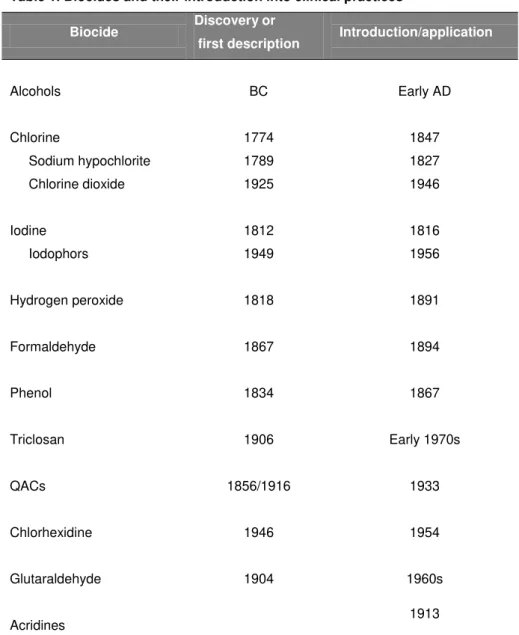
developed; it compared various salt solutions with a standard (sea salt) for their capacity of preservation. Alcohol has been used for over 2000 years as an antimicrobial agent, although it was not recognized as such until recently. Wine was used to heal all kind of injuries, but its concentration was very low to be used as an antiseptic. Time passed by and the concentration of this compound in wine and other alcoholic drinks increased. There are some reports of brandy used to clean and disinfect wounds. The Portuguese surgeon A. M. Barbosa, reported a mortality rate of only 10% for 243 amputations using alcohol in a Lisbon hospital during 1855 and 1866 and only one death in 14 tight operations. Later in 1903, Harrington and Walker showed that a 60% to 70% alcohol solution was the most effective, although it did not kill bacterial spores (4).
Table 1. Biocides and their introduction into clinical practices
Biocide Discovery or
first description Introduction/application
Alcohols BC Early AD
Chlorine
Sodium hypochlorite Chlorine dioxide
1774 1789 1925
1847 1827 1946
Iodine Iodophors
1812 1949
1816 1956
Hydrogen peroxide 1818 1891
Formaldehyde 1867 1894
Phenol 1834 1867
Triclosan 1906 Early 1970s
QACs 1856/1916 1933
Chlorhexidine 1946 1954
Glutaraldehyde 1904 1960s
Acridines 1913
Adapted from (52); BC - Before Christ; AD - Anno Domini (After Christ)
Nowadays their used is generalized in the hospital environment, food industry, veterinary medicine and even in our homes.
1.3 Chemical groups and active molecules in biocidal products
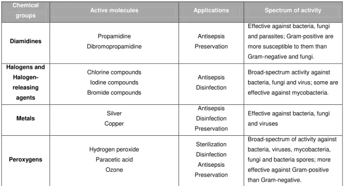
The number of biocides in use is large. They can be used alone or in combination with a variety of products. During the research work for the elaboration of this thesis, the three biocides studied were benzalkonium chloride, chlorhexidine and triclosan. Their chemical group will be described in more detail; other important biocide chemical groups are summarised in Table 2.
1.3.1 Biguanides
Biguanides are compounds that contain the C2HN7 ligand. The most common active molecules are:
Chlorhexidine Alexidine
Polymeric biguanides
1.3.1.1 Chlorhexidine
Chlorhexidine (CHX) is the most commonly used biguanide with a broad range of application: antimicrobial soaps, wound dressings, mouth washes, hair care products, surface disinfectants and preservatives (for example, contact lens storage solution). It is used for surface disinfection at 0.5% to 4%, for antisepsis at 0.02% to 4% and for preservation at 0.0025% to 0.01% (31, 33). Chlorhexidine has a broad-spectrum of activity, produces minimal skin irritation and its substantivity on skin and mucous membranes. Although in few cases, some sensitivity to chlorhexidine has been described. Chlorhexidine plays an important role in the control of hand transmission pathogens in hospital. It has the ability to remain present at a bacteriostatic concentration on skin even after hand washing. Chlorhexidine and other biguanides can be inactivated by nonionic surfactants, in some soaps, hand creams and inorganic water contaminants (33).
precipitation of proteins and nucleic acids (31, 33). This biocide shows a rapid action against Gram-positive and Gram-negative bacteria. It is less effective against fungi, including yeast and molds. It has no sporicidal activity, although it can inhibit (but not the germination of) spores. Mycobacteria are generally less susceptible to chlorhexidine,
Mycobacterium avium-intracelluare is considerably more tolerant than
other mycobacteria. The antiviral activity of chlorhexidine is variable; it is more effective against lipid-enveloped viruses and cannot inactivate nonenveloped virus, such as rotaviruses, hepatitis A virus or polioviruses. It is believed that the outer coat of virus is the major target of chlorhexidine (31, 33).
1.3.2. Phenolics
Phenolics are a class of alcohol compounds with one or more hydroxyl groups attached to an aromatic hydrocarbon ring. A wide variety of phenolics are used for disinfection, antisepsis and preservation (31):
Coal tar Cresol Phenol Xylenols Naphthols
Non-coal tar phenols 2-Phenylphenol 4-Hexylresorcinol
Chlorophenol
4-Chloro-3,5-dimethylphenol (chloroxylenol; para-chloro-meta-xylenol; PCMX)
2-Nitrophenol 4- Nitrophenol
Bisphenols
Hexachlorophene
Triclosan (2,4,4’-trichloro-2’-hydroxydiphenyl ether)
Other phenol derivatives Salicylic acid 2,3-Diaminophenol
1.3.2.1 Triclosan
Triclosan (TCS) is an antimicrobial agent used for several purposes for more than 20 years (57). It is commonly used in antiseptic soaps, hand rinses, lotions, cleaners, shampoos, deodorants, gels and antiacne washes. Concentrations range between 0.1 to 2%, but higher concentrations can be used in higher-risk applications (33). It has a cumulative and persistent effect on skin and is neither toxic nor irritant to skin and mucous membranes. Triclosan is also used in mouth rinses and antibacterial toothpastes. It is also used as a preservative in cosmetic and other products, usually in combination with other biocides. Due to its thermal and chemical stability, triclosan has also been incorporated into plastics and fabrics.
Triclosan as antiseptic can penetrate into and through skin without any toxic, allergenic or mutagenic effect. Its stability and activity is preserved in the presence of organic matter and high temperatures, allowing triclosan to be used as an antimicrobial barrier in textiles and plastics. Due to its stability and extensive use, triclosan has been found in the environment, raising some concern about its ecological effects (33, 36).
Triclosan has a broad-spectrum of activity, it is particularly efficient against Gram-positive bacteria, but it is also active against Gram-negative bacteria and yeasts. Lower activity is observed against some enveloped viruses, fungi and some pseudomonads. Pseudomonas aeruginosa is
low concentrations it has more specific targets, such as the fatty acid biosynthesis. Triclosan blocks lipid synthesis by interacting specifically with the substrate binding site of FabI enoyl reductase and simulating enzyme’s natural substrate. It was proven that in Escherichia coli and
mutations in or overexpression of the gene fabI (encoding for enoyl
reductase) prevents this blockage (28, 34). This has also been observed
in S. aureus and Mycobacteria.
1.3.3 Quaternary ammonium compounds
QACs are cationic surfactants (or “surface-active agents”), containing one quaternary nitrogen associated with at least one major hydrophobic substituent. The most common used are:
Benzalkonium chloride Cetrimide
QACs are used for disinfection, antisepsis, preservation and cleaning (31). From an antimicrobial perspective, cationic surfactants, especially QACs, are the most commonly used.
1.3.3.1 Benzalkonium chloride
chains range between C8 to C18. It is used as household, industrial and health care general surface disinfectant, in high-level surgical instrument sterilizing and disinfecting solutions. Benzalkonium chloride can also be used as preservative (e.g. contact lens solutions and cosmetics at
concentration of 0.001-0.01%) (36), fabric and laundry deodorization or softening.
Benzalkonium chloride provides a good cleaning and disinfection, being noncorrosive and nonstaining on surfaces. It has a “clean” odor and is regarded as nontoxic under typical conditions of use. At high concentrations it can be irritant to skin and mucous membranes (33).
Table 2: Biocides chemical groups and characteristics. Chemical
groups Active molecules Applications Spectrum of activity
Alcohols
Ethyl alcohol (ethanol) Isopropyl alcohol (isopropanol)
Methyl alcohol (methanol) Benzyl alcohol
Phenylethanol (phenylethyl alcohol) Bronopol (2-bromo-2-nitro-1,3-diol) Phenoxyethanol (phenoxetol) Chlorbutanol (chlorbutol) 2,4-Dichlorobenzyl alcohol Antisepsis Disinfection Preservation
Broad-spectrum activity against vegetative bacteria (including mycobacteria), viruses and fungi. Not sporicidal, however inhibit sporulation and spore germination. Aldehydes Glutaraldehyde (pentanedial) Formaldehyde (methanol) Ortho-phthlaldehyde Sterilization Disinfection Preservation Virucidal, bactericidal,
mycobactericidal, fungicidal and also sporicidal (at low
concentrations sporistatic). Anillides Triclocarban Salicylanilide Diphenylureas (carbanilides) Tribromsalan Antisepsis Preservation
Table 2: Continued.
Adapted from (31, 33, 36 54, 60).
Chemical
groups Active molecules Applications Spectrum of activity
Diamidines Propamidine
Dibromopropamidine
Antisepsis Preservation
Effective against bacteria, fungi and parasites; Gram-positive are more susceptible to them than Gram-negative and fungi.
Halogens and Halogen-releasing agents Chlorine compounds Iodine compounds Bromide compounds Antisepsis Disinfection
Broad-spectrum activity against bacteria, fungi and virus; some are effective against mycobacteria.
Metals Silver
Copper
Antisepsis Disinfection Preservation
Effective against bacteria, fungi and viruses Peroxygens Hydrogen peroxide Paracetic acid Ozone Sterilization Disinfection Antisepsis Preservation
1.4 Biocide activity
1.4.1 Mechanisms of action
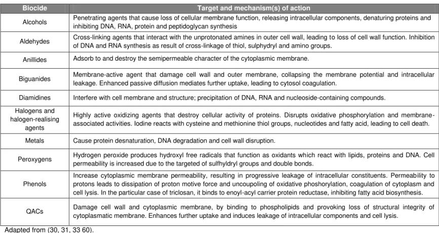
The use of biocides has increased in the past years in very different environments, such as health-care and veterinary facilities, food- and pharmaceutical industries and even in our homes. Biocides may act at one or several generalized targets within the cell, while antibiotics have a specific target. This non-specificity of biocides reduces their selectivity and negates their use in therapy (23). Some years ago, it was discovered a biocide with a single target site (triclosan) (34). Used concentrations of biocides are generally high, so they cause membrane disruption or inactivate a broad range of enzymes. Lower concentrations (like residual concentrations that may be left after cleaning and disinfection of surfaces) may act in the same way as antibiotics, specifically affecting one or two cell targets (23). Biocides vary greatly in their chemical structures, as already mentioned. The precise mechanism(s) of action may reflect this variety, although the final damage may seem quite similar (when high or lethal concentrations are used) (Table 3).
The action of a biocide may be defined according to the bacterial structure which it is acting. Therefore, three levels of interaction can be described: a) interaction with outer cell components (cell wall); b) interaction with cytoplasmic membrane and c) interaction with cytoplasmic constituents (14, 27, 30).
1.4.1.1 Interaction with outer cell components
Table 3. Biocides: targets and mode of action.
Biocide Target and mechanism(s) of action
Alcohols Penetrating agents that cause loss of cellular membrane function, releasing intracellular components, denaturing proteins and inhibiting DNA, RNA, protein and peptidoglycan synthesis
Aldehydes Cross-linking agents that interact with the unprotonated amines in outer cell wall, leading to loss of cell wall function. Inhibition of DNA and RNA synthesis as result of cross-linkage of thiol, sulphydryl and amino groups.
Anillides Adsorb to and destroy the semipermeable character of the cytoplasmic membrane.
Biguanides Membrane-active agent that damage cell wall and outer membrane, collapsing the membrane potential and intracellular leakage. Enhanced passive diffusion mediates further uptake, leading to cytosol coagulation.
Diamidines Interfere with cell membrane and structure; precipitation of DNA, RNA and nucleoside-containing compounds. Halogens and
halogen-realising agents
Highly active oxidizing agents that destroy cellular activity of proteins. Disrupts oxidative phosphorylation and membrane-associated activities. Iodine reacts with cysteine and methionine thiol groups, nucleotides and fatty acid, leading to cell death.
Metals Cause protein desnaturation, DNA degradation and cell wall disruption.
Peroxygens Hydrogen peroxide produces hydroxyl free radicals that function as oxidants which react with lipids, proteins and DNA. Cell permeability is increased due to the targeted of sulfhyldryl groups and double bonds.
Phenols
Increase cytoplasmic membrane permeability, resulting in progressive leakage of intracellular constituents. Permeability to protons leads to dissipation of proton motive force and uncoupoling of oxidative phoshorylation, coagulation of cytoplasm and cell lysis. In the particular case of triclosan, it binds to enoyl-acyl carrier protein reductase, inhibiting fatty acid biosynthesis.
1.4.1.2 Interactions with cytoplasmic membrane
The so called “membrane active agents” is applied to biocides active at the cytoplasmic membrane level. These include phenols, biguanides, QACs, alcohols, parabens and polymxyxins, and due to their different chemical structures, their effect against cytoplasmic membrane must be different. We can group the effects of these biocides in three different categories: cytoplasmic membrane, proton motive force (PMF) and enzymatic systems.
Cytoplasmic membrane. Disruption of the cytoplasmic membrane usually
implies leakage of intracellular components. Phenols, cresols and their chlorinated derivates induce leakage of intracellular materials in bacteria. Chlorhexidine initially causes a high rate of leakage of intracellular components, but in higher concentrations it coagulates the cytosol. Polyhezamethylene biguanide causes domain formation of the acidic phospholipid, causing cytoplasmic membrane disruption. QACs, as well as chlorhexidine, also induce the leakage of intracellular components, and it is believed that they combine with the membrane phospholipids, thus causing cytoplasmic membrane disruption. Anionic agents, organic acids and esters, may also induce leakage of intracellular components, by causing membrane disruption. Ethanol causes release of intracellular components and disorganizes the membranes (14, 27, 31, 30, 60).
Proton motive force. Several agents cause dissipation of the proton motive
acids and esters, by accelerating the movement of protons, inhibiting the active uptake of amino and oxo acids, changing pH and denaturing proteins. TCS and other cationic compounds also discharge the membrane potential component (14, 27, 31, 30, 60).
Enzymatic systems. Some biocides interact with enzymes embedded in
the cytoplasmic membrane. Ethanol inhibits enzymes involved in glycolysis, fatty acid and phospholipid synthesis and solute uptake, causing membrane disruption. Other compounds such as metals (copper and silver), interact with the thiol group of proteins, producing cell inactivation or inhibition. High concentrations of chlorhexidine inhibit membrane-bound adenosine triphosphatase. Triclosan (in low concentrations) has effect upon fatty acid biosynthesis, by targeting the enoyl-acyl carrier protein redutase (FabI) in E. coli and Mycobacterium
smegmatis. (28, 27, 30, 34).
1.4.1.3 Interactions with the cytoplasmic constituents
1.4.2 Factors influencing biocide activity
Generally speaking, several factors influence the activity of biocides: period of contact, nature, number, location and condition of microorganism (bacteria, spores, yeast and molds, protozoa) or entity (prions, viruses), temperature, formulation effects, presence of organic matter or other interfering or enhancing materials/compounds, concentration and pH (51, 54). Other important aspects to take into account when studying the activity of biocides are related to the pre-treatment, during and post-treatment factors (6, 55).
1.4.2.1 Pre-treatment factors
Pre-treatment factors are related to culture growth conditions. Bacterial cell walls are variable structures which change in response to growth environment. In a batch-grown culture, there are some aspects to take into consideration (6, 55):
- growth medium: may influence the subsequent sensitivity of cells to biocides;
- growth phase: latter phase cultures contain a high proportion of dead cells that will protect the viable ones;
- pH of culture medium: cell walls of bacteria may be different according to the pH in which they were grown and this may lead to variations in response to biocides;
- incubation temperature: different temperatures may lead to changes in phospholipid content or composition of spores, thus leading to different responses to biocides;
- presence/absence of oxygen: few information is available on this parameter.
factors into account. The cultures must be always incubated in the same conditions and the growth medium used must be the same (even from the manufacture).
1.4.2.2 During treatment factors
Several factors may affect the activity of an antibacterial agent during treatment. Their effects maybe on the bacterial cell itself or may result from direct effects on the agents. Herein, will be described the most important ones to take into account under experimental conditions.
Concentration
This is a major factor in biocidal activity. The concentration exponent (n or ) measures the effect of concentration, or dilution, on the activity of the biocide. To determine , it is necessary to measure the time required to produce a comparable degree of death of a bacterial suspension at two different concentrations of the biocide.
= (log t2– log t1) / (log C1– log C2)
Temperature
The activity of a biocide usually increases when the temperature at which it is acting is increased. The effect of temperature can be calculated from the following formula:
(T
2- T1) = k2 / k1
Where k2 and k1 are the death rate constants at temperatures T2 and T1, respectively. The temperature coefficient () refers to the effect of temperature per 1 ºC increments, and is nearly always between 1.0 and 1.5. Due to that, is more common to use 10 (or Q
10) value, which is the change of activity per 10 ºC rise. Some temperature coefficients (Q10) are: 2.9 to 5.8 for benzalkonium chloride; 3 to 16 for chlorhexidine; 45 for ethanol and 5 for phenol (15). Biocides respond differently to temperature variations, and this is an important factor to take into account when performing experimental assays or any cleaning and disinfection procedure (6, 55).
Number of microorganism cells
The activity of a biocide decreases when the inoculum size increases. This is dependent of the type of microorganism used and is particularly important in the production of various types of pharmaceutical and cosmetic products (6, 55).
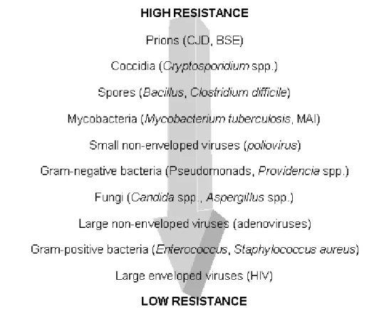
Type of microorganism
envelope, are more sensitive to biocides than Gram-negative and prions are on the top of the list in what concerns biocide low susceptibility (Figure 1).
Bacteria can be surrounded by a capsule and secreted materials. These may play an important role in decrease susceptibility to biocides. The outer membrane of Gram-negative and the cell wall of mycobacteria act as permeability barriers and are responsible for intrinsic resistance of these bacteria to some biocides (6, 30, 55, 49).
Environmental pH
Figure 1. Classification of microorganisms according to their sensitivity to biocides.
CJD – Creutzefeld-Jacb disease agent; BSE – bovine spongiform encephalopathy agent; MAI –Mycobacterium avium intracellulare; HIV – human immunodeficiency virus. Adapted from (30, 49).
Interfering substances
The presence of non-ionic agents can also reduce the activity of some biocides (e.g. QACs). Significant increases in concentration of the biocide are required in order to inhibit microorganism in the presence of these agents (6).
Humidity
Relative humidity has a great influence on the activity of gaseous disinfectants (e.g. formaldehyde). It is the most important factor influencing the activity of vapour-phase disinfectants (6, 55).
1.4.2.3 Pos-treatment factors
There are several factors that influence the recovery of microorganisms exposed to a biocide: composition and pH of recovery medium, removal of the biocide, temperature and period of incubation and composition of the diluents used for serial dilutions in viable cells counts (6).
1.5 Mechanisms of bacterial resistance to biocides
Bacterial response to biocides is usually dependent of its chemical nature and other factors already mentioned above. Mechanisms of bacterial resistance are divided in two main types: intrinsic and acquired. Intrinsic resistance is due to some inherent characteristic of the cells, while acquired resistance is obtained after a previous exposure of the sensitive cell to the antimicrobial agent (31, 47, 50).
1.5.1 Intrinsic resistance
mycobacteria and bacterial spores (46, 47). This intrinsic resistance has contributions from several cell compartments, namely the outer layers of the cell and lipopolysaccharides, acting as a permeability barrier; charge property of the cell surface, causing bacterial resistance to positively charge biocides; the presence of efflux pumps, which decreases the intracellular concentration of the biocides; and enzymatic transformations. Gram-Positive bacteria such as enterococci, staphylococci and streptococci are generally more sensitive to biocides than Gram-negative bacteria. Enterococci are generally less sensitive to biocides than are staphylococci (31); although the exact mechanism remains unclear, but could involve reduced uptake of biocides. The outer membrane of Gram-negative bacteria acts as a barrier limiting or preventing the entry of several biocides, thus being responsible for confer intrinsic resistance.
Pseudomonas are particularly resistant to some biocides (31). They
tolerate high concentrations of QACs, chlorhexidine and triclosan. These bacteria have differences in the lipopolysaccharide composition and in the cationic content of the outer membrane. Members of the genus Proteus
have high resistance to chlorhexidine, QACs, EDTA and diamides due to the presence of a less acidic type of outer membrane lipopolysacharides (31).
1.5.1.1 Physiological (phenotypic) adaptation
Biofilms
Bacteria residing in bioflims are 10- to 100-fold more resistant to biocides than planktonic bacteria. There are several reasons that may justify this: (i) difficult access of the biocide to the inner part of the biofilm; (ii) different physiology of bacteria at different parts of the biofilm due to different nutritional conditions; and consequently (iii) different growth rates within the depths of the biofilm (iv) chemical interaction between the biocide and the biofilm itself; (v) modulation of the microenvironment; (vi) production of degradative enzymes (and neutralizing chemicals); and (vii) genetic exchange between cells in a biofilm. However, biofilms can change according to the microorganism and biocide in question. Bacteria removed from a biofilm and recultured in a culture media, are generally as susceptible as the “normal” planktonic cells of that species (31, 47).
Biofilm formation and consequent decreased susceptibility to biocides, has important implication in clinical environment (biofilm formation in catheters, artificial joints and other medical devices) leading to patient infections, and in several industries (biofilm formation in surfaces of storage tanks, pipelines, filtrations systems and other machineries), leading to product contamination.
Biofilms provide an important example of how physiological adaptation can play a role in conferring intrinsic resistance.
1.5.2 Acquired resistance
1.5.2.1 Plasmid-mediated resistance to biocides
There are some possible plasmid-mediated resistance mechanisms to biocides.
Inactivation
Biocide inactivated as result of an enzymatic modification. The most common example of biocide inactivation involves mercury and other organomercury compounds. Although, mercurials are no longer used as disinfectants, phenylmercuric salts and thiomersal are still used in some types of pharmaceutical products. The mechanism of resistance to mercury involves detoxifying reductase and hydrolase enzymes. Resistance to formaldehyde has also been reported in some bacteria as associated with formaldehyde dehydrogenase (provoking aldehyde degradation) (47, 48).
Impermeability and cell surface alterations
Biocide uptake and accumulation is reduced in case of impermeability and cell surface alterations. One example is the plasmid-mediated resistance to silver salts (still used as topical antimicrobial agents). Although, it is not totally understood, it is believed that the mechanism is by reducing the accumulation rather than silver reduction. This reduced uptake as also been studied for QACs and chlorhexidine, but results are not conclusive. There are some reports of plasmid-encoded changes in the outer membrane proteins of some Gram-negative bacteria leading to reduced susceptibility to formaldehyde and other industrial biocides (31, 48).
Efflux
biological systems. Those proteins act like bilge pumps and decrease the intracellular concentration of the drug. In prokaryotic organism the drug efflux process is largely conferred by pumps in which the drug extrusion is coupled to the influx of a proton (H+). The pumps can be categorized into several families: major facilitator (MF) superfamily, the small multidrug resistance (SMR) family, resistance/nodulation/cell division (RND) family, and drug/metabolite efflux (DME) family. In other pumps, the drug efflux is coupled to Na+ influx – multidrug and toxic compound extrusion (MATE) family; or gets energy from the hydrolysis of ATP – ATP-binding cassette (ABC) family (7, 29, 43).
Staphylococci are one of the most well studied bacteria concerning the genetic aspect of plasmid-mediated biocide resistance mechanisms. In these bacteria, known biocide exporters are members of the MF and SMR super families and include several determinants, namely: qacA, which
encodes resistance to QACs, acridine, ethidium bromide and low-level resistance to chlorhexidine, qacB, which is similar but specifies resistance
to QACs and intercalating dyes, qacC and qacD, which specify resistance
to QACs and low-level resistance to ethidium bromide (47, 48). Biocide exporters in Gram-negative bacteria are in general chromosomally encoded, with the exception of qacE, qacE1 and qacF genes (acting
against compounds such as QACs and ethidium bromide) (10, 44).
1.5.3 Possible link between biocide use and antibiotic resistance
have multiple target sites and the mechanism of resistance to them is often concentration dependent. At high concentrations, multiple structural and metabolic targets are involved, but at low concentrations fewer targets are affected. In contrast, antibiotics usually have distinct structural and/or metabolic targets.
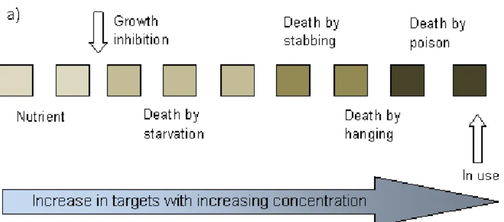
When bacteria are killed by biocides at in use concentrations, several target sites are involved (Figure 2a)). If very low concentrations of biocide are used, they may work as a nutrient. This can be useful in case the objective is biodegradation of the biocide. As concentration increases, bacteria will be affected in several targets and killed by various ways. This means, that at in use concentration, in order to become resistant, several different mechanisms would have to be involved and multiple targets resistance is unlikely to occur. If the biocide is diluted, as its concentration gets lower than its in use concentration, the susceptible targets will also be reduced (Figure 2b)). This may happen when a surface is cleaned or disinfected, as there will be areas in which bacteria will be exposed to sub inhibitory concentrations of biocide (22). This also happens in biofilms, as already mention. A gradient of concentrations is created and at some point there will probably be a selection pressure on a single target. If this single target happens to be shared with another antimicrobial agent, such as an antibiotic, cross resistance may occur.
QACs, get into bacterial cells by the so called “self-promoted uptake”, the same mechanism of cell entry of aminoglycoside antibiotics. Changes in the permeability of the cell wall also occur in the presence of some antibiotics and biocides. Therefore, adaptations against these agents may lead to cross-resistance. Filament formation induced in Gram-negative bacteria by some antibiotics (e.g. -lactams and fluoroquinolones) is also
induced by biocides (such as acridines, phenoxyethanol and phenylethyl alcohol). The autolysis provoked by low concentrations of phenolics and inorganic and organic mercury is said to be also induced by penicillin (53). There is also possibility of genetic linkage between genes for biocide resistance and antibiotic resistance when biocide and antibiotic resistance genes are part of the same genetic element (44).
Figure 2. Each box represents a different target site. a) biocide mechanism: at in use
1.5.4 Some resistance and cross-resistance described
Biocide resistance was first recognized in 1936 by Heathman et al.
(18) who identified Salmonella Typhi resistant to chlorine. In 1952, the
isolation of Serratia marcescens resistant to QACs was reported by
Chaplin (9). Dance et al. (13) reported the outbreak of Proteus mirabilis
resistant to chlorhexidine in a general hospital in the United Kingdom. These strains were also resistant to some antibiotics, like gentamicin, trimethoprim and ampicillin.
Some of the outbreaks described are related to contaminated antiseptics and disinfectants. These outbreaks usually occurred due to Gram-negative bacteria and mycobacteria. Some of outbreaks reported were related to chlorhexidine; most of them related to contaminated water used to prepared the solutions and/or reuse of bottles to dispense the biocide without adequate disinfection. There are also reports of outbreaks related to the use of QACs, especially benzalkonium chloride. Many of these were linked to the storage of this biocide with cotton or gauze, and others with improper dilution of its solution. Few outbreak reports are related to triclosan, but one was due to intrinsic contamination of the antiseptic soap. Pseudomonas, Burkholderia and Ralstonia were the
genera usually involved in these outbreaks (63).
Triclosan, as already mentioned, inhibits the growth of some bacteria by targeting an enoyl reductase enzyme (FabI gene) in E. coli; for
Mycobacteria, this enzyme is also the major target for the anti-tubercular
drug isoniazid, hexachlorophene and some new antibiotics, the diazoborines. The high degree of homology between the enoyl reductase
of E.coli and S. aureus might have implications in a future emergence of
triclosan-resistant staphylococci; mutations in the FabI gene of
happens in the real world is still unknown, namely how triclosan affects the microbial flora in all environments in which it is used. The widespread of this biocide, could lead to environments where Pseudomonas and other
triclosan-resistant bacteria may prevail (58). Another mechanism for triclosan resistance occurs through multidrug efflux pumps in E. coli
(AcrAB) and P. aeruginosa (Mex proteins); these pumps confer both
resistance to triclosan and antibiotics.
Resistance to QACs, chlorhexidine, acridines and diamidines has been observed in staphylococcal strains, Pseudomonas spp. and
Enterobacteriaceace. The resistance is encoded by several multidrug resistance determinants, of which qacA, B, C, D and E genes are included
(10, 18, 48). These multidrug efflux pumps can efflux a variety of antibiotics (trimethoprim, sulphonamids, oxacillin and aminoglycosides) and biocides, as well as dyes such as ethidium bromide (18). Having in mind that several S. aureus are methicillin-resistant (MRSA), this could
become a problem, especially in the hospital environment. Significant difference between the efficacy of these agents against MRSA and methicillin-susceptible S. aureus (MSSA) has already been
described/found (31).
Enterococci are the second most frequently reported cause of surgical wound and nosocomial urinary tract infections and the third most frequently reported cause of bacteraemia (39). Little is known about their resistance to biocides.
2. The genus Enterococcus
1906 Andrewes and Horder classified an organism isolated from a patient with endocarditis as Streptococcus faecalis; as it was very similar to other
strains isolated from human intestine (19, 37). In 1933, Lancefield developed a serological typing system for streptococci, those of “faecal origin” had the group D antigen. Later Sherman proposed a classification scheme which separated streptococcus into four divisions: pyrogenic, viridians, lactic and enterococcus. The latter was used for those organisms able to grow at temperatures from 10 to 45 ºC and most of them can survive for 30 min at 60 ºC, in the presence of 6.5 % of NaCl and at pH 9.6 and are able to hydrolyze esculin. The enterococcus group included S. faecalis, S. faecium, S. bovis and S. equinus; Streptococcus
durans was accepted either as a separate species or as a subspecies of
S. faecium. As years passed by, more species were isolated, such as S.
avium, S. casseliflavus and S. gallinarum. The use of nucleic acid
relatedness helped clarified and expanded the classification of enterococci. In 1984, Schleifer and Kilpper-Bälz showed, by DNA-DNA and DNA-rRNA hybridization studies, that S. faecalis and S. faecium were
distantly related to streptococci that they should be transfered to a new genus, the Enterococcus. There are more than 30 different species
included in this genus (19, 37).
Enterococci are robust Gram-positive, catalase-negative, facultative anaerobic bacteria. They are ovoid in shape and grow in short chains, in pairs or single cells. They are able to grow in the presence of 40 % of bile salts (esculin hydrolyzation) at pH from 4.6 to 9.6 (37, 39). Due to these characteristics, they are able to grow/survive in many different environments, such as humans and animal intestinal tract, sand, plants, water, hospitals, food industry, cheese, meat and even in insects. In the human and other animal intestine, the most common species found are E.
faecalis and E. faecium. These species are also found in plants and
Since the beginning of the last century, enterococci have been recognized as opportunistic human pathogens. The earliest reports associating human diseases with enterococci were published in the beginning of the 20th century. Probably, many of these strains were not truly enterococci or could have been contaminated. As already mentioned, enterococci are the second most frequently cause of surgical wound and nosocomial urinary tract infections and the third most frequently cause of bacteraemia. Enterococci can also cause neonatal infections, central nervous system infections and intraabdominal and pelvic infections (39). They can also cause opportunistic infections in animals, such as septicaemia in chickens, bovine mastitis, endocarditis in cattle and lambs, and urinary-tract infections in dogs and cats (45). Enterococcal infections are predominantly caused by E. faecalis followed by E. faecium, E.
durans, E. gallinarum, E. casseliflavus and E. raffinosus have also been
responsible for infections (39).
strains are still isolated from farm animals and from the environment (8, 21, 26, 35). Enterococci also have virulence factors which are involved in attachment both to host cells and to extracellular matrix proteins (AS – aggregation substance; Esp – Enterococcal surface protein; EfaA – E.
faecalis antigen A), in resistance to macrophages (AS; HypR – hydrogen
peroxide regulator), in cell and tissue damage (Cyl - cytolysin; GelE – gelatinase; SprE – serine protease) and in immune system evasion (capsular polysaccharides) (39). Some of these virulence factors are plasmid encoded which can be transferred between enterococci.
Enterococci are not just “problematic” bacteria. They play an important role in the food industry as fermented foods and as probiotics. They are much used in several Southern European countries (Portugal included) to produce cheeses, contributing to the final organoletic properties of the product. Enterococci have a positive influence in the traditional cheese ripening and the ability to produce bacteriocins (especially E. faecalis and E. faecium) against some bacteria (such as
Listeria monocytogenes, S. aureus, Clostridium botulium and Clostridium
perfringens) (11, 12). These technological and metabolic characteristics
turn enterococci into good starter cultures for the cheese industry. Enterococci can also be used as probiotic for humans and farm animals. However, their use as such is still controversial, due to risk of transference of antibiotic resistance and virulence genes to human strains (19, 20, 42, 59).
3. Enterococci and biocides
enterococci play a critical role due to their ability to cause nosocomial infections. This environment is the most frequently studied and usually the susceptibility of enterococci is evaluated by measuring Minimal Inhibitory Concentration (MIC) or Minimal Bactericidal Concentration (MBC), either using surface carrier tests and/or suspension tests. The presence of enterococci in the human mouth and their ability to survive after some endodontic treatments is also well documented. Enterococci together with lactobacilli, actinomyces and streptococcus, constitute the best invaders of dentine and root canals. E. faecalis has been recovered from tooth canals
when endodontic treatments failed. Several studies were performed on this area testing the efficacy of the most commonly used biocides in endodontic treatments (such as chlorhexidine, calcium hydroxide and sodium hypochlorite) against enterococci (3, 24, 40). Concentrations of 2 % for chlorhexidine and 5.25 % for sodium hypochlorite were able to eliminate enterococci. Although some studies have been performed in order to understand the susceptibility of enterococci to biocides, the way they respond to these compounds and which mechanisms are involved are question that still need to be addressed.
3.1 Thesis outline
in the susceptibility to benzalkonium chloride and its dissemination through enterococci of the three different environments above mention.
E. faecalis V583 was the first enterococcal strain fully sequenced
(41) and its EFA0010 was described as a putative multidrug resistance protein. The specific role of this gene was not clarified and to which drugs it confers “resistance” to was an open question. In chapter II it is discussed the role of EFA0010 in the susceptibility to benzalkonium chloride and the distribution of this gene in enterococci from different origins (clinical, veterinary and dairy isolates), as well as the susceptibility pattern of the same isolates to the two different biocides benzalkonium chloride and chlorhexidine.
the presence of biocides and fitness assays, in order to understand if one took advantage to the other under the tested conditions.
As previously mentioned, enterococci also play an important role in the veterinary environment, causing animal diseases and threating bio-security by entering into the human food chain. Animal facilities and instruments are cleaned and disinfected using biocides (e.g. chlorhexidine and benzalkonium chloride), but few or nothing is known about how these compounds are affecting or selecting bacteria present in these environments. Chapter III presents a complete study of enterococci isolated from the first time from breeding pig facilities dust. The isolates were identified to species level, characterized for their susceptibility to benzalkonium chloride and chlorhexidine. The presence of VRE isolates and their identification and characterization was investigated, as well as their susceptibility to the same biocides. Understand if there was any association between their presence and the use of these compounds was also discussed.
References
1. Aarestrup FM, Agerso Y, Gerner-Smidt P, Madsen M and Jensen LB, 2000. Comparasion of antimicrobial resistance phenotypes and resistance genes in Enterococcus faecalis and Enterococcus faecium from humans
in the community, broilers, and pigs in Denmark. Diagnostic Microbiology and Infectious Disease, 37: 127-137.
3. Berber VB, Gomes BPFA, Sena NT, Vianna ME, Ferraz CCR, Zaia AA and Souza-Filho FJ, 2006. Efficacy of various concentrations of NaOCl and instrumentation techniques in reducing Enterococcus faecalis within
root canals and dentinal tubules. International Endodontic Journal, 39: 10-17.
4. Block SS, 2001. Chapter 1 Historical Review, p.3-16. In Seymour S.
Block (ed), Disinfection, Sterilization, and Preservation, 5th ed, Lippincott Williams and Wilkins, Philadelphia, USA.
5. Block SS, 2001. Chapter 2 Definition of terms, p. 22. In Seymour S.
Block (ed), Disinfection, Sterilization, and Preservation, 5th ed, Lippincott Williams and Wilkins, Philadelphia, USA.
6. Bloomfield SF, 1991. Methods for Assessing Antimicrobial Activity, p 4-7. In S.P. Denyer and W.B. Hugo (ed), Mechanisms of action of chemical
biocides: Their Study and Exploitation, 1st ed, Blackwell Scientific Publications, Oxford, UK.
7. Borges-Walmsley MI, McKeegan KS and Walmsley AR, 2003. Structure and function of efflux pumps that confer resistance to drugs. Biochemical Journal, 376: 313-338.
8. Bywater R, McConville M, Phillips I and Shryock T, 2005. The susceptibility to growth-promoting antibiotics of Enterococcus faecium
isolates from pigs and chickens in Europe. Journal of Antimicrobial Chemotherapy, 56: 538-534.
10. Chapman JS, 2003. Biocide resistance mechanisms. International Biodeterioration & Biodegradation, 51: 133-138.
11. Cintas LM, Rodriguez JM, Fernandez MF, Sletten K, Nes IF, Hernandez PE and Holo H, 1995. Isolation and charactertization of pediocin L50, a new bacteriocin from Pediococcus acidilactici with a broad
inhibitory spectrum. Applied and Environmental Microbiology, 61: 2643-2648.
12. Cintas LM, Casaus P, Holo H, Hernandez PE, Nes IF and Håvarstein LS, 1998. Enterocins L50A and L50B, two novel bacteriocins from
Enterococcus faecium L50, are related to staphylococcal hemilysins.
Journal of Bacteriology, 180: 1988-1994.
13. Dance DAB, Person AD, Seal DV and Lowes JA, 1987. A hospital outbreak caused by chlorhexidine and antibiotic resistant Proteus
mirabilis. Journal of Hospital Infection, 10: 10-16.
14. Denyer SP and Stewart GSAB, 1998. Mechanisms of action of disinfectants. International Biodeterioration & Biodegradation, 41: 261-268.
15. Denyer SP, 2007. Antimicrobial Preservatives and Their Properties p 327, 329. In Denyer S.P. and R.M. Baird (ed.), Guide to Microbiological
Control in Pharmaceuticals and Medical Devices, 2nd ed, CRC Press, Boca Rotan, USA.
17. Directive 98/8/EC of the European Parliament and of the Council of 16 February 1998 concerning the placing of biocidal products on the market. Official Journal of the European Communities, 1998.
18. Fraise AP, 2002. Biocide abuse and antimicrobial resistance – a cause of concern? Journal of Antimicrobial Chemotherapy, 49: 11-12.
19. Franz CMAP, Holzapfel WH and Stiles ME, 1999. Enterococci at the crossroads of food safety? International Journal of Food Microbiology, 47: 1-24.
20. Franz CMAP, Stiles ME, Schleifer KH and Holzapfel WH, 2003. Enterococci in food – a conundrum for food safety. International Journal of Food Microbiology, 88: 105-122.
21. Garcia-Migura L, Pleydell E, Barnes S, Davies RH and Liebana E, 2005. Characterization of vancomycin-resistant Enterococcus faecium
isolated from broiler poultry and pig farms in England and Wales. Journal of Clinical Microbiology, 43: 3283-3289.
22. Gilbert P and McBain AJ, 2001. Biocide usage in the domestic setting and concern about antibacterial and antibiotic resistance. Journal of Infection, 43: 85-91.
24. Haapasalo M, Quian W, Portenier I and Waltino T, 2007. Effects of dentine on the antimicrobial properties of endodontic medicaments. Journal of Endodontics, 33: 917-925.
25. Heath RJ, Roland GE and Rock CO, 2000. Inhibition of the
Staphylococcus aureus NADPH-dependet enoyl-acyl carrier protein
reductase by triclosan and hexachlorophene. Journal of Biological Chemistry, 275: 4654-4659.
26. Herrero IA, Teshager T, Garde J, Moreno MA, Domínguez L, 2000. Prevalence of vancomycin-resistant Enterococcu faecium (VREF) in pig
faeces from slaughterhouses in Spain. Preventive Veterinary Medicine, 47: 255-262.
27. Lambert PA, 2004. Chapter 5 Mechanisms of action of biocides, p. 139-148. In Fraise A.P, P.A. Lambert and J.-Y. Maillard (ed.), Principals
and Practice of Disinfection, Preservation, and Sterilization, 4th ed., Blackwell Science, Oxford, UK.
28. Larson DW, Matthers U, Gerrath JA, Gerrath JM, Nekola JC, Walker GL and Larson NWK, 1999. Molecular basis of triclosan activity. Nature, 398: 383-384.
29. Levy SB, 2002. Active efflux, a common mechanism for biocide and antibiotic resistance. Journal of Applied Microbiology Symposium Supplement, 92: 65S-71S.
31. McDonnell GE and Russell AD, 1999. Antiseptics and Disinfectants: Activity, Action, and Resistance. Clinical Microbiology Reviews, 12: 147-179.
32. McDonnell GE, 2007. Chapter 1 Introduction: Definitions, p. 2. Antisepsis, disinfection, and sterilization: types, action, and resistance, 1st ed. ASM Press, Washington, DC., USA.
33. McDonnell GE, 2007. Chapter 3 Chemical Disinfection, p. 79-148. Antisepsis, disinfection, and sterilization: types, action, and resistance, 1st ed. ASM Press, Washington, DC., USA.
34. McMurray LM, Oethinger M, Levy SB, 1998. Triclosan targets lipid synthesis. Nature, 394: 531-532.
35. Messi P, Guerrieri E, Niederhäusern S, Sabia C and Bondi M, 2006. Vancomycin-resistant enterococci (VRE) in meat and environmental samples. International Journal of Food Microbiology, 107: 218-222.
36. Moore SL, Payne DN. Chapter 2 Types of antimicrobial agents, p. 21-36. In Fraise A.P, P.A. Lambert and J.-Y. Maillard (ed.), Principals and
Practice of Disinfection, Preservation, and Sterilization, 4th ed., Blackwell Science, Oxford, UK.
37. Murray BE, 1990. The life and times of the Enterococcus. Clinical
Microbiology Reviews, 3: 46-65.
39. Ogier J-C and Serror P, 2008. Safety assessment of dairy microorganisms: The Enterococcus genus. International Journal of Food
Microbiology, 126: 291-301.
40. Oliveira DP, Barbizan JVB, Trope M and Teixeira FB, 2007. In vitro antibacterial efficacy of endodontic irrigants against Enterococcus faecalis.
Oral Surgery, Oral Medicine, Oral Pathology, Oral Radiology and Endodontology, 103: 702-706.
41. Paulsen IT, Banerjei L, Myers GSA, Nelson KE, Seshadri R, Read TD, Fouts DE, Eisem JA, Gill SR, Heidelberg JF, Tettelin H, Dodson RJ, Umayam L, Brinkac L, Beanan M, Daugherty S, DeBoy RT, Durkin S, Kolonay J, Madupu R, Nelson W, Vamathevan J, Tran B, Upton J, Hanse T, Shetty J, Khouri H, Utterback T, Radune D, Getchum KA, Dougherty BA and Fraser CM, 2003. Role of mobile DNA in the evolution of vancomycin-resistant Enterococcus faecalis. Science, 299: 2071-2074.
42. Pimentel LL, Semedo T, Tenreiro R, Crespo MT, Pintado MM and Malcata FX, 2007. Assessment of safety of enterococci isolated throughout traditional Terrincho chessemaking: virulence factors and antibiotic susceptibility. Journal of Food Protection, 70: 2161-2167.
43. Poole K, 2002. Mechanism of bacterial biocide and antibiotic resistance. Journal of Applied Microbiology Symposium Supplement, 92: 55S-64S.
44. Poole K, 2004. Bacterial resistance. Acquired resistance, p.170-178.
Practice of Disinfection, Preservation, and Sterilization, 4th ed., Blackwell Science, Oxford.
45. Quinn PJ, Carter ME, Markey B and Carter GR, 1999. The
Streptococci and related Cocci, p. 130. In: Clinical Veterinary
Microbiology, Elsevier Health Sciences, Spain.
46. Russell AD, 1990. Bacterial spores and chemical sporicidal Agents. Clinical Microbiology Review, 3: 99-119.
47. Russell AD, 1995. Mechanisms of bacterial resistance to biocides. International Biodeterioration & Biodegradation, 36: 247-265.
48. Russell AD, 1997. Plasmids and bacterial resistance to biocides. Journal of Applied Microbiology, 82: 255-265.
49. Russell AD, Furr JR and Maillard J-Y, 1997. Microbial susceptibility and resistance to biocides: an understanding. ASM News, 63: 481-487.
50. Russell AD, 1998. Bacterial resistance to disinfectants: present knowledge and future problems. Journal of Hospital Infection, 43: S57-S68.
51. Russell AD and McDonnell G, 2000. Concentration: a major factor in studying biocidal action.
53. Russell AD, 2002. Mechanisms of antimicrobial action of antiseptics and disinfectants: an increasingly important are of investigation. Journal of Antimicrobial Chemotherapy, 49: 597-599.
54. Russell AD, 2003. Biocide use and antibiotic resistance: the relevance of laboratory findings to clinical and environmental situations.
55. Russell AD, 2004. Bacterial adaptation and resistance to antiseptics, disinfectants and preservatives is not a new phenomenon. Journal of Hospital Infection, 57: 97-104.
56. Russell AD 2004. Chapter 3 Factors influencing the efficacy of antimicrobial agents, p. 98-118. In Fraise A.P, P.A. Lambert and J.-Y.
Maillard (ed.), Principals and Practice of Disinfection, Preservation, and Sterilization, 4th ed., Blackwell Science, Oxford.
57. Russell AD, 2004. Wither triclosan? Journal of Antimicrobial Chemotherapy, 53: 693-695.
58. Schweizer HP, 2001. Triclosan: a widely used biocide and its link to antibiotics. FEMS Microbiology Letter, 202: 1-7.
59. Semedo T, Santos MA, Lopes MF, Figueiredo Marques JJ, Barreto Crespo MT and Tenreiro R, 2003. Virulence factors in food, clinical and reference Enterococci: A common trait in the genus? Systematic and Applied Microbiology, 26: 13-22.