243
Case Report
Short Communication
Revista da Sociedade Brasileira de Medicina Tropical 47(2):243-246, Mar-Apr, 2014http://dx.doi.org/10.1590/0037-8682-0007-2013
Address to: Dra Raquel da Silva Pacheco. Laboratório de Sistemática Bioquímica/ FIOCRUZ. Av. Brasil 4365, Manguinhos, 21040-360 Rio de Janeiro, RJ, Brasil. Phone: 55 21 3865-8182; Fax: 55 21 3865-8200
e-mail: rpacheco@ioc.fi ocruz.br Received 29 January 2013 Accepted 25 March 2013
Cutaneous and visceral leishmaniasis co-infection in
dogs from Rio de Janeiro, Brazil: evaluation by
specifi c PCR and RFLP-PCR assays
Marize Quinhones Pires
[1],
Maria de Fátima Madeira
[2],
Vânia Rita Elias Pinheiro Bittencourt
[3]and Raquel da Silva Pacheco
[1][1]. Laboratório de Sistemática Bioquímica, Instituto Oswaldo Cruz, Fundação Oswaldo Cruz, Rio de Janeiro, RJ. [2]. Laboratório de Vigilância em Leishmanioses, Instituto de Pesquisa Clínica Evandro Chagas, Fundação Oswaldo Cruz, Rio de Janeiro, RJ. [3].Departamento de Parasitologia Animal, Universidade Federal Rural do Rio de Janeiro, Seropédica, RJ.
ABSTRACT
Introduction: During a diagnostic evaluation of canine visceral leishmaniasis (VL), two of seventeen dogs were found to be co-infected by Leishmania (Viannia) braziliensis and Leishmania (Leishmania) chagasi. Methods: Specifi c polymerase
chain reaction (PCR)and restriction fragment length polymorphism-PCR (RFLP-PCR) assays were performed. Results: PCR
assays for Leishmania subgenusidentifi cation followed by RFLP-PCR analysis in biopsies from cutaneous lesions and the
spleen confi rmed the presence of Leishmania (Viannia) braziliensis and Leishmania (Leishmania) chagasi in those fragments. Conclusions: This report reinforces the importance of using serological and molecular techniques in the epidemiological surveillance of canine populations in endemic areas in which both diseases are known to co-exist. In such cases, a reassessment of the control measures is required.
Keywords: Visceral leishmaniasis. Tegumentary leishmaniasis. Restriction fragment length polymorphism-polymerase chain reaction.
Visceral leishmaniasis (VL) and tegumentary leishmaniasis (TL) are zoonoses of great importance for public health. In the
State of Rio de Janeiro, Leishmania (Viannia) braziliensis is the most prevalent species implicated in the epidemiological cycle of TL. Its transmission occurs in periurban areas in which primitive rain forest vegetation is being depredated due to disorderly human occupation1, and infections in man,
dogs and horses have been reported2,3. Canine VL, which is
caused by Leishmania (Leishmania) chagasi, is endemic in the Municipality of Rio de Janeiro. Dogs represent one of the main reservoirs in urban areas in which the disease has been observed
due to several factors that infl uence the natural epidemiological
scenario4. The overlapping transmission of TL caused by L. (V.) braziliensis and VL caused by L. (L.) chagasi has been reported in certain areas of the Municipality of Rio de Janeiro. Mixed infection with both parasites has already been reported in a patient5 and in a dog6. Fortunately, according to Marzochi
et al.7, no new human cases of co-infection have been registered
since, but there is concern about the persistence of canine seroprevalence. Control measures are based on interrupting the
transmission cycle, which involves the diagnosis and treatment of human cases and vector control through insecticides and serological screening, with the subsequent culling of dogs found to be seropositive4.
The present article discusses the detection of mixed TL
and VL infections in two of seventeen dogs from endemic areas of Rio de Janeiro, Brazil, which tested seropositive by indirect immunofl uorescence (IIF) analysis of serum samples.
The occurrence of canine cases with both diseases in the same geographic area impairs the diagnosis and implementation of control measures.
The animals included in this study were referred to the Zoonosis Service of the Instituto de Pesquisa Clínica Evandro Chagas-Fundação Oswaldo Cruz (IPEC-FIOCRUZ) with an
indication for euthanasia according to the recommendations of
the Brazilian Program for the Control of Leishmaniasis4 after
serological tests by IIF on serum samples, which were performed by the Epidemiology Service of the Municipality of Rio de Janeiro. This study was approved by the Ethics Committee on Animal Experimentation of the Fundação Oswaldo Cruz
(CEUA/FIOCRUZ; program no L-023/06).
All studied dogs were from urban and periurban areas of the Municipality of Rio de Janeiro and presented IIF titers ranging from 1:80 to 1:1,280. In certain animals, clinical symptoms of
VL were evident, whereas others were asymptomatic. Cutaneous
lesions were frequent (Table 1).
244
Pires MQ et al - Cutaneous and visceral leishmaniasis co-infection in dogs
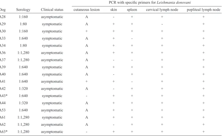
TABLE 1 - The serological titers, clinical status and PCR results of the 17 dogs from Rio de Janeiro.
PCR with specifi c primers for Leishmania donovani
Dog Serology Clinical status cutaneous lesion skin spleen cervical lymph node popliteal lymph node
A28 1:160 asymptomatic A - + + +
A29 1:80 symptomatic A + + - +
A30 1:160 symptomatic + + + + +
A33 1:640 symptomatic A + - + +
A34 1:80 symptomatic A + + + +
A36 1:1,280 asymptomatic A + + + +
A37 1:1,280 asymptomatic A - - + +
A39 1:640 symptomatic A + + + +
A40 1:640 symptomatic A - + + +
A41 1:640 asymptomatic + + - + +
A42 1:320 asymptomatic A - + + +
A43* 1:640 symptomatic - + + + +
A44 1:320 symptomatic A + + + +
A53 1:640 asymptomatic A + + + +
A61 1:1,280 symptomatic A + + + +
A62 1:1,280 asymptomatic A - + + +
A63* 1:1,280 asymptomatic - + + + +
PCR: polymerase chain reaction; A: absent; *mixed infection.
all animals were collected after thiopental-overdose euthanasia
and submitted for polymerase chain reaction (PCR) analyses
using primers for the variable regions of kinetoplast DNA
(kDNA) minicircles. Specifi c primers for the L. braziliensis
complex (5’-GGGGTTGGTGTAATATAGTGG-3’ and
5’-CTAATTGTGCACGGGGAGG-3’)8 and for the Leishmania
donovani complex (5’-CCAGTTTCCCGCCCCG-3’ and
5’-GGGGTTGGTGTAAAATAG-3’)9 were adopted, as
previously described10,11. Then, the amplifi ed PCR products
were visualized on agarose gels. Restriction fragment length polymorphism-polymerase chain reaction (RFLP-PCR) analyses were performed with a panel of four restriction enzymes (Msp I, Rsa I, Hinf I and Mbo I) to confi rm the specifi city of the amplifi ed kDNA minicircle products.
All of the canine biopsy fragments, except for two from cutaneous lesions, produced the expected 800bp diagnostic bands after PCR with primers D1/D2. Those fragments of cutaneous lesions that tested negative were submitted to PCR assays with the primers B1/B2, and the expected 750bp diagnostic bands were observed (Figure 1A).
PCR was performed in combination with RFLP-PCR to
confi rm the presence of L. (V.) braziliensis deoxyribonucleic
acid (DNA) in the cutaneous lesion biopsies and L. (L.) chagasi
DNA in the spleen and lymph node fragments from two dogs (dogs A43 and A63, Table 1). In Figures 1B, 1C, 1D and 1E, the
PCR/RFLP results confi rming the mixed infection are observed.
Because previous results from our group12,13 have demonstrated
that the restriction enzymes Msp I, Rsa I, Hinf I and Mbo I
are the most appropriate for typing Leishmania species from the subgenera Viannia and Leishmania, these enzymes were
adopted in the present study. The Msp I restriction enzyme linearizes the kDNA minicircles, displaying a major band of
approximately 750bp in the case of Leishmania (V.) species. In
contrast, a polymorphic restriction profi le is always observed
in L (L.) chagasi12.
Herein, the Hinf I restriction patterns of the amplifi ed
parasitic DNA from cutaneous lesions were similar to the patterns of the L. (V.) braziliensis (MHOM/BR/75/M2903)
reference strain kDNA. The Rsa I restriction patterns obtained
with the amplifi ed products from the spleen and lymph node
fragments were also similar compared with the patterns of the
L. (L.) chagasi (MHOM/BR/74/PP75) reference strain kDNA
(Figure 1C). Both subgenera were confi rmed after digestion with Msp I (Figure 1E). RFLP-PCR analysis with the restriction
enzymes Msp I, Rsa I and Mbo I corroborated the PCR results, justifying the use of such a technique in Leishmania species
identifi cation.
In clinical-epidemiological surveillance, emphasizing the
245
Rev Soc Bras Med Trop 47(2):243-246, Mar-Apr, 2014
M 1 2 3 4 M 5 6 7 8 9 10 bp
bp
bp
bp 750
500
500
500
500 800
800
800
200
200 750
500
800 M 1 2 3 4 5 6 7 8
200
200 M 1 2 3 4 5 1 2 3 4 5 M M 1 2 3 4 5 6
800
A
B
C
D
E
FIGURE 1 - A: Specifi c PCR products in 1.5% agarose gel: line M: DNA ladder, 100bp; primers B1/B2: line 1: dog A43, cutaneous lesion; line 2: dog A63, cutaneous lesion; line 3: Leishmania (Viannia) braziliensis reference strain; lines 4 and 10: negative controls; primers D1/D2: lines 5-6: dog A43, spleen fragment and cervical lymph node, respectively; lines 7-8: dog A63, spleen fragment and cervical lymph node, respectively; line 9: Leishmania (Leishmania) chagasi reference strain. B: RFLP analyses in high-resolution 1.8% agarose gel after digestion with the enzymes Rsa I and Hinf I: lines 1 and 4: dog A43, cutaneous lesion; lines 2 and 5: dog A63, cutaneous lesion; lines 3 and 6: Leishmania (Viannia) braziliensis reference strain. C and D: RFLP analyses with Rsa I and Mbo I: lines 1-2: dog A43, spleen and cervical lymph node, respectively; lines 3-4: dog A63, spleen and cervical lymph node, respectively; line 5: Leishmania (Leishmania) chagasi reference strain. E: RFLP analyses with Msp I: line 1: dog A43, cutaneous lesion; line 2: dog A63, cutaneous lesion; line 3: Leishmania (Viannia) braziliensis reference strain; lines 4-5: dog A43, spleen and cervical lymph node, respectively; line 6-7: dog A63, spleen and cervical lymph node, respectively; line 8: Leishmania
246
both diseases overlap, such as certain rural areas in Rio de Janeiro. In these areas, where closely related etiological groups are present, the interpretation of serological data is a limiting aspect due to possible serological cross-reactions. Although Trypanosoma cruzi
infection is unknown in the Municipality of Rio de Janeiro, a new species, Trypanosoma caninum, was recently described in dogs14.
In this scenario, a reassessment of control measures is required. The control of leishmaniasis is relatively complex, particularly in areas where both the tegumentary and visceral forms of the
disease co-exist. As a control measure, the Brazilian government
usually culls seropositive dogs4. However, according to a recent
review15, the strategy of killing dogs is hampered for several
reasons, including the low accuracy of the methods used to assess the infectivity of dogs and the high replacement rate of
these animals. In this scenario, a search for sensitive and specifi c
molecular tools is needed to distinguish dogs infected with
L. (V.) braziliensis, thus preventing unnecessary sacrifi ce. The results
presented here show the usefulness of specifi c PCR assays and the
RFLP technique for differentiating between L. (V.) braziliensis and
L. (L.) chagasi and may contribute to providing support for control
programs. Conversely, dogs with TL and VL co-infection would be subjected to euthanasia according to the guidelines of the Ministry
of Health. Concerning control measures, including the detection
and treatment of human cases, the disposal of dogs with VL, the
efforts to control vectors with systematic indoor and outdoor spraying and the use of collars and mosquito nets impregnated with
insecticides, the latter measure alone would likely be more effi cient than the fi rst two measures together. Finally, the development
of human vaccines should also be cons idered as a high priority.
The authors declare that there is no confl ict of interest. CONFLICT OF INTEREST
FINANCIAL SUPPORT
This work received fi nancial support from Conselho Nacional
de Desenvolvimento Científi co e Tecnológico (CNPq), Fundação Carlos Chagas de Apoio a Pesquisa do Estado do Rio de Janeiro
(FAPERJ) and Instituto Kinder do Brasil (IKB). RS Pacheco is
a Brazilian CNPq investigator.
REFERENCES
1. Marzochi MCA, Marzochi KBF. Tegumentary and visceral leishmaniases in Brazil: emerging anthropozoonosis and possibilities for their control. Cad Saude Publica 1994; 10 (Suppl II):359-375.
2. Marzochi MCA, Coutinho SG, Souza WJ, Toledo LM, Grimaldi Jr G, Momen H, et al. Canine visceral leishmaniasis in Rio de Janeiro, Brazil. Clinical, parasitological, therapeutical and epidemiological fi ndings (1977-1983). Mem Inst Oswaldo Cruz1985; 80:349-357.
3. Barbosa-Santos EGO, Marzochi MCA, Urtado W, Queirós F, Chicarino J, Pacheco RS. Leishmaniasis disseminated by Leishmania braziliensis in a mare (Equus cabalus) immunotherapy and chemotherapy assays. Mem
Inst Oswaldo Cruz 1994; 89:217-220.
4. Ministério da Saúde: Manual de vigilância e controle da leishmaniose visceral. Brasília: Editora MS; 2006.
5. Oliveira-Neto MP, Marzochi MCA, Grimaldi Jr G, Pacheco RS, Toledo LM, Momen H. Concurrent human infection with Leishmania donovani
chagasi and Leishmania braziliensis braziliensis. Ann Trop Med Parasitol
1986; 80:587-592.
6. Madeira MF, Schubach A, Schubach TPM, Pacheco RS, Oliveira FS, Pereira SA, et al. Mixed infection with Leishmania (Viannia) braziliensis
and Leishmania (Leishmania) chagasi in a naturally infected dog from
Rio de Janeiro. Trans R Soc Trop Med Hyg 2006; 100:442-445.
7. Marzochi MCA, Fagundes A, Andrade MV, Souza MB, Madeira MF, Mouta-Confort E, et al. Visceral leishmaniasis in Rio de Janeiro, Brazil: eco-epidemiological aspects and control. Rev Soc Bras Med Trop 2009; 42:570-580.
8. De Bruijn MH, Barker DC. Diagnosis of New World leishmaniasis: specifi c detection of species of the Leishmania braziliensis complex by amplifi cation of kinetoplast DNA. Acta Trop 1992; 52:45-58.
9. Smyth AJ, Ghosh A, Hassan MQ, Basu D, De Bruijn MH, Mallik KK, et al. Rapid and sensitive detection of Leishmania kinetoplast DNA from spleen and blood samples of Kala-azar patients. Parasitol 1992; 105: 183-192.
10. Oliveira FS, Pirmez C, Pires MQ, Brazil RP, Pacheco RS. PCR-based diagnosis for detection of Leishmania in skin and blood of rodents from an endemic area of cutaneous and visceral leishmaniasis in Brazil. Vet Parasitol 2005; 129:219-227.
11. Silva ES, Gontijo CMF, Pacheco RS, Brazil RP. Diagnosis of human visceral leishmaniasis by PCR using blood samples on fi lter paper. Genet Mol Res 2004; 3:251-257.
12. Lopes UG, Momen H, Grimaldi Jr G, Marzochi MCA, Pacheco RS, Morel CM. Schizodeme and zymodeme characterization of Leishmania
in the investigation of foci of visceral and cutaneous leishmaniasis. J Parasitol 1984; 70:89-98.
13. Pacheco RS, Lopes UG, Morel CM, Grimaldi Jr G, Momen H. Schizodeme analysis of Leishmania isolates and comparison with some phenotypic techniques. In: Rioux JA, editor. Leishmania, Taxonomie et Phylogenése. Applications éco-épidémiologiques. Montpellier: IMEEE; 1986. p.57-65.
14. Barros JH, Almeida AB, Figueiredo FB, Sousa VR, Fagundes A, Pinto AG, et al. Occurrence of Trypanosoma caninum in areas overlapping with leishmaniasis in Brazil: what is the real impact of canine leishmaniasis control? Trans R Soc Trop Med Hyg 2012; 106:419-423.
15. Costa CHN. How effective is dog culling in controlling zoonotic visceral leishmaniasis? A critical evaluation of the science, politics and ethics behind this public health policy. Rev Soc Bras Med Trop 2011; 44: 232-242.