INTRODUCTION
Address to: Dra Janaína Luz Narciso-Schiavon. Deptº Clínica Médica/HU Polydoro Ernani de São Thiago/UFSC. R. Professora Maria Flora Pausewang s/no/3º Andar, Trindade, 88040-900 Florianópolis, SC, Brasil.
Phone: 55 48 3721-9014
e-mail: [email protected] Received 21 January 2014 Accepted 10 April 2014
Clinical and laboratory characteristics associated with
dyslipidemia and liver steatosis in chronic HBV carriers
Angélica Luciana Nau
[1],
Júlia Cristina Soares
[1],
Maria Beatriz Cacese Shiozawa
[2],
Esther Buzaglo Dantas-Corrêa
[1],
Leonardo de Lucca Schiavon
[1]and
Janaína Luz Narciso-Schiavon
[1][1]. Núcleo de Estudos em Gastroenterologia e Hepatologia, Departamento de Clínica Médica, Universidade Federal de Santa Catarina, Florianópolis, SC. [2]. Departamento de Patologia, Universidade Federal de Santa Catarina, Florianópolis, SC.
ABSTRACT
Introduction: Chronic hepatitis B virus (HBV) infection and liver steatosis (LS) are the most common causes of chronic liver disease, and their coexistence is frequently observed in clinical practice. Although metabolic syndrome is the main cause of LS, it has not been associated with HBV infection. The aims of this study wereto describe the lipid profi le and prevalence of LS among HBV carriers and to identify the characteristics associated with LS in this group. Methods: This retrospective cross-sectional study included hepatitis B surface antigen (HBsAg)-positive patients evaluated during 2011 and 2012. Results: Of the 83 patients included, the mean age was 46.4±12.5 years, 53% were men, and 9.1% were hepatitis B e antigen (HBeAg) -positive. These patients
exhibited the following lipid profi le: total cholesterol = 175.4±38.8mg/dL, low-density lipoprotein (LDL) = 113.0±32.7mg/dL, and triglycerides = 91.1±45.2mg/dL. Their fasting glucose was 95.3±14.5g/dL, and fasting insulin was 6.1±5.9µIU/mL. Liver
steatosis was observed on abdominal ultrasound in 11.3% of individuals. Factors associated with the presence of LS included
higher levels of total cholesterol, prothrombin activity, fasting insulin, and body mass index (BMI) as well as lower levels of
aspartate aminotransferase (AST). Conclusions: These fi ndings suggest that LS in patients with chronic HBV appears to be a
consequence of metabolic alterations and insulin action rather than of viral factors.
Keywords: HBV. Hepatitis B. Fatty liver. Lipids. Cholesterol. Triglycerides.
Chronic hepatitis B virus (HBV) infection and liver steatosis (LS) are the most common causes of chronic liver disease. An estimated 350 million individuals are chronically infected with HBV worldwide1. The lack of specifi c and sensitive noninvasive diagnostic tests for LS limits the ability to reliably detect the disease and determine its real prevalence. Often, non-alcoholic fatty liver disease (NAFLD) is diagnosed presumptively when imaging studies suggest the presence of LS or when elevated liver enzymes are noted in overweight or obese individuals with
no other identifi able cause of liver disease2. For many patients, LS is indolent; however, approximately one-third of patients progress to cirrhosis and, in some cases, liver failure. Patients with simple LS may progress to steatohepatitis and cirrhosis3.
It has been established that chronic hepatitis C is closely
associated with LS, insulin resistance, and an increased risk
of type 2 diabetes. Although these associations may be a consequence of metabolic factors, the hepatitis C virus itself has the capacity to promote steatosis and insulin resistance4. However, metabolic syndrome has not been associated with HBV infection5,6.
It has been demonstrated that HBV carriers have decreased
levels of total cholesterol, high-density lipoprotein, and low-density lipoprotein, suggesting that HBV infection counteracts dyslipidemia7-10. Moreover, LS in chronic HBV patients is associated with changes in anthropometric indices and metabolic factors but not HBV itself11.
Factors affecting the development of LS in patients with chronic HBV infection remain obscure, although clinical observations report the common coexistence of both diseases12,13. Additionally, data are lacking on this subject in Brazil. This study aimed to evaluate the prevalence of LS and examine the
lipid profi les of chronic HBV carriers, as well as to compare the clinical features and lipid profi les among individuals with
chronic HBV infection with and without LS.
METHODS
RESULTS
Hepatology Outpatient Clinic of the University Hospital of the Federal University of Santa Catarina between August 2011 and September 2012, and provided their written informed consent to participate in the study. Patients with incomplete clinical or laboratory data in their medical records were excluded from the study. A diagnosis of hepatocellular carcinoma was also a cause for exclusion.
Clinical, laboratory, and histological fi ndings were collected
from data contained in the medical records. The patient records were analyzed for the following clinical and demographic
characteristics: gender, age, comorbidities (diabetes mellitus,
dyslipidemia, and hypertension), current antiviral therapy, and
body mass index (BMI). The laboratory variables analyzed included the following: hepatitis B e antigen (HBeAg), hepatitis
B virus-deoxyribonucleic acid (HBV-DNA), alpha-fetoprotein (AFP), creatinine, hemoglobin, platelets, prothrombin activity, albumin, direct bilirubin, aspartate aminotransferase (AST), alanine aminotransferase (ALT), alkaline phosphatase (ALP), gamma-glutamyltransferase (GGT), total cholesterol, high-density lipoprotein (HDL), low-density lipoprotein (LDL), triglycerides, glycemia, fasting insulin, and glycated hemoglobin. Biochemical test results were expressed as absolute values. The hepatic biochemistry tests, including the levels of AST, ALT, ALP, and GGT, were expressed as a multiple of the upper limit of normal (×ULN). Laboratory tests performed within 6 months of the date of the ultrasound were used for this study. The levels of HBV DNA were measured using an in-house real-time quantitative polymerase chain reaction (qPCR) assay with a lower limit
of detection of 20IU/ml. The most recent level of HBV-DNA
was considered for inclusion in the study. Some patients were taking nucleos(t)ide analogues when their levels of HBV-DNA were tested.
Ultrasound data were collected from the medical records. An upper abdominal ultrasound is routinely performed for all patients positive for HBsAg to screen for hepatocellular carcinoma. A liver biopsy is indicated for all patients positive for HBeAg, regardless of their ALT levels. Patients negative for HBeAg with elevated ALT and/or a high viral load
(≥ 2,000.0IU/mL) are also subjected to a liver biopsy. According to the National Consensus on the Classifi cation of Chronic
Hepatitis14, the following histological characteristics were
examined during the liver biopsies: advanced fi brosis (defi ned as structural changes of stage 3 or 4) and marked infl ammatory activity (defi ned as periportal activity of stage 3 or 4).
Continuous variables were compared using Student’s t test or the Mann-Whitney U test when appropriate. Categorical variables were compared using the chi-square test or Fisher’s exact test. Univariate analysis was used to identify characteristics associated with the presence of liver steatosis (either on abdominal ultrasound or from the liver biopsy). The correlation between the liver biochemistry results and lipid
profi le (total cholesterol, HDL, LDL, and triglycerides) was assessed using Pearson’s correlation coeffi cient. The level of statistical signifi cance adopted was 5% (p-value < 0.05). All tests were two-tailed and conducted using the statistical software
Statistical Package for the Social Sciences (SPSS) version 17.0
(SPSS Inc., Chicago, Illinois, USA).
Ethical considerations
The study protocol conformed to the ethical guidelines of the 1975 Helsinki Declaration and was approved by the review board of the Federal University of Santa Catarina under the number 131.513.
Patient characteristics
From August 2011 to September 2012, 112 patients infected with HBV were considered for enrollment. Twenty-two individuals were excluded from the study due to lack of a lipid
profi le; four patients had no record of an abdominal ultrasound;
and three had hepatocellular carcinoma (Figure 1).
The characteristics of the 83 consecutive patients fulfi lling
the entry criteria are summarized in Tables 1 and 2. The mean age was 46.4±12.5 years; 53% of the patients were men; and 9.1% of the patients were HBeAg positive. The laboratory values, expressed as mean ± standard deviation (median), were ALT 0.9±0.1 (0.6) ×ULN, albumin 3.9±0.4 (3.9)g/dL, prothrombin activity 83.3%±14.9% (85.2%), and platelets 192,036.1±68,939.8 (182,000.0)/mm3. The lipid profi le results, expressed as mean ± standard deviation (median), were total cholesterol 175.4±38.8 (176.0)mg/dL, HDL 47.1±12.9 (45.0) mg/dL, LDL 113.0±32.7 (112.0)mg/dL, and triglycerides 91.1±45.2 (79.0)mg/dL. The fasting glucose was 95.3±14.5
(93.0)g/dL, fasting insulin 6.1±5.9 (4.1)µIU/mL, and glycated
hemoglobin 6.0%±1.3% (5.7%). Steatosis of the liver was observed on abdominal ultrasound in 11.3% of the individuals. Among 39 individuals subjected to liver biopsy, steatosis was
present in 41% (n = 16).
Correlation between liver biochemistry and lipid profi le test results
A positive correlation was observed between the platelet count
and total cholesterol (r=0.284; p=0.01) and LDL (r=0.35; p<0.01).
Chronic hepatitis B n = 112
Patients enrolled n = 83
lack of a lipid profile (n = 22)
no abdominal ultrasound (n = 4) hepatocelullar carcinoma (n = 3) Excluded
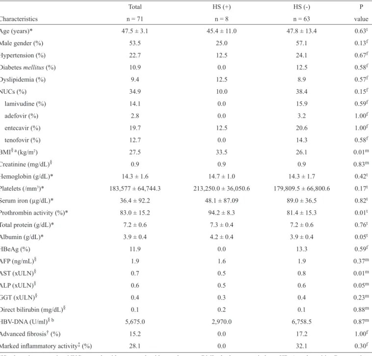
TABLE 1 - Clinical and laboratory characteristics of 71 patients with hepatitis B infection according to the presence of hepatic steatosis on ultrasonography.
Total HS (+) HS (-) P
Characteristics n = 71 n = 8 n = 63 value
Age (years)* 47.5 ± 3.1 45.4 ± 11.0 47.8 ± 13.4 0.63t
Male gender (%) 53.5 25.0 57.1 0.13f
Hypertension (%) 22.7 12.5 24.1 0.67f
Diabetes mellitus (%) 10.9 0.0 12.5 0.58f
Dyslipidemia (%) 9.4 12.5 8.9 0.57f
NUCs (%) 34.9 10.0 38.4 0.15f
lamivudine (%) 14.1 0.0 15.9 0.59f
adefovir (%) 2.8 0.0 3.2 1.00f
entecavir (%) 19.7 12.5 20.6 1.00f
tenofovir (%) 12.7 0.0 14.3 0.58f
BMI§a(kg/m2) 27.5 33.5 26.1 0.01m
Creatinine (mg/dL)§ 0.9 0.9 0.9 0.83m
Hemoglobin (g/dL)* 14.3 ± 1.6 14.7 ± 1.0 14.3 ± 1.7 0.42t
Platelets (/mm3)* 183,577 ± 64,744.3 213,250.0 ± 36,050.6 179,809.5 ± 66,800.6 0.17t
Serum iron (µg/dL)* 36.4 ± 92.2 48.1 ± 87.09 89.0 ± 36.5 0.82t
Prothrombin activity (%)* 83.0 ± 15.2 94.2 ± 8.3 81.4 ± 15.3 0.01t
Total protein (g/dL)* 7.2 ± 0.6 7.3 ± 0.4 7.2 ± 0.6 0.76t
Albumin (g/dL)* 3.9 ± 0.4 4.2 ± 0.4 3.9 ± 0.4 0.05t
HBeAg (%) 11.9 0.0 13.3 0.59f
AFP (ng/mL)§ 1.9 1.6 1.9 0.37m
AST (xULN)§ 0.7 0.5 0.8 0.01m
ALP (xULN)§ 0.6 0.5 0.6 0.05m
GGT (xULN)§ 0.4 0.3 0.4 0.23m
Direct bilirubin (mg/dL)§ 0.1 0.2 0.1 0.88m
HBV-DNA (U/ml)§ b 5,675.0 2,970.0 6,758.5 0.87m
Advanced fi brosis† (%) 15.2 0.0 17.2 1.00f
Marked infl ammatory activity‡ (%) 28.1 0.0 32.1 0.30f
HS: hepatic steatosis; NUCs: nucleoside or nucleotide analogues; BMI: body mass index; HBeAg: hepatitis B e antigen; AFP: alpha-fetoprotein; AST: aspartate aminotransferase; ALT: alanine aminotransferase; ALP: alkaline phosphatase; GGT: gamma-glutamyltransferase; xULN: times the upper limit of normal; HBV-DNA: hepatitis B virus-deoxyribonucleic acid. *mean ± standard
deviation; §median; tStudent’s t test; mMann-Whitney U test; fFisher's exact test; †advanced fi brosis = structural changes of stage
3 or 4; ‡marked infl ammatory activity = periportal activity of stage 3 or 4; aavailable in 19 patients; bavailable in 64 patients.
A positive correlation was also observed between serum albumin
and total cholesterol (r=0.257; p=0.03) and LDL (r=0.34;
p<0.01). Prothrombin activity was positively correlated with the
total cholesterol (r=0.355; p<0.01) and triglycerides (r=0.296; p=0.02).
Negative correlations were observed between AST and
total cholesterol (r=-0.314; p<0.01), HDL (r=-0.246; p=0.03),
and LDL (r=-0.264; p=0.03). Negative correlations were
also observed between direct bilirubin and total cholesterol
(r=-0.396; p<0.01), LDL (r=-0.391; p<0.01) and triglycerides (r=-0.285; p=0.02).
No correlation was observed between ALT, ALP, GGT, and
TABLE 2 - Lipid profi le of 71 patients with hepatitis B infection according to the presence of hepatic steatosis on ultrasonography.
Total HS (+) HS (-) P
Characteristics n = 71 n = 8 n = 63 value
Total cholesterol (mg/dL)* 173.8 ± 39.7 217.5 ± 41.2 168.2 ± 36.2 <0.01t
HDL (mg/dL)* 47.6 ± 3.5 45.6 ± 5.4 47.9 ± 14.2 0.40t
LDL (mg/dL)§ 109.5 135.0 104.0 0.01m
Triglycerides (mg/dL)§ 77.5 116.5 86.9 0.05m
Fasting glucose (mg/dL)§a 93.0 98.0 93.0 0.86m
Fasting insulin (µIU/mL)§ b 3.5 9.2 3.3 0.03m
Glycated hemoglobin (%)§ c 5.8 5.8 5.7 0.75m
HS: of hepatic steatosis; HDL: high-density lipoprotein; LDL: low-density lipoprotein; * mean ± standard deviation; §median; tStudent’s t test;
mMann-Whitney U test; aavailable in 69 patients; bavailable in 31 patients; cavailable in 36 patients.
Factors associated with the presence of liver steatosis on abdominal ultrasound
Patients with LS, compared to those without steatosis on abdominal ultrasound, exhibited a higher mean total cholesterol (201.7±50.4 vs. 171.8±35.9mg/dL, respectively; p=0.02),
greater prothrombin activity (94.1±7.9 vs. 81.6±15.1mg/dL; p<0.01), a higher median fasting insulin (9.2 vs. 3.3mg/dL;
p= 0.04), and a higher median BMI (33.5 vs. 26.3kg/m2; p=0.01). Patients with LS also showed lower median AST levels on abdominal ultrasound compared to those without steatosis (0.5 vs. 0.8 ×ULN, respectively; p=0.02) (Tables 1 and 2).
No differences were found on ultrasound between individuals with or without LS with regard to age, gender, skin color, history of hypertension, diabetes or dyslipidemia, antiviral therapy, HBeAg, HBV-DNA, AFP, creatinine, hemoglobin, platelets, direct bilirubin, ALT, ALP, GGT, HDL, LDL, triglycerides, or fasting glucose (Tables1 and 2). Neither the presence nor the absence of LS on abdominal ultrasound was correlated with
advanced fi brosis or marked periportal infl ammatory activity.
Factors associated with the presence of steatosis on liver biopsy
Only 39 individuals were subjected to liver biopsy. Only two of these patients were HBeAg-positive, and 16 (41%) presented LS on biopsy. When patients with steatosis were compared to those without steatosis on liver biopsy, no differences were
observed with regard to age (p=0.59), gender (p=0.24), skin color (p=0.63), history of hypertension (p=1.00), diabetes (p=1.00), dyslipidemia (p=0.62), antiviral therapy (p=0.43), BMI (p=0.12), HBeAg (p=0.51), HBV-DNA (p=0.53), platelets (p=0.54), direct bilirubin (p=0.43), AST (p=0.19), ALT (p=0.82), ALP (p=0.23), GGT (p=0.93), total cholesterol (p=0.95), HDL (p=0.23), LDL (p=0.75), triglycerides (p=0.33), and fasting glucose (p=0.69). Neither the presence nor the absence of LS on biopsy was correlated with advanced fi brosis (p=0.37) and marked periportal infl ammatory activity (p=0.94).
200,000.000
Liver steatosis (abdominal US)
100,000.000
50,000.000
0
0 1
HB
V
-DNA
(IU/mL)
150,000.000
FIGURE 2 - Dispersion diagram illustrating the relationship between the HBV viral load and liver steatosis. HBV-DNA; hepatitis B virus-deoxyribonucleic acid; US: ultrasound.
The relationship between the HBV viral load and liver steatosis is illustrated in Figure 2.
Among those individuals with steatosis according to abdominal ultrasound, liver biopsy failed to confirm the diagnosis in one case (Table 3). Among those without steatosis
according to abdominal ultrasound, fi ve steatosis cases were
confirmed during the liver biopsy. Abdominal ultrasound
demonstrated the following accuracy parameters: accuracy = 0.692, prevalence = 0.410, sensitivity = 0.313, specifi city = 0.957, positive
TABLE 3 - Distribution of 39 patients according to the presence of hepatic steatosis (HS) on ultrasonography and on liver biopsy.
Characteristics HS (+) Bx (n = 16) HS (-) Bx ( n = 23) Total (n = 39)
n % n % n %
HS (+) on ultrasonography 5 31.3 1 4.3 6 15.4
HS (-) on ultrasonography 11 68.8 22 95.7 33 84.6
HS: hepatic steatosis; Bx: liver biopsy. p-value = 0.033.
DISCUSSION
Due to the global burden of obesity and increased body weight15, LS has become a common medical problem. In Brazil, the prevalence of obesity has increased over time, from 2.2% for men and 7.4% for women in 1975 to 8.8% for men and 13% for women in 200316. In 2010, 16% of the Brazilian population was obese. The prevalence of obesity is predicted to increase to 46% by 205017. In the United States, the prevalence of obesity is higher than in Brazil, involving 32% of adults and 17% of young persons18.
Among a sample of 90 Brazilian obese adolescents, the prevalence of LS on ultrasound was 15.5%19. Hepatic ultrasound is a simple, noninvasive technique that is widely used in clinical
practice to detect fatty infi ltration of the liver20. Several studies
have assessed the sensitivity and specifi city of ultrasound for detecting LS. In these studies, the sensitivity ranged from 60% to 94% and the specifi city from 84% to 97%21-24. In a sample of 94 Brazilian individuals with elevated ALT, 40% presented LS
on ultrasound, and both the BMI and history of diabetes were
independently associated with the presence of LS25. Almost one-third of a sample of 2,287 American subjects evaluated by Browning et al. presented with LS according to magnetic resonance imaging (31%). LS was associated not only with metabolic syndrome but also particularly with obesity and insulin resistance26.
In the present study, the presence of LS on abdominal
ultrasound was associated with fasting insulin and total
cholesterol, similar to fi ndings previously reported by other
authors in non-HBV patients25,27-29, indicating that LS is related to disordered metabolism of blood glucose and lipids. Although
BMI data were available only in a small subset of patients (n = 19), these data revealed a signifi cant difference between the two study groups. Insulin resistance plays a central role in the pathogenesis of NAFLD. It has been demonstrated that obesity is
associated with insulin resistance, leading to hyperinsulinemia, increased free fatty acid concentrations, and hyperglycemia.
Insulin resistance leads to increased delivery of free fatty acids
to the liver, increased fatty acid synthesis, and impaired release
of triglycerides from the liver. These modifi cations cause
triglycerides to accumulate in the hepatocyte30.
A recent experimental study revealed that adenovirus containing the HBV genome (Ad-HBV) up-regulated the expression of genes related to cholesterol metabolism in
HepG2 cells31, suggesting that HBV itself infl uences cholesterol
metabolism. Nevertheless, the lipid profi les among individuals
with chronic HBV remain a matter of debate in the literature. When HBV carriers were compared to HBsAg-negative
individuals, the HBV carriers exhibited signifi cantly lower odds
ratios for hypercholesterolemia and hypertriglyceridemia, as well as higher LDL cholesterol levels9.
Biopsy-confi rmed LS has been described in 18%-76% of
chronic HBV patients32-36. The wide variation in the prevalence of LS in HBV carriers cannot be explained easily, as the patient characteristics do not appear to differ substantially among studies. No association was demonstrated between
the histological fi ndings of steatosis and dyslipidemia in the
present study, similar to previous findings33-37. A possible limitation of the present study is that patients did not undergo
biopsy specifi cally for this study. The data collection was
retrospective, and the indication for liver biopsy was based on the viral load and ALT levels of the patients. Although this study design may indicate a source of bias, it represents a real-life approach to these cases. Nevertheless, as previously mentioned,
abdominal ultrasound possesses high sensitivity and specifi city
for detecting LS, and in a sample of patients presenting more
than 30% LS, the sensitivity and the specifi city were shown to
increase to 89.7% and 100%, respectively24.
Among a sample of 350 individuals with chronic HBV
in India, only the serum triglyceride level was found to be
independently associated with LS according to multivariate analysis36. Other studies have indicated that patients with chronic
HBV infection and LS had signifi cantly higher BMI and higher
levels of fasting glucose, triglycerides, and total cholesterol than did those without steatosis11,13,32,35. Based on these fi ndings, we
can surmise that it is important to closely monitor the BMI, insulin resistance, and lipid profi le in patients with chronic HBV,
as well as in the general population, to prevent the occurrence of LS. However, considering that a substantial amount of fatty
infi ltration in the liver may contribute to the seroclearance of
HBsAg, it has been shown that HBsAg carriers with mild LS did not present an increased likelihood of HBsAg seroclearance. However, moderate-to-severe LS has been associated with 3- to 4-fold increased odds of HBsAg seroclearance compared to those with no evidence of LS38.
No association was demonstrated between steatosis and either HBV DNA viral load or HBeAg status. Given the relatively small number of patients with viral load data
HBV in mediating LS, and further studies that examine a larger cohort are needed to address this question. Some authors have found an association between viral load and the absence of steatosis11,32, and an inverse relationship between metabolic syndrome and chronic HBV has been demonstrated6. The reason why HBsAg-positive subjects are less prone to developing metabolic syndrome remains unclear. One possible explanation
is that individuals with a high BMI and moderate-to-intense
ultrasound grading of LS tend to clear HBsAg from their serum.
In the present study, the level of AST was associated with
the presence of LS on abdominal ultrasound. Aminotransferases are serum markers of liver damage that are usually altered in the presence of LS39. Despite observing lower levels of AST among patients with steatosis, both study groups presented altered AST
levels. The association between LS and advanced fi brosis was
not evaluated in the present study; nevertheless, it has been
shown that LS is less common in patients with advanced fi brosis,
and lower AST levels have been observed in individuals with
advanced fi brosis35.
Other possible limitations of the present study should be mentioned. Although the total number of patients included in the cohort was representative of the overall population, we examined possible associations between certain factors and LS, considerably reducing the number of subjects presenting particular variable combinations and, consequently, the power of the statistical tests. Although it is not a strict rule, maintaining a minimum of ten events per variable is recommended during logistic regression analysis. This recomendation is based on studies that showed increasing
bias and variability, unreliable coverage of confi dence intervals,
and problems with model convergence as the events per variable declined below ten40-42. For this reason, these results need to be
confi rmed in a larger set of patients. Additionally, the study design
was cross-sectional and did not include a longitudinal follow-up. The ultrasounds may not have been uniform in terms of operator
and evaluation method, as this was not a prospective study. Indeed, the ultrasound results refl ect a day-to-day medical practice in which one relies on ultrasound to initially defi ne whether the patient
presents with LS. As we have demonstrated, ultrasonography does not always match biopsy results perfectly. Data on alcohol ingestion, diet quality, and routine exercise were also lacking; therefore, an analysis of the effect of these variables on metabolic disease in this population was not possible. Another limitation of our study was the absence of a control group; nevertheless, our findings are comparable to previously published data.
The causes and signifi cance of LS in HBV-related liver disease continues to be investigated. Individuals with LS present lower AST levels than do those without steatosis. The fi ndings
of the present study indicate that LS in HBV carriers appears to be a result of metabolic factors attributable to the host and related to insulin action rather than to viral factors; however,
larger studies are needed to validate these fi ndings. Therefore,
it may be surmised that to prevent LS in patients with chronic
HBV, AST and BMI should be periodically monitored and the blood glucose and lipid profi les should be controlled, in addition to educating these patients about healthy lifestyle, scientifi c
dieting, and physical exercise.
REFERENCES
The authors declare that there is no confl ict of interest.
CONFLICT OF INTEREST
1. Lavanchy D. Hepatitis B virus epidemiology, disease burden, treatment, and current and emerging prevention and control measures. J Viral Hepat
2004; 11:97-107.
2. Clark JM, Brancati FL, Diehl AM. Nonalcoholic fatty liver disease.
Gastroenterology 2002; 122:1649-1657.
3. Harrison SA, Torgerson S, Hayashi PH. The natural history of nonalcoholic
fatty liver disease: a clinical histopathological study. Am J Gastroenterol 2003; 98:2042-2047.
4. Machado MV, Cortez-Pinto H. Insulin resistance and steatosis in chronic
hepatitis C. Ann Hepatol 2009; 8 (suppl I):67-75.
5. Li WC, Lee YY, Chen IC, Sun C, Chiu FH, Chuang CH. Association
between the hepatitis B and C viruses and metabolic diseases in patients
stratifi ed by age. Liver Int 2013; 33:1194-1202.
6. Jan CF, Chen CJ, Chiu YH, Chen LS, Wu HM, Huang CC, et al. A population-based study investigating the association between metabolic syndrome and hepatitis B/C infection (Keelung Community-based
Integrated Screening study No. 10). Int J Obes (Lond) 2006; 30:794-799.
7. Kanel GC, Radvan G, Peters RL. High-density lipoprotein cholesterol and
liver disease. Hepatol 1983; 3:343-348.
8. Cicognani C, Malavolti M, Morselli-Labate AM, Zamboni L, Sama C, Barbara L. Serum lipid and lipoprotein patterns in patients with liver
cirrhosis and chronic active hepatitis. Arch Int Med 1997; 157:792-796.
9. Su TC, Lee YT, Cheng TJ, Chien HP, Wang JD. Chronic hepatitis B virus infection and dyslipidemia. J Formosan Med Assoc (Taiwan yi zhi) 2004;
103:286-291.
10. Liu PT, Hwang AC, Chen JD. Combined effects of hepatitis B virus infection and elevated alanine aminotransferase levels on dyslipidemia.
Metabolism 2013; 62:220-225.
11. Zheng RD, Chen JN, Zhuang QY, Lu YH, Chen J, Chen BF. Clinical and virological characteristics of chronic hepatitis B patients with hepatic
steatosis. Int J Med Sci 2013; 10:641-646.
12. Fan JG, Chitturi S. Hepatitis B and fatty liver: causal or coincidental?
J Gastroenterol Hepatol 2008; 23:679-681.
13. Zheng RD, Xu CR, Jiang L, Dou AX, Zhou K, Lu LG. Predictors of hepatic steatosis in HBeAg-negative chronic hepatitis B patients and their
diagnostic values in hepatic fi brosis. Int J Med Sci 2010; 7:272-277.
14. Gayotto LCC. Visão histórica e consenso nacional sobre a classifi cação
das hepatites crônicas. Gastroenterol Endosc Digest 2000; 19:4.
15. Kopelman PG. Obesity as a medical problem. Nature 2000; 404:635-643.
16. Monteiro CA, Conde WL, Popkin BM. Income-specifi c trends in obesity
in Brazil: 1975-2003. Am J Public Health 2007; 97:1808-1812.
17. Rtveladze K, Marsh T, Webber L, Kilpi F, Levy D, Conde W, et al. Health
and economic burden of obesity in Brazil. PloS One 2013; 8:e68785.
18. Ogden CL, Carroll MD, Curtin LR, McDowell MA, Tabak CJ, Flegal KM. Prevalence of overweight and obesity in the United States, 1999-2004.
JAMA 2006; 295:1549-1555.
19. Fernandes MT, Ferraro AA, Azevedo RA, Fagundes Neto U. Metabolic differences between male and female adolescents with non-alcoholic fatty liver disease, as detected by ultrasound. Acta Paediatr 2010;
99:1218-1223.
20. Mehta SR, Thomas EL, Bell JD, Johnston DG, Taylor-Robinson SD. Non-invasive means of measuring hepatic fat content. World J Gastroenterol
2008; 14:3476-3483.
21. Joseph AE, Saverymuttu SH, al-Sam S, Cook MG, Maxwell JD. Comparison of liver histology with ultrasonography in assessing diffuse
22. Foster KJ, Dewbury KC, Griffi th AH, Wright R. The accuracy of ultrasound in the detection of fatty infi ltration of the liver. Br J Radiol 1980; 53:440-442.
23. Saverymuttu SH, Joseph AE, Maxwell JD. Ultrasound scanning in the detection of hepatic fi brosis and steatosis. Br Med J (Clin Res Ed) 1986; 292:13-15.
24. Palmentieri B, de Sio I, La Mura V, Masarone M, Vecchione R, Bruno S, et al. The role of bright liver echo pattern on ultrasound B-mode examination in the diagnosis of liver steatosis. Dig Liver Dis 2006; 38:485-489.
25. Narciso-Schiavon JL, Schiavon LL, Carvalho-Filho RJ, Hayashida DY, Wang JH, Souza TS, et al. Clinical characteristics associated with hepatic steatosis on ultrasonography in patients with elevated alanine aminotransferase. Sao Paulo Med J 2010; 128:342-347.
26. Browning JD, Szczepaniak LS, Dobbins R, Nuremberg P, Horton JD, Cohen JC, et al. Prevalence of hepatic steatosis in an urban population in the United States: impact of ethnicity. Hepatol 2004; 40:1387-1395. 27. Cassader M, Gambino R, Musso G, Depetris N, Mecca F, Cavallo-Perin P,
et al. Postprandial triglyceride-rich lipoprotein metabolism and insulin sensitivity in nonalcoholic steatohepatitis patients. Lipids 2001; 36: 1117-1124.
28. Hsiao PJ, Kuo KK, Shin SJ, Yang YH, Lin WY, Yang JF, et al. Signifi cant correlations between severe fatty liver and risk factors for metabolic syndrome. J Gastroenterol Hepatol 2007; 22:2118-2123.
29. Venturi C, Zoppini G, Zamboni C, Muggeo M. Insulin sensitivity and hepatic steatosis in obese subjects with normal glucose tolerance. Nutr Metab Cardiovasc Dis 2004; 14:200-204.
30. Meek SE, Nair KS, Jensen MD. Insulin regulation of regional free fatty acid metabolism. Diabetes 1999; 48:10-14.
31. Li YJ, Zhu P, Liang Y, Yin WG, Xiao JH. Hepatitis B virus induces expression of cholesterol metabolism-related genes via TLR2 in HepG2 cells. World J Gastroenterol 2013; 19:2262-2269.
32. Minakari M, Molaei M, Shalmani HM, Mohammad Alizadeh AH, Jazi AH, Naderi N, et al. Liver steatosis in patients with chronic hepatitis B
infection: host and viral risk factors. Eur J Gastroenterol Hepatol 2009; 21:512-516.
33. Gordon A, McLean CA, Pedersen JS, Bailey MJ, Roberts SK. Hepatic steatosis in chronic hepatitis B and C: predictors, distribution and effect on fi brosis. J Hepatology 2005; 43:38-44.
34. Altlparmak E, Koklu S, Yalinkilic M, Yuksel O, Cicek B, Kayacetin E, et al. Viral and host causes of fatty liver in chronic hepatitis B. World J Gastroenterol 2005; 11:3056-3059.
35. Thomopoulos KC, Arvaniti V, Tsamantas AC, Dimitropoulou D, Gogos CA, Siagris D, et al. Prevalence of liver steatosis in patients with chronic hepatitis B: a study of associated factors and of relationship with fi brosis. Eur J Gastroenterol Hepatol 2006; 18:233-237.
36. Rastogi A, Sakhuja P, Kumar A, Hissar S, Jain A, Gondal R, et al. Steatosis in chronic hepatitis B: prevalence and correlation with biochemical, histologic, viral, and metabolic parameters. Indian J Pathol Microbiol 2011; 54:454-459.
37. Zamin Jr I, Mattos AA, Zettler CG. Nonalcoholic steatohepatitis in nondiabetic obese patients. Canadian J Gastroenterol 2002; 16:303-307. 38. Chu CM, Lin DY, Liaw YF. Does increased body mass index with
hepatic steatosis contribute to seroclearance of hepatitis B virus (HBV) surface antigen in chronic HBV infection? Int J Obes (Lond) 2007; 31:871-875.
39. Paschos P, Paletas K. Non alcoholic fatty liver disease and metabolic syndrome. Hippokratia 2009; 13:9-19.
40. Concato J, Peduzzi P, Holford TR, Feinstein AR. Importance of events per independent variable in proportional hazards analysis. I. Background, goals, and general strategy. J Clin Epidemiol 1995; 48:1495-1501. 41. Peduzzi P, Concato J, Feinstein AR, Holford TR. Importance of events
per independent variable in proportional hazards regression analysis. II. Accuracy and precision of regression estimates. J Clin Epidemiol 1995; 48:1503-1510.