DOI 10.1007/s00296-015-3287-0
Rheumatology
INTERNATIONALORIGINAL ARTICLE - OBSERVATIONAL RESEARCH
Vitamin D levels in juvenile idiopathic arthritis from an
equatorial region
Sâmia Araújo de Sousa Studart1 · Ana Caroline Rocha Melo Leite1 ·
Aryana Lushese Lima Feitosa Marinho1 · Ana Carolina Matias Dinelly Pinto1 · Carlos Nobre Rabelo Júnior2 · Rodolfo de Melo Nunes1 ·
Hermano Alexandre Lima Rocha3 · Francisco Airton Castro Rocha1,4
Received: 21 February 2015 / Accepted: 11 May 2015 / Published online: 20 May 2015 © Springer-Verlag Berlin Heidelberg 2015
the first study on 25OHD levels in JIA patients living in a low-latitude region, showing the lowest prevalence of vita-min D deficiency ever reported. Serum 25OHD was similar in JIA and controls and did not vary regardless of JIA cat-egory or severity.
Keywords Juvenile idiopathic arthritis · Vitamin D · Parathyroid hormone · Autoimmunity · Epidemiology
Introduction
Juvenile idiopathic arthritis (JIA) is the most frequent chronic arthropathy in childhood, affecting one or more joints in children <16 years old, being more common among girls. JIA prevalence in white Caucasians varies from 0.07 to 4.01/1000 children [1]. In São Paulo, Brazil, JIA accounts for more than 30 % of new cases seeking pediatric rheumatologists [2], and JIA prevalence was esti-mated at 0.34/1000 children [3].
There is controversy as to whether serum vitamin D levels influence both the prevalence and outcome of JIA [4, 5]. Low vitamin D has been associated with an increased risk of osteoporosis-related fractures [6] as well as with increased prevalence and/or sever-ity of multiple sclerosis [7], rheumatoid arthritis (RA) [8–10], and systemic lupus erythematosus [11]. The mechanisms of action of vitamin D are multiple and include modulation of transcription pathways leading to cytokine production, regulation of the expression of cyclooxygenase 2, and lipoxygenase genes, and it may directly interfere with cytokine gene expression and signaling, favoring a change from a T helper (Th)-1 into a Th-2 cytokine profile, as well as a decrease in Th-17 response [12].
Abstract We aimed to describe the serum levels of 25-hydroxyvitamin D (25OHD) in juvenile idiopathic arthritis (JIA) patients living in a low-latitude (3°43′S) region. Fifty JIA patients, 31 (62 %) female, seen between May 2012 and April 2013 in the northeast of Brazil had clinical data and serum collected for determination of 25OHD and parathyroid hormone (PTH) using a chemi-luminescent ELISA; 20 age- and sex-matched controls were used for comparison. Mean age was 13.4 ± 4 years. Twenty-five (50 %), 15 (30 %), 4 (8 %), 4 (8 %), and 2 (4 %) patients were of the polyarticular, oligoarticular, systemic, enthesitis-related, and undifferentiated cat-egories, respectively. Mean 25OHD was 31.6 ± 10 and 30.4 ± 5.7 ng/mL in patients and controls (P > 0.05), respectively; PTH was normal in JIA and controls; 25OHD was similar regardless of JIA category, disease activity, or severity measured by JADAS-27, CHAQ, or presence of joint deformities. Twenty-six (52 %), 20 (40 %), and 4 (8 %) patients were considered to have optimal, suffi-cient, and deficient 25OHD levels, respectively, whereas 11 (52 %) and 10 (48 %) controls had optimal and sufficient 25OHD. Ethnicity, body mass index, seasonal variation, and use of steroids did not influence 25OHD levels. This is
* Francisco Airton Castro Rocha arocha@ufc.br
1 Department of Internal Medicine, Faculty of Medicine,
Universidade Federal do Ceará, Fortaleza, CE, Brazil
2 Rheumatology Service, Pediatric Rheumatology Division,
Hospital Geral de Fortaleza, Fortaleza, CE, Brazil
3
Department of Public Health, Faculty of Medicine, Universidade Federal do Ceará, Fortaleza, CE, Brazil
4 Rua Coronel Nunes de Melo, 1315, Rodolfo Teofilo,
Since there are no clearly defined normal vitamin D levels for children, cutoff levels proposed for adults have been used in studies with children. With this strategy, up to 82 % of JIA children were reported to have insufficient vitamin D and an association with disease outcome could not be found [13]. A previous study conducted in JIA children from Boston, MA, found low vitamin D in over 50 % of the patients. In that study, age, ethnicity, body mass index (BMI), and seasonal variation were shown to influence serum vitamin D, but there was no association with disease activity [14]. In JIA children from Morocco, hypovitaminosis D, which was found in 75 % of the patients, was also not associated with disease activity [15].
In adult patients with inflammatory polyarthritis, higher vitamin D metabolite levels were associated with a decreased Health Assessment Questionnaire (HAQ) score [16]. Despite finding similar levels in RA patients and controls, another study reported an inverse association of 25-OH vitamin D levels and RA disease activity [17]. A recent study with RA patients from China living in a 32° latitude region found not only lower serum vitamin D levels, as compared to controls, but also a negative association of vitamin D with RA disease activity and serum levels of the interleukin 17/23 cytokines [9].
Latitude differences, sunshine exposure, diet, BMI, and use of dietary supplements influence serum vitamin D [11, 18, 19]. A previous study that collected data from all continents but Africa showed a large variation of vitamin D levels across different countries [19]. However, in that study, the Latin-American region was represented solely by Mexico and Argentina, although the mileage distance of their capitals is around 4600 miles. In fact, less exposure to sunlight in the extreme south of Argentina was associated with lower vitamin D3 levels and rickets, and the in vitro photoconversion of provitamin D3 to previtamin D3 was absent during winter in southern latitudes in Argentina, as compared to the same levels obtained in Buenos Aires [20].
We live in Fortaleza, capital of the state of Ceará, Brazil, with a low-income population of 8,779,000 inhabitants in the state and 3,597,000 living in Fortaleza. Our geographic coordinates are 3°43′S and 38°32′W. According to the offi-cial Brazilian tourism agency, among the largest cities in Brazil, Fortaleza is the one with more sunny days with a mean of 239 days per year (http://en.wikipedia.org/wiki/ Fortaleza#Climate). To our knowledge, there are no stud-ies evaluating the possible influence of vitamin D in auto-immune diseases in children from a region as close to the equator line. Our aim was to report serum vitamin D levels in JIA patients living in the state of Ceará as well as its pos-sible association with JIA categories and disease activity.
Methods
Patients with a diagnosis of JIA according to the Interna-tional League of Associations for Rheumatology (ILAR) [21] seen at the rheumatology outpatient clinics of the Hospital Universitário Walter Cantídio of the Faculty of Medicine of the Universidade Federal do Ceará and Hospi-tal Geral de ForHospi-taleza, between May 2012 and April 2013, comprising 50 children, were cross-sectionally evaluated.
Considering that deficient vitamin D levels were found in up to 82 % of JIA patients [13] and a conservative esti-mation of a 60 % prevalence of deficient vitamin D levels in our JIA sample with 5 % level of significance, the sam-ple size had to be of at least 41 participants.
The clinical protocol was submitted and approved by our local ethics regulatory committee that follows the rules of the Brazilian National Ethics Committee on Clinical Research (CONEP). All patients or their responsible rela-tives signed an informed consent before any intervention.
A clinical chart was filled for each patient registering demographic data, clinical evaluation, and current treat-ment. A complete physical examination was performed, and samples collected for blood cell counts, biochemistry (glucose, creatinine, urea, aspartate aminotransferase, ala-nine aminotransferase), C-reactive protein and erythrocyte sedimentation rate (ESR), and type I urinalysis. Testing for the presence of rheumatoid factor using nephelometry and anti-nuclear antibodies using indirect immunofluorescence was also done.
Vitamin D levels were measured as the serum concen-tration of 25-hydroxyvitamin D (25OHD), in addition to serum calcium and phosphate, alkaline phosphatase, and parathyroid hormone (PTH) levels; 25OHD and PTH lev-els were determined using a chemiluminescence ELISA (Immulite 2000 system analyzer™), as instructed by the manufacturer (normal range for PTH = 12–65 pg/mL; (Siemens Ltda., SP, Brazil™). The intra- and inter-assay coefficients of variation (CV) were 9.7 and 14.7 % for 25OHD and 3 and 4 % for PTH, respectively. Most stud-ies have a special attention to seasonal variations regard-ing determination of serum vitamin D levels. However, our low latitude means no defined seasons across the year. Therefore, we collected serum samples regardless of the month of the year. Exclusion criteria were history or pre-sent evidence of any other disease, including bone-related diseases, but JIA. There were no pregnancies during the study period. Patients on medications for osteoporosis, except for calcium supplements, were excluded. No patient was on vitamin D supplementation. BMI was calculated as weight/squared height (kg/m2). A group of 20 age- and
control, was specifically evaluated for BMI and determina-tion of serum 25OHD and PTH levels.
Since there are no defined 25OHD normal levels for children, we considered values higher than 30 ng/mL as optimal, those between 20 and 30 ng/mL as sufficient, and levels below 20 ng/mL as deficient [22].
Disease activity measurement was done using the Juve-nile Arthritis Disease Activity Score (JADAS)-27 [23] that considers physician global assessment of disease activ-ity [(0–10 cm visual analog scale (VAS)], parent global assessment of child’s well-being (0–10 cm; VAS), number of joints (0–27) with active disease, and ESR, providing a total score of 0–57 where high scores mean high disease activity. A Portuguese version of Childhood Health Assess-ment Questionnaire (CHAQ) was used for evaluation of functional status [24].
Patients were considered to have clinically inactive dis-ease if the following was achieved, as described previously: (1) no joints with active arthritis; (2) no fever, rash, serosi-tis, splenomegaly, or generalized lymphadenopathy attrib-utable to JIA; (3) no active uveitis; (4) ESR in the normal
range; and (5) a physician’s global assessment of disease activity score of zero [25]. Using recent previously defined cutoff levels, JIA patients with the oligoarticular and pol-yarticular presentations were defined as having high dis-ease activity if JADAS-27 > 4.2 and 8.5, respectively [26]. The number of children with joint deformities, defined as an irreversible damage to the anatomic structure and/ or function that interfered with joint range of motion that could be attributed to the JIA, was also recorded.
Statistics used descriptive analysis for demographics and outcomes using mean ± SD or medians, as appropri-ate. Assessment of normality of continuous data was done using the Kolmogorov–Smirnov test. Dichotomous vari-ables were expressed as absolute and percent values. Dif-ferences between means were evaluated using Student’s “t” test; dichotomous data were evaluated using Chi-square or Fisher’s exact (when needed) tests. Pearson’s correlation coefficient was applied for variables with normal distribu-tion. The level of significance was set at 0.05. Sample size, entering of data, and data analysis were done using SPSS v17, SPSS Inc.
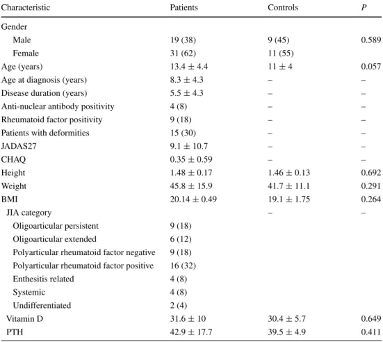
Table 1 Demographic, clinical, and laboratory data of patients with juvenile idiopathic arthritis
Data are n (%) and mean ± SD, JADAS Juvenile Arthritis Disease Activity Score, CHAQ Childhood Health Assessment Questionnaire, BMI body mass index, PTH parathyroid hormone
Characteristic Patients Controls P
Gender
Male 19 (38) 9 (45) 0.589
Female 31 (62) 11 (55)
Age (years) 13.4 ± 4.4 11 ± 4 0.057
Age at diagnosis (years) 8.3 ± 4.3 – –
Disease duration (years) 5.5 ± 4.3 – –
Anti-nuclear antibody positivity 4 (8) – –
Rheumatoid factor positivity 9 (18) – –
Patients with deformities 15 (30) – –
JADAS27 9.1 ± 10.7 – –
CHAQ 0.35 ± 0.59 – –
Height 1.48 ± 0.17 1.46 ± 0.13 0.692
Weight 45.8 ± 15.9 41.7 ± 11.1 0.291
BMI 20.14 ± 0.49 19.1 ± 1.75 0.264
JIA category – –
Oligoarticular persistent 9 (18) Oligoarticular extended 6 (12) Polyarticular rheumatoid factor negative 9 (18) Polyarticular rheumatoid factor positive 16 (32)
Enthesitis related 4 (8)
Systemic 4 (8)
Undifferentiated 2 (4)
Vitamin D 31.6 ± 10 30.4 ± 5.7 0.649
Results
Demographic data are given in Table 1. Among the 50 patients included, 31 (62 %) were of the female gender. The mean (SD) age was 13.4 ± 4 years, and the mean (SD) age at diagnosis was 8.3 ± 4.3 years. Mean disease duration was 5.5 years, and there were 15 (30 %) patients with joint deformities. Among those, 10 (20 %) were of the polyarticular, 3 (6 %) oligoarticular, and one (2 %) each of the systemic and undifferentiated categories. The polyarticular presentation was the most common category, representing 25 (50 %) patients comprising 16 (32 %) and 9 (18 %) that were positive and negative for the rheuma-toid factor, respectively. All patients that tested positive for the rheumatoid factor had the polyarticular presentation. Among the 15 (30 %) patients classified with the oligoar-ticular presentation, 9 (18 %) were considered as persis-tent and 6 (12 %) as extended. Four (8 %) had the systemic presentation, 4 (8 %) others were classified as enthesitis related, and 2 (4 %) were considered undifferentiated. Four (8 %) patients, two each of the polyarticular and oli-goarticular categories, tested positive for the presence of anti-nuclear antibodies (dense fine speckled pattern).
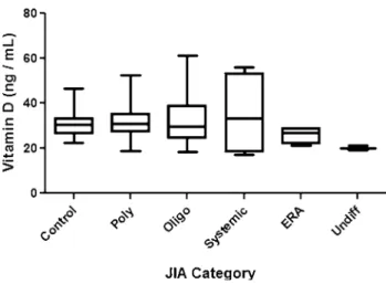
Serum calcium, phosphorus, and alkaline phos-phatase were within normal limits in all patients (data not shown). Mean serum 25OHD was 31.6 ± 10 ng/mL in JIA patients (Table 1). There was no difference when com-paring 25OHD levels among the various JIA categories (Fig. 1). Specifically, mean 25OHD levels in JIA patients of the polyarticular category (31.9 ± 8.2 ng/mL) were very similar to those of patients categorized as oligoarticular (33.17 ± 11.3 ng/mL) (Fig. 1). As shown in Fig. 2, 25OHD levels were similar in patients of the polyarticular and oli-goarticular categories considered to be on high or not-high disease activity level.
Twenty-six (52 %), 20 (40 %), and 4 (8 %) JIA patients were considered to have optimal, sufficient, and deficient 25OHD levels, respectively, whereas 11 (52 %) and 10 (48 %) controls had optimal and sufficient 25OHD levels, respectively, meaning no difference among those groups (P= 0.649). Nobody in the control group and only 4 (8 %) JIA patients had deficient (<20 ng/mL) 25OHD levels. Serum PTH was 42.9 ± 17.7 and 39.5 ± 4.9 pg/mL in JIA patients and controls, respectively, so that all samples had normal PTH levels (Table 1).
The mean JADAS-27 score was 9.1 ± 10.7. Having a 4.2 or 8.5 JADAS-27 score, as said above, was considered a minimum for a high disease activity for the oligoarticular and polyarticular JIA categories, respectively [26]. Using those cutoff values, 22 (44 %) patients were considered with high activity, comprising 14 (28 %) polyarticular and 8 (16 %) oligoarticular. Table 2 illustrates the data of JIA patients concerning the presence of deformities. Those with
deformities had a significantly higher CHAQ, whereas the number of active joints at the time of evaluation was simi-lar between both groups. Those with deformities also had a tendency to display a higher JADAS-27, although not reaching statistical significance (Table 2).
Twenty-three (46 %) patients were using either ibu-profen (65 %) or naproxen (35 %) as non-steroidal anti-inflammatory drugs (NSAIDS). Fourteen patients (30 %) were using low-dose oral steroids, comprising 8 (16 %), 4 (8 %), and 2 (4 %) of the polyarticular, oligoarticular, and systemic categories, respectively, with a 0.16 mg/kg mean daily prednisone dose. There was one patient of the sys-temic category on a 1 mg/kg prednisone daily dose due to disease exacerbation. Forty-one (82 %) patients were using non-biologic DMARDS (Table 3). Methotrexate, combined or isolated, was being used in 38 (76 %) children, lefluno-mide in 10 (20 %), comprising seven (14 %) combined to methotrexate, 2 (4 %) as isolated, and 1 (2 %) combined
Fig. 1 Serum levels of 25-hydroxyvitamin D in JIA patients accord-ing to categories
to a biologic DMARD. Two patients (4 %) that presented liver enzyme alterations while on methotrexate were also using azathioprine. Twenty-three (46 %) patients were using biologic DMARDs (Table 3) as follows: Etanercept was used in 16 (32 %), adalimumab in 4 (8 %), and inf-liximab in 1 (2 %) patient. Two patients classified as sys-temic received tocilizumab and abatacept each. We should mention that 9 (56 %) out of the 16 patients that received biologic DMARDs had deformities, meaning refractory severe disease. The mean JADAS-27 and CHAQ scores in those patients with deformities were 10.6 ± 12.1 and 0.49 ± 0.69, respectively. Eleven (69 %) patients on etaner-cept used it combined to methotrexate, 3 (19 %) as mono-therapy, and 2 (12 %) combined to leflunomide.
Discussion
Our data show that mean serum 25OHD levels in a cohort of JIA patients living in a low-latitude region are within normal limits and similar to those found in a con-trol disease-free population. Different from what has been
published so far in this issue, the majority (52 %) of our JIA patients had 25OHD levels considered optimal, another major part, comprising 40 % of our patients, had sufficient vitamin D levels, and a minority (8 %) of our JIA patients had what has been currently considered deficient 25OHD serum levels [13]. A recent systematic literature review and meta-analysis focusing on 14 reports that measured 25OHD levels in JIA patients, as done in our study, revealed a mean of 24.56 ng/mL (range 11.5–56.4 ng/mL) level in 529 chil-dren, being thus lower as compared to 31.5 ± 10.1 ng/mL mean levels in our patients [13]. Two more recent studies, not included in that meta-analysis, found mean vitamin D levels of 21.8 ± 0.8 ng/mL and 22.1 ± 10.87 ng/mL in JIA patients living in Florence, Italy, and Rabat, Morocco [15,
27]. It has to be pointed out that all those studies were done in high latitudes, including those on Florence and Rabat, meaning 42° and 32°N [15, 27], respectively, as compared to the present study. Perhaps more striking, with regard to the importance of geographical variation, 25OHD levels in children from the state of São Paulo, Brazil (23°S), were reported as 25.7 ± 8.65 ng/mL [28] that are also lower to those found in our study, focusing on people living in a 4°S region of Brazil.
Blood samples in the present study were collected throughout the year, as opposed to all those studies, with samples collected at the end of the winter or in spring, as a strategy to minimize bias associated with less sun expo-sure. Hence, mean serum vitamin D levels in those stud-ies, if collected throughout the year, could possibly be even lower to the values reported. This aspect further strengthens the importance of geographical variation when analyzing serum vitamin D levels [5, 19].
As we mentioned above, there are no defined normal parameters of serum 25OHD for children. Thus, similar to what was done in all previous studies, we used adult levels in our data. However, guidelines from the American Acad-emy of Pediatrics defined the minimum required 25OHD level in infants and children as 20 ng/mL [29]. If we were to use that cutoff level, only 3 (6 %) out of our 50 patients could be considered as presenting deficient 25OHD.
Mean serum PTH levels in our sample were also simi-lar to PTH levels in our controls. In the meta-analysis mentioned above, mean PTH in JIA patients of the pauci-articular, polypauci-articular, and systemic categories were 26.28 (range 20.6–52 pg/mL), 28.7 (range 14.5–77 pg/mL), and 20.3 (range 14.4–51 pg/mL), respectively [13], whereas in the Italian study, mean PTH levels were 46.8 ± 26.9 pg/ mL [27]. Attempts to associate 25OHD levels to markers of bone metabolism, mostly PTH, do not completely rule out controversies regarding what can be considered nor-mal. This inconsistency does also hold true for adults [12,
17, 22]. The fact that our PTH levels were similar to those of JIA patients from higher latitudes despite our patients
Table 2 Comparison of JIA patients considering the presence of deformities
Data are mean ± SD, JADAS Juvenile Arthritis Disease Activity Score, CHAQ Childhood Health Assessment Questionnaire
Characteristic No deformity Deformity P
Active joints 2.54 ± 1.16 3.53 ± 1.19 0.3 JADAS-27 7.59 ± 1.88 12.54 ± 2.29 0.067
CHAQ 0.22 ± 0.08 0.68 ± 0.19 0.005
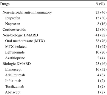
Table 3 Drugs used in JIA patients
Drugs N (%)
Non-steroidal anti-inflammatory 23 (46)
Ibuprofen 15 (30)
Naproxen 8 (16)
Corticosteroids 15 (30)
Non-biologic DMARD 41 (82)
Oral methotrexate (MTX) 38 (76)
MTX isolated 31 (62)
Leflunomide 10 (20)
Azathioprine 2 (4)
Biologic DMARD 23 (46)
Etanercept 16 (32)
Adalimumab 4 (8)
Infliximab 1 (2)
Tocilizumab 1 (2)
presenting an apparently lower prevalence of 25OHD defi-ciency lead us to question the use of increased PTH as an indication of 25OHD deficiency.
Serum 25OHD levels did not vary across different JIA categories in our patients, which is similar to what has been reported in most studies [13]. Moreover, using recently proposed cutoff levels for definition of high activity in JIA [26], there was no difference of 25OHD levels in our patients, regardless of being considered with high activity. Likewise, levels were similar both in those with or without joint deformities and with active/inactive JIA, strongly sug-gesting a lack of association between serum 25OHD and disease outcome even in the long term.
Criticism to the cross-sectional analysis done in our study can be minimized since the presence of joint deform-ities would reflect more severe and long-standing disease. Further, CHAQ values, which reflect ongoing disease severity at various domains, were also similar regardless of serum 25OHD levels in our patients. In fact, despite having an increased proportion of polyarticular category patients, who are believed to carry a risk of worse prognosis, as compared to the more prevalent oligoarticular presentation in other studies in Caucasians [30], CHAQ values in our patients reveal an apparently milder disease. For instance, in the Italian study mentioned above, there were 63.2 % oligoarticular and 23 % polyarticular patients, with a mean CHAQ-DI of 0.71 ± 0.58, as compared to 30 and 50 % patients in those categories, respectively, in our cohort, with a considerably lower CHAQ (0.35 ± 0.59).
Low-income populations are expected to display more severe disease that can be related to less access to medical treatment [31] as well as to an increased prevalence of the polyarticular category [30]. More than 90 % of our patients have a family (mean 6 people/family) monthly income of <U$ 900.00 (data not shown). Although we have no limi-tations concerning prescription of high-cost medications in our reference services, difficulties of patients to attend the outcare clinic, irregular adherence to prescriptions, and very limited access to non-pharmacological therapy may adversely impact disease severity [31, 32]. We cannot rule out that underdiagnosis of less severe oligoarticular presen-tations accounts for our low numbers of this JIA category [5]. However, despite the economic burden, our patients had an apparently less than expected joint damage in the long term. Coupled to a greater prevalence of polyarticular patients, the low CHAQ, vis-à-vis other series from devel-oped countries, lead us to speculate that normal or near-normal 25OHD levels increased prevalence in our region could positively influence the evolution of JIA.
Among the factors that could influence serum 25OHD levels, we may include ethnicity, seasonal variation, BMI, and medications [5, 11]. As said above, samples were col-lected throughout the year, as the number of sunny days in
our region is very stable, thus virtually ruling out this varia-tion as an influence to our data. BMI can also be excluded as a relevant variable since it was normal in the whole majority of both our JIA patients and in all controls so that there were no underweight or obese patients, and only 8 % of our JIA patients were classified as mildly overweight. Prednisone use has been associated to reduction in serum vitamin D [11], but the fact that all but one of our patients used very low-dose prednisone makes this possibility as influencing our results very unlikely. Ethnic variation is also not a major issue in our sample. Our local population is mostly formed from native Brazilian and white Portuguese descendants, as neither did we receive significant number of Africans during the period of black slavery in Brazil nor white people from other regions but Portugal, which is different from southern regions of Brazil.
Limitations of our study include the relatively low num-ber of patients, as compared to other previous reports. In fact, this limitation could have influenced our finding of no differences regarding 25OHD levels among patients in the various JIA categories in our sample. However, the inclu-sion of 50 patients was more than what was required for the sample size, based on a conservative estimation of a 60 % worldwide prevalence of deficient 25OHD levels. Although we did not analyze dietary vitamin D intake related to regular food ingestion, we should stress that our patients were not taking vitamin D supplementation. Thus, the low prevalence of 25OHD deficiency is probably associated with increased sun exposure, as compared to people living in higher latitudes. The cross-sectional nature of the study precludes analyzing the effect of 25OHD in long-term dis-ease activity. However, the relatively low CHAQ values obtained despite a predominance of patients of the polyar-ticular category argues for a better disease outcome in our cohort. In keeping with this assumption, we have recently shown that up to two-thirds of our JIA patients, despite the social economic burden and the more prevalent polyarticu-lar presentation, achieve clinical disease control without the use of biological DMARDs [33].
Taken together, we report a series of JIA patients living in a low-latitude region that presents the lowest prevalence of serum 25OHD deficiency yet published, thus strengthen-ing the relevance of geographical variation when analyzstrengthen-ing serum 25OHD levels.
Conflict of interest The authors declare no conflicts of interest to disclose.
References
2. Terreri MT, Campos LM, Okuda EM, Silva CA, Sacchetti SB, Marini R et al (2013) Profile of paediatric rheumatology special-ists and services in the state of São Paulo. Rev Bras Reumatol 53:346–351
3. Yamashita E, Terreri MT, Hilário MO, Len CA (2013) Preva-lence of juvenile idiopathic arthritis in children aged 6 to 12 years in Embu das Artes, state of Sao Paulo, Brazil. Rev Bras Reumatol 53:542–545
4. Shapira Y, Agmon-Levin N, Shoenfeld Y (2010) Geoepidemi-ology of autoimmune rheumatic diseases. Nat Rev Rheumatol 6:468–476
5. Ellis JA, Munro JE, Ponsonby AL (2010) Possible environmen-tal determinants of juvenile idiopathic arthritis. Rheumatology 49:411–425
6. Cauley JA, Danielson ME, Boudreau R, Barbour KE, Horwitz MJ, Bauer DC et al (2011) Serum 25-hydroxyvitamin D and clinical fracture risk in a multiethnic cohort of women: the Wom-en’s Health Initiative (WHI). J Bone Miner Res 26:2378–2388 7. Munger KL, Levin LI, Hollis BW, Howard NS, Ascherio A
(2006) Serum 25-hydroxyvitamin D levels and risk of multiple sclerosis. JAMA 296:2832–2838
8. Rossini M, Maddali Bongi S, La Montagna G, Minisola G, Malavolta N, Bernini L et al (2010) Vitamin D deficiency in rheumatoid arthritis: prevalence, determinants and associations with disease activity and disability. Arthritis Res Ther 12:R216 9. Hong Q, Xu J, Xu S, Lian L, Zhang M, Ding C (2014)
Associa-tions between serum 25-hydroxyvitamin D and disease activity, inflammatory cytokines and bone loss in patients with rheuma-toid arthritis. Rheumatology 53:1994–2001
10. Cutolo M, Otsa K, Laas K, Yprus M, Lehtme R, Secchi ME et al (2006) Circannual vitamin d serum levels and disease activity in rheumatoid arthritis: Northern versus Southern Europe. Clin Exp Rheumatol 24:702–704
11. Pelajo CF, Lopez-Benitez JM, Miller LC (2010) Vitamin D and autoimmune rheumatologic disorders. Autoimmun Rev 9:507–510
12. Wöbke TK, Sorg BL, Steinhilber D (2014) Vitamin D in inflam-matory diseases. Front Physiol 5:244
13. Nisar MK, Masood F, Cookson P, Sansome A, Ostör AJ (2013) What do we know about juvenile idiopathic arthritis and vitamin D? A systematic literature review and meta-analysis of current evidence. Clin Rheumatol 32:729–734
14. Pelajo CF, Lopez-Benitez JM, Kent DM, Price LL, Miller LC, Dawson-Hughes B (2012) 25-hydroxyvitamin D levels and juvenile idiopathic arthritis: Is there an association with disease activity? Rheumatol Int 32:3923–3929
15. Bouaddi I, Rostom S, El Badri D, Hassani A, Chkirate B, Abouqal R et al (2014) Vitamin D concentrations and disease activity in Moroccan children with juvenile idiopathic arthritis. BMC Musculoskelet Disord 15:115
16. Patel S, Farragher T, Berry J, Bunn D, Silman A, Symmons D (2007) Association between serum vitamin D metabolite levels and disease activity in patients with early inflammatory polyar-thritis. Arthritis Rheum 56:2143–2149
17. Turhanoğlu AD, Güler H, Yönden Z, Aslan F, Mansuroglu A, Ozer C (2011) The relationship between vitamin D and disease activity and functional health status in rheumatoid arthritis. Rheumatol Int 31:911–914
18. LeBlanc ES, Zakher B, Daeges M, Pappas M, Chou R (2015) Screening for Vitamin D deficiency: a systematic review for the US preventive services task force. Ann Intern Med 162:109–122 19. Lips P, Duong T, Oleksik A, Black D, Cummings S, Cox D et al
(2001) A global study of vitamin D status and parathyroid func-tion in postmenopausal women with osteoporosis: baseline data from the multiple outcomes of raloxifene evaluation clinical trial. J Clin Endocrinol Metab 86:1212–1221
20. Ladizesky M, Lu Z, Oliveri B, San Roman N, Diaz S, Holick MF et al (1995) Solar ultraviolet B radiation and photoproduction of vitamin D3 in central and southern areas of Argentina. J Bone
Miner Res 10:545–549
21. Petty RE, Southwood TR, Manners P, Baum J, Glass DN, Gold-enberg J et al (2004) International League of Associations for Rheumatology classification of juvenile idiopathic arthritis: sec-ond revision, Edmonton, 2001. J Rheumatol 31:390–392
22. Holick MF (2007) Vitamin D deficiency. N Engl J Med 357:266–281
23. Consolaro A, Ruperto N, Bazso A, Pistorio A, Magni-Manzoni S, Filocamo G et al (2009) Development and validation of a composite disease activity score for juvenile idiopathic arthritis. Arthritis Rheum 61:658–666
24. Machado CS, Ruperto N, Silva CH, Ferriani VP, Roscoe I, Cam-pos LM, Oliveira SK, Kiss MH, Bica BE, Sztajnbok F, Len CA, Melo-Gomes JA (2001) The Brazilian version of the Child-hood Health Assessment Questionnaire (CHAQ) and the Child Health Questionnaire (CHQ). Paediatric Rheumatology Inter-national Trials Organisation. Clin Exp Rheumatol 19(4 Suppl 23):S25–S29
25. Wallace CA, Ruperto N, Giannini E (2004) Preliminary criteria for clinical remission for select categories of juvenile idiopathic arthritis. J Rheumatol 31:2290–2294
26. Consolaro A, Ruperto N, Bracciolini G, Frisina A, Gallo MC, Pistorio A et al (2014) Defining criteria for high disease activ-ity in juvenile idiopathic arthritis based on the juvenile arthritis disease activity score. Ann Rheum Dis 73:1380–1383
27. Stagi S, Bertini F, Cavalli L, Matucci-Cerinic M, Brandi ML, Falcini F (2014) Determinants of vitamin D levels in children, adolescents, and young adults with juvenile idiopathic arthritis. J Rheumatol 41:1884–1892
28. Munekata RV, Terreri MT, Peracchi OA, Len C, Lazaretti-Castro M, Sarni RO et al (2013) Serum 25-hydroxyvitamin D and bio-chemical markers of bone metabolism in patients with juvenile idiopathic arthritis. Braz J Med Biol Res 46:98–102
29. Wagner CL, Greer FR (2008) Prevention of rickets and vitamin D deficiency in infants, children, and adolescents. Pediatrics 122:1142–1152
30. Ravelli A, Martini A (2007) Juvenile idiopathic arthritis. Lancet 369:767–778
31. Rocha FA, Leite AK, Pompeu MM, Cunha TM, Verri WA Jr, Soares FM et al (2008) Protective effect of an extract from Ascaris suum in experimental arthritis models. Infect Immun 76:2736–2745
32. Quinn MA, Emery P (2005) Potential for altering rheumatoid arthritis outcome. Rheum Dis Clin North Am 31:763–772 33. Alcântara AC, Leite CA, Leite AC, Sidrim JJ, Silva FS Jr, Rocha