*Corresponding author. E‑mail: h.chong@uol.com.br (H.J. Chong‑Neto).
aUniversidade Federal do Paraná, Curitiba, PR, Brazil. bFaculdades Integradas do Brasil, Curitiba, PR, Brazil.
cCentro Universitário Autônomo do Brasil, Curitiba, PR, Brazil.
Received on April 12, 2017; approved on September 22, 2017; available online on July 10, 2018.
VITAMIN D RECEPTOR GENE
MUTATIONS AND VITAMIN D SERUM
LEVELS IN ASTHMATIC CHILDREN
Mutações do gene do receptor de vitamina D
e níveis séricos de vitamina D em crianças com asma
Hevertton Luiz Bozzo Silva Santos
a, Silvia de Souza e Silva
b, Estela de Paula
b,
Lilian Pereira-Ferrari
c, Liya Mikami
c, Carlos Antônio Riedi
a,
Herberto José Chong-Neto
a,*, Nelson Augusto Rosário
aObjective: To verify the relationship between polymorphisms of the vitamin D receptor gene (VDR), clinical findings, and serum vitamin D (VD) levels in asthmatics.
Methods: A cross sectional study of 77 children aged 7 to 14 years old, who were attended at a specialized clinic. The children were divided into 3 groups: asthmatics who had been using inhaled corticosteroids (ICS) for more than one year; asthmatics who had not been using ICS; non‑asthmatics, and children without allergies (according to the International Study of Asthma and Allergies in Childhood – ISAAC). Spirometry, skin prick tests, the presence of a VDR promoter CDX2 polymorphism from an allele‑specific polimerase chain reaction (PCR), exons 2 and 3 polymorphisms genotyping by PCR‑SSCA (single-strand conformational analysis), total immunoglobulin E (IgE) and specific IgE to mites and grass were evaluated in these three groups. Levels of 25‑hydroxyvitamin D were determined in asthmatics only. Results: The mean age of the children was 10.8±2.0 years old, 57% were male, 38 were asthmatic and using ICS, 22 were asthmatic and not using ICS, and 17 were non‑asthmatic. Allergic rhinitis was present in 90% of asthmatics. Homozygous CDX2 was detected in 23% of the patients and absent in the control group (p=0.03). Lower forced expiratory volume in 1 second (FEV1%) values were observed in CDX2 homozygous asthmatics (p=0.001). Variations in the exon 2 and 3 sequences were not related to asthma or the other tests. VD deficiency or insufficiency was detected in 98% of asthmatics. There was no association between VD levels and genetic polymorphisms from exons 2 and 3.
Objetivo: Verificar a relação dos polimorfismos do gene do receptor de vitamina D (RVD) com sinais clínicos e níveis de vitamina D (VD) em asmáticos.
Métodos: Estudo transversal com 77 crianças de 7 a 14 anos de um ambulatório especializado, divididas em 3 grupos: asmáticos, em uso de corticoide inalatório (ICS) por mais de um ano; asmáticos sem necessidade de ICS; não asmáticos e não alérgicos (de acordo com oInternational Study of Asthma and Allergies in Childhood – ISAAC. Foram avaliados: espirometria, testes alérgicos, presença do polimorfismo CDX2 do promotor do RVD por reação em cadeia da polimerase (PCR) e genotipagem de polimorfismos dos éxons 2 e 3 por PCR‑SSCA (single-strand conformational analysis), imunoglobulina E (IgE) total e IgE específica para ácaros e gramíneas nos três grupos estudados. Níveis de 25‑hidroxivitamina D foram dosados nos asmáticos.
Resultados: A média de idade foi 10,8±2,2 anos, 57% masculinos,
38 asmáticos com ICS, 22 sem ICS e 17 não asmáticos. Rinite alérgica esteve presente em 90% dos asmáticos, polimorfismo CDX2 em 23% dos asmáticos e ausente nos controles (p=0,03). Menores níveis de volume expiratório forçado no primeiro segundo (VEF1%) foram observados nos asmáticos homozigotos para CDX2 (p=0,001). Variações nas sequências dos éxons 2 e 3 não foram relacionadas com a asma ou demais testes. Deficiência ou insuficiência de VD foi diagnosticada em 98% dos asmáticos. Não houve associação entre níveis de VD e polimorfismos genéticos dos éxons 2 e 3.
INTRODUCTION
Asthma is the most common childhood chronic illness.1 It arises
from the interaction between genetic factors, exposure to the environment, and other specific factors that lead to the devel-opment and continuation of symptoms.
Vitamin D (VD) deficiency is related to a higher inci-dence of asthma and other allergic diseases.2-4 Dietary intake
accounts for a small part of the daily requirement of VD. As populations become more prosperous and westernized, people spend more time indoors, have less exposure to sun-light, and use sunscreen. As such, the prevalence of hypovi-taminosis D increases.
Perinatal VD deficiency is capable of affecting lung and fetal immune system development, and may be exacerbated by post-natal VD deficiency.5 Low VD levels were associated
with an increased frequency of asthma,6 particularly in boys,7
exacerbated asthma symptoms and more severe seizures,2 a
greater severity of atopic dermatitis8 and a higher frequency
of anaphylaxis.9 However, the exact relationship between VD
and allergic diseases remains unknown. Although VD supple-ments may reduce the risk of seizures in asthma patients,10 it
is not currently possible to recommend the use of VD in the prevention and treatment of allergic diseases.11,12
In order for VD to properly function, its receptor must be produced and work correctly. The vitamin D receptor (VDR) is a nuclear protein, which is composed of 437 amino acids, encoded by the VDR gene located on chromosome 12. The VDR is composed of 11 exons and spans 75 kb.13 More
than 900 single nucleotide polypmorphisms (SNP) were iden-tified in this gene, including coding and non-coding regions. Most of these are concentrated in exons 2 and 3, which are responsible for encoding the DNA binding domain. Alterations in these two exons modify the zinc finger that binds to DNA, generating a deformation at the receptor, which prevents the vitamin from binding.14 Changes in the 5’ region of the VDR
gene promoter can alter expression patterns and messenger ribonucleic acid (mRNA) levels, whereas variations in the 3’ region (UTR) may affect mRNA stability and the efficiency
of protein translation.15 The CDX2 polymorphism is derived
from the substitution of an adenine (A) with a guanine (G), in a 1e portion of the VDR gene promoter region at position 4913, altering VDR transcription levels and the overall activ-ity of nuclear receptor transcription, which is responsible for modulating the genes that affect inflammation, immunoregu-lation, and the remodeling of airways.16
Knock-out mice for VDR have high levels of interleukin (IL) 13 and immunoglobulin E (IgE).17 Mutations in the VDR
gene have been associated with colorectal cancer, precocious puberty, atopic dermatitis, and other allergic diseases in certain populations.18 Changes in this gene modify its mechanisms,
thus preventing or hindering the activity of the VD, even in individuals with normal levels of the vitamin.
The objectives of this study were: to identify the frequency of the CDX2 polymorphisms of the promoter region and the polymorphisms of the exons 2 and 3 of the VDR gene in a sample of asthmatic patients, as well as to determine if there is any relationship between these polymorphisms and the diag-nosis of asthma, VD and calcium serum levels, and the need for corticosteroids.
METHOD
The study had an observational, analytical and cross-sec-tional design, in which individuals aged 7 to 14 years old participated. A convenience sample was recruited on the day of the consultation in a specialized outpatient clinic. Two groups were identified: individuals diagnosed with persistent asthma according to the Global Initiative for Asthma (GINA 2010), using inhaled corticosteroid (ICS) ≥ 400 mcg / day of beclomethasone or an equivalent for a period of more than 1 year, and individuals with intermit-tent asthma, who did not use ICS. A third group with the following characteristics were included in order to serve as a comparison group for the presence of VDR gene polymor-phisms in non-asthmatics: patients with no history of asthma and/or atopy, patients considered to be non-asthmatic and
Conclusions: There was a positive association between homozygous CDX2 polymorphism, asthma and lower FEV1% values. CDX2 is capable of modifying cell interaction between VDR and VD, and it could be associated with the prevalence of asthma, and the difficulty in controlling the disease.
Keywords: Asthma; Vitamin D; Genetic polymorphism; CDX2 transcription factor; Pediatrics.
Conclusões: Observou‑se associação positiva entre polimorfismo CDX2 em homozigoze com asma e menores valores de VEF1%. O CDX2 pode modificar a interação celular do RVD com a vitamina, bem como pode estar associado com a asma e com a dificuldade de controle da doença.
non-atopic, and patients that age-matched the asthmatics. These patients were randomly selected according to when they arrived to collect exams requested by other outpatient clinics of the Hospital de Clínicas of the Universidade de Paraná (HC-UFPR). We excluded individuals with a clinical diagnosis of associated chronic diseases, as well as non-asth-matic patients who answered affirmatively to a question on the International Study of Asthma and Allergies in Childhood (ISAAC) questionnaire.
Clinical data on asthmatics were obtained from standardized medical charts. Spirometry [percent predicted forced expira-tory volume in the first second (% FEV1)] was obtained using a Koko®
Spirometer (NSPIRE Health, Longmont, CO, USA), with reference spirometric values from Polgar and Promadhat19
and allergic skin tests using a skin prick for Dermatophagoides pteronyssinus (DP), Blomia tropicalis (BT) and Lolium perene
(LP) with IPI ASAC extracts from Brazil (São Paulo, Brazil) were registered.
Levels of total and specific IgE for DP, BT, and LP were obtained from all of the individuals. For statistical purposes, total IgE values above 5,000 international units per mL (IU/mL) were considered in ascending order as 5.001, 5.002, 5.003 etc. IU/ mL; and specific IgE values above 100 KU / L, in the same manner, were considered in ascending order for 101, 102, 103 etc. KU / L.
Levels of 25 hydroxyvitamin D [25 (OH) VD], morning cortisol, parathyroid hormone (PTH), calcium, phosphorus and alkaline phosphatase levels were determined in asthmat-ics. The reference values for VD were: levels <30 ng / mL = insufficiency; levels <15 ng / mL = deficiency; levels ≥30 ng / mL = normal.20,21
To evaluate the promoter region of the VDR gene, we used the allele-specific polymerase chain reaction (PCR) with the fol-lowing primers: A-for 5’TCCTGAGTAAACTAGGTCACAA3’, A-rev 5’ACGTTAAGTTCAGAAAGATTAATTC3’, G-for 5’AGGATAGAGAAAATAATAGAAAACATT3’ e G-rev 5’AACCCATAATAAGAAATAAGTTTTTAC3’.
Reactions were performed at a final volume of 20 mcL and contained 18 mcL of PCR supermix (Invitrogen™, Carlsbad, CA, USA), 150 ng DNA, 16.4 pM of primer A-for, 20.3 pM of primer A-ver, 18.8 pM of primer G-for, and 18.7 pM of primer G-ver. The PCRs were performed in a thermal cycler and the amplification cycles were the same for each pair of primers, using the following programming:
1. 94ºC for 5 minutes;
2. 94ºC for 45 seconds;
3. 56ºC for 45 seconds;
4. 72ºC for 45 seconds; repeat steps 2 to 4 28 times;
5. 72ºC for 5 minutes.
The set of primers G-for and G-rev specifically amplify the G allele, generating a 110 bp fragment. A-for and A-rev specifically amplify the A allele that is 235 bp in size. Primers G-for and A-rev amplify the internal control fragment that is 297 bp in size.22 The fragments were separated by size in 2.5%
agarose gel electrophoresis at 125 V for 1 h. Gel staining was then performed with ethidium bromide and was revealed in an ultraviolet transilluminator.
For the evaluation of polymorphisms in exons 2 and 3, a PCR-SSCA (single-strand conformational analy-sis) was used. For exon 2, the primers used were a 2a sense (5’AGCTGGCCCTGGCACTGACTCTGCTCT3’) and a 2a antisense (5’ATGGAAACACCTTGCTTCTTCTCCCTC3’) a n d f o r e x o n 3 , 3 a s e n s e p r i m e r s (5’AGGGTGAAGGAGCCGGAAGTTCAGTGAC3’) and 3a anti-sense primers (5’CTTTCCCTGACTCCACTTCAGGCCCAA3’) were used.
The PCR reaction for each exon was 20 mcL, and contained 18 mcL PCR Supermix (Invitrogen), 150 ng DNA and 18 pM of each primer. The PCR reaction for exon 2 was performed by 35 touchdown cycles starting with an annealing tempera-ture at 60 ° C and ending at 50 °C. For exon 3, 35 amplifica-tion cycles were used, as described: 94°C for 1 minute, 48°C for 1 minute, and 72°C for 1 minute.
The samples, after the amplification reaction, generated, for exon 2, a fragment of 267 bp and, for exon 3, a fragment of 220 bp. After PCR, the amplified product was subjected to 10% polyacrylamide gel electrophoresis, and the run was per-formed at 250 V for 2 hours and 30 minutes. Gel staining was performed with silver nitrate.23
Clinical characteristics, frequency of VDR gene polymorphisms according to the presence of asthma, and a diagnosis of asthma according to genotype, were presented in frequency distribution and proportions, and for comparison, Fisher’s exact test was used. Dosages of specific and total IgE were presented in medians and, to make comparison between groups, the Kruskal Wallis test was used. The data were stored in Microsoft Excel®
(MicrosoftTM Redmond, USA) and were processed with Action® software (Estatcamp®, São
Carlos, Brazil). Univariate relationships between serum levels of 25 (OH) VD and patient demographic characteristics were determined using the Pearson correlation coefficient, with p values being ≤0.05.
The study was approved by the Ethics Committee on Human Research of the HC-UFPR, and a free and informed consent form was signed by all of the parents.
RESULTS
individuals. Forty-four (57%) patients were males and their mean age was 10.8 ± 2.2 years. The mean body mass index (BMI) among the boys was 18.2 ± 3.0 kg/m2 and, among the
girls, it was 18.6 ± 3.5 kg/m2.
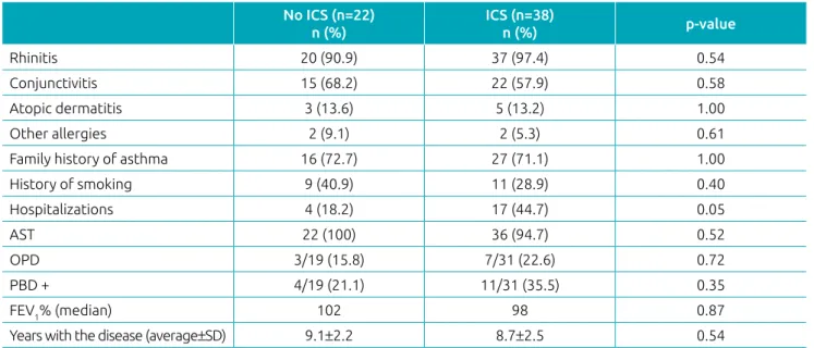
When assessed individually, the asthmatics group using ICS, the asthmatics not using ICS, and non-asthmatics group were similar with regard to personal and family history of atopy. Pulmonary function tests were performed on 50 asthmatics. There were no differences in clinical characteristics among the asthmatics (Table 1).
Almost 98% of the individuals tested had inadequate levels of VD, and there was no difference between groups of asth-matics that were or were not using ICS.
Total and specific IgE levels for PD, BT, and LP were higher in the asthmatic groups using and not using corticosteroids, when compared to the control group (p <0.001). The total and specific IgE median values were verified from the distribution of the patients in the three groups, according to the genotype in the promoter region (Table 2).
There was no significant correlation between serum lev-els of VD and other laboratory tests performed on 60 asth-matics. There was also no significant correlation between serum levels of VD, values of FEV1, and the number of pos-itive allergens obtained from the allergic skin test (47 and 60, respectively, of asthmatics). The Pearson correlation test was applied to variables with normal distribution (calcium, phosphorus, cortisol, parathormone, alkaline phosphatase, specific IgE for PD, BT, LP and total IgE) and did not show any significance.
For an analysis of the VDR gene, three genotypes from the promoter region were evaluated in the 77 individuals. CDX2 polymorphism was present in 71.4% of the population eval-uated (71.6% of asthmatics and 70.5% of non-asthmatics). The data point to a trend (p = 0.06) of an association between genotype of the promoter region and the diagnosis of asthma (Table 3). Also in Table 3, the presence of exon 3 mutations in 7 of the 69 individuals tested was observed. There was no association between the presence of the mutation and the diag-nosis of asthma. No individuals were identified with genetic variations in exon 2.
In order to better evaluate the association between the pres-ence of the CDX2 polymorphism and a diagnosis of asthma, the subjects were compared as follows: homozygous group for the A allele (AA) versus the Gx group [homozygous for the G allele (GG) allele and heterozygotes (GA)]. Subsequently, the
Table 1 Clinical characteristics of the asthmatic group.
No ICS (n=22)
n (%)
ICS (n=38)
n (%) p‑value
Rhinitis 20 (90.9) 37 (97.4) 0.54
Conjunctivitis 15 (68.2) 22 (57.9) 0.58
Atopic dermatitis 3 (13.6) 5 (13.2) 1.00
Other allergies 2 (9.1) 2 (5.3) 0.61
Family history of asthma 16 (72.7) 27 (71.1) 1.00
History of smoking 9 (40.9) 11 (28.9) 0.40
Hospitalizations 4 (18.2) 17 (44.7) 0.05
AST 22 (100) 36 (94.7) 0.52
OPD 3/19 (15.8) 7/31 (22.6) 0.72
PBD + 4/19 (21.1) 11/31 (35.5) 0.35
FEV1% (median) 102 98 0.87
Years with the disease (average±SD) 9.1±2.2 8.7±2.5 0.54
ICS: inhaled corticosteroid use; AST: allergy skin test; OPD: obstructive pulmonary disease; PBD: Post bronchodilator test; FEV1%: forced expiratory volume in the first second; SD: standard deviation.
Table 2 Medium immunoglobulin E (kU/L) values
according to the genotype of the promoter region of the gene.
IgE GG GA AA p‑value*
DP 44.7 33.2 101.0 0.01
BT 16.5 3.2 27.3 0.08
LP 0.3 0.1 0.6 0.06
Total 775.0 498.0 1108.5 0.06
GG group versus the Ax group, which is composed of AA and GA, was compared. From this analysis, 46 asthmatics and 17 controls had the Gx genotype, while 14 asthmatics and no con-trols had the AA genotype. An analysis of the contingency table shows an association between the homozygous for the A allele and the diagnosis of asthma (p = 0.03) (Table 4). The evalua-tion of individuals with Ax versus GG genotypes showed no association with the diagnosis of asthma (p = 1.00). There was also no association between inhaled corticosteroid therapy and the polymorphisms being studied.
Asthmatics were divided into 2 groups (usual genotype and variant genotype) according to the presence of mutations presented for exon 3. The promoter region was divided into three groups, according to the genotype (GG, GA and AA). There was no significant difference between groups for VD, calcium, phosphorus, PTH, cortisol, and number of positive allergic tests. A significant difference (p = 0.004) was observed for the mean percentage of FEV1 predictors among asthmatics when grouped according to promoter region genotypes (GG: 98.2%, GA: 102.5% and AA: 79.5%). When comparing the FEV1% of the Gx (100.8%) versus the AA (79.4%) groups, a significant difference was observed (p = 0.001). This relation was not observed in the Ax (96.0%) versus GG (98.2%) anal-ysis (p = 0.71).
DISCUSSION
The occurrence of hypovitaminosis D has been widely reported, both among healthy individuals and those with allergic diseases.24
A possible explanation for the low serum levels of VD found in the studies may be due to the inadequacy of the reference val-ues used.20 Initially, the reference values were based on health
individuals, who had little sun exposure. This method is not very accurate, given that VD levels are related to sun exposure, diet, latitude of residence, skin color, lifestyle and age.4 Since
VD levels are dependent on multiple variables, there is a need
to establish reference values for each population. Nevertheless, the serum level of VD that is associated with the incidence of diseases was established to be 30 ng / dL.20,21
In the evaluated sample, the promoter polymorphism, 4913G> A, known as CDX2, was frequent in the tested pop-ulation (71.4%) and was positively associated with the diagno-sis of asthma, when it was homozygous. In the heterozygotes, for any mutation, including the CDX2 mutation, the expres-sion of two different proteins can occur. However, the struc-tural changes of the variant protein on phenotype expression (which are sufficient to cause clinical consequences) determine the pattern of dominance.25 In this case, similar to
heterozy-gotes, where 50% of the protein is normally expressed, it can be inferred that, in asthma, the G allele has a dominant pattern, because it is associated with the lower prevalence of the disease.
In addition to being associated with a higher prevalence of asthma, the presence of the A allele also demonstrated a rela-tionship with lower values of FEV1 observed among asthmatics. Although worse pulmonary function values were associated with a greater severity of the disease, the presence of the CDX2 mutation in this group did not result in a greater severity of the disease, as it was similarly distributed among both asthmatics treated with inhaled corticosteroids (persistent asthma) and asthmatics that did not require treatment (intermittent asthma).
Genetic variations of exon 3 were uncommon in the study population and did not influence the diagnosis, treatment or laboratory parameters.
Table 3 Frequency of the polymorphism of the vitamin D receptor genes in accordance with the presence of asthma.
Region Genotype Asthma Control p‑value*
n % n %
Promoter
GG 17/60 28.3 5/17 29.4
0.06
GA 29/60 48.3 12/17 70.6
AA 14/60 23.3 0 0
Exon 2 Usual 51/51 100 14/14 100
Exon 3 Variant 7/52 13.5 0 0 0.18
Usual 45/52 86.5 17/17 100
GG: homozygotes for the G allele; GA: heterozygotes for the G and A alleles; AA: homozygotes for the A allele; *Fisher’s exact test.
Table 4 Number of individuals diagnosed with asthma
according to genotype.
Genotype Asthma (n=60)
n (%)
Controls (n=17)
n (%) p‑value*
AA 14 (23.3) 0 (0)
0.03
Gx 46 (76.6) 17 (100)
Although it is associated with the diagnosis of asthma, the CDX2 polymorphism has been shown to have a protective effect in other studies. It was related to the promoter’s higher transcription rate and, consequently, to a possible increase in the intestinal absorption of calcium.26 It has also been
associ-ated with a lower risk of fractures, osteoporosis and increased bone density.27
Other polymorphisms have been investigated and are, in fact, associated with an increased frequency of asthma and other allergic diseases in population studies. However, the influence of these polymorphisms on how the VDR and other genes function remains unclear.28 It is not yet clear how these
muta-tions relate to asthma and other allergies.
Deficiency or insufficiency of VD was verified in 98% of the evaluated individuals. There was no relationship between VD levels with any of the polymorphisms studied. Similarly, there was no relationship between VD values and the use of ICS. There was no correlation between VD and FEV1 values, which is in disagreement with the literature, which showed an inverse association between VD levels and FEV1% values.29
Lower VD serum levels were related to a higher frequency of allergic diseases or asthma. Although less stringent refer-ence values, which considered values greater than 20 ng / dL, were used as artifice in this group, 30 more than
three-quar-ters of the evaluated subjects did not reach adequate levels of the vitamin.
Low levels of cortisol serum were found in 17 (20%) asth-matic patients. However, this frequency was similar among those treated with ICS. Thus, it is not possible to state that such levels are due to the use of the drug. It is probable that the use of nasal topical corticosteroids to control rhinitis (pres-ent in approximately 90% of this group) is related to this result. Another hypothesis is that corticoids could have been prescribed to treat acute illnesses or crises in other centers and, therefore, was not recorded in the medical record, and may have been omitted, forgotten or even be unknown to the interviewees.
Although the results were not presented, changes in cal-cium, phosphorus, alkaline phosphatase and PTH levels were uncommon and they occurred in a similar fashion among asth-matics, with no statistical significance.
Serum Ige levels are inversely associated with serum VD levels.31 However, there is no evidence of a linear relationship
between serum IgE levels and 25(OH)VD.32 When there are
VD levels, sensitivity to home allergens like dust mites is com-mon in asthmatic children.6 Natural exposure to allergens leads
to increased inflammatory reactions and increased bronchial responsiveness, as well as to elevated eosinophil counts in the
bronchoalveolar lavage, just like hypovitaminosis D.29 Although
VD is correlated to asthma in a manner similar to IgE, there is a lack of experimental studies that demonstrate how this inter-action actually occurs.
This study provided information about the association between CDX2 polymorphism, and the VD receptor gene promoter region, with the diagnosis of asthma, and lower FEV1 values. However, there are some limitations to consider, including the use of a convenience sample and the possible undeclared use of corticosteroids in the group of asthmatics supposedly not using ICS to control the disease. Another lim-itation exists with regard to the comparison group, which was selected to serve only as a control for the mutations. Therefore, it is suggested that an intervention study with asthmatics be performed in order to demonstrate if there is a decrease in the need for corticosteroids to control the disease after the use of VD supplements.
Since hypovitaminosis D has been linked to an increased frequency of asthma and other allergic diseases, the vitamin D receptor may be involved in the mechanism of the disease. Similarly to CDX2, it is possible that other polymorphisms or mutations in the VD receptor gene, which are capable of modifying cellular interaction with the vitamin, are associated with the prevalence of asthma and, perhaps, with the difficulty of controlling the disease.
It was concluded that the presence of CDX2 polymorphism was frequent (71.4%). The unspecified polymorphisms of exons 2 and 3 occurred discretely (7 and 2%, respectively). CDX2 polymorphism was associated with the diagnosis of asthma, as well as with lower FEV1% results in spirometry. There was no correlation between VD levels and the polymorphisms stud-ied, nor was there a correlation between serum calcium levels and these polymorphisms. There was no significant associa-tion between inhaled corticosteroid therapy for the control of asthma and the polymorphisms studied.
ACKNOWLEDGEMENTS
We thank Professor Mônica Nunes Lima for her contribution with the statistical analyzes, and Dr. Marco Antônio Largura, Dr. Álvaro Largura and Alisson Aparecido Pereira Trespach for the laboratory support.
Funding
This study did not receive funding.
Conflict of interests
REFERENCES
1. Sociedade Brasileira de Pneumologia e Tisiologia. Diretrizes da Sociedade Brasileira de Pneumologia e Tisiologia para o Manejo da Asma ‑ 2012. J Bras Pneumol. 2012;38(Supl 1):S1‑46. 2. Camargo CA, Rifas‑Shiman SL, Litonjua AA, Rich‑Edwards JW, Weiss ST, Gold DR, et al. Maternal intake of vitamin D during pregnancy and risk of recurrent wheeze in children at 3 y of age. Am J Clin Nutr. 2007;85:788‑95.
3. Devereux G, Litonjua AA, Turner SW, Craig LC, Mcneill G, Martindale S, et al. Maternal vitamin D intake during pregnancy and childhood wheezing. Am J Clin Nutr. 2007;85:853‑9. 4. Holick MF. Vitamin D Deficiency. N Engl J Med. 2007;357:266‑81. 5. Sandhu MS, Casale TB. The role of vitamin D in asthma. Ann
Allergy Asthma Immunol. 2010;105:191‑9.
6. Wawro N, Heinrich J, Thiering E, Kratzsch J, Schaaf B, Hoffmann B, et al. Serum 25(OH)D concentrations and atopic diseases at age 10: results from the GINIplus and LISAplus birth cohort studies. BMC Pediatr. 2014;14:286. 7. Hollams EM, Hart PH, Holt BJ, Serralha M, Parsons F, Klerk NH,
et al. Vitamin D and atopy and asthma phenotypes in children: a longitudinal cohort study. Eur Respir J. 2011;38:1320‑7. 8. Peroni DG, Piacentini GL, Cametti E, Chinellato I, Boner AL.
Correlation between serum 25‑hydroxyvitamin D levels and severity of atopic dermatitis in children. Br J Dermatol. 2011;164:1078‑82.
9. Camargo CA, Clark S, Kaplan MS, Lieberman P, Wood RA. Regional differences in EpiPen prescriptions in the United States: the potential role of vitamin D. J Allergy Clin Immunol.2007;120:131‑6. 10. Majak P, Olszowiec‑Chlebna M, Smejda K, Stelmach I. Vitamin D supplementation in children may prevent asthma exacerbation triggered by acute respiratory infection. J Allergy Clin Immunol. 2011;127:1294‑6.
11. Giustina AD, Landi M, Bellini F, Bosoni M, Ferrante G, Onorari M, et al. Vitamin D, allergies and asthma: focus on pediatric patients. World Allergy Organ J. 2014;7:27.
12. Castro M, King TS, Kunselman SJ, Cabana MD, Denlinger L, Holguin F, et al. Effect of vitamin D3 on asthma treatment failures in adults with symptomatic asthma and lower vitamin D levels the VIDA randomized clinical trial. JAMA. 2014;311:2083‑91.
13. Ranganathan, P. Genetics of bone loss in rheumatoid arthritis ‑ role of vitamin D receptor polymorphisms. Rheumatology (Oxford). 2009;48:342‑6.
14. Mechica JB, Leite MO, Mendonca BB, Frazzatto ES, Borelli A, Latronico AC. A novel nonsense mutation in the first zinc finger of the vitamin D receptor causing hereditary 1,25‑dihydroxyvitamin D3‑resistant rickets. J Clin Endocrinol Metab. 1997;82:3892‑4.
15. Valdivielso JM, Fernandez E. Vitamin D receptor polymorphisms and diseases. Clin Chim Acta. 2006;371:1‑12.
16. Bossé Y, Hudson TJ. Toward a comprehensive set of asthma susceptibility genes. Annu Rev Med. 2007;58:171‑84. 17. Wittke A, Chang A, Froicu M, Harandi OF, Weaver V,
August A, et al. Vitamin D receptor expression by the lung microenvironment is required for maximal induction of lung inflammation. Arch Biochem Biophys. 2007;460:306‑13.
18. Ferrarezi DA. Variações alélicas no gene do receptor da vitamina D (VDR) e risco de doença arterial coronariana em pacientes diabéticos tipo 2 [PhD thesis]. São Paulo (SP): USP; 2011.
19. Polgar G, Promadhat V. Pulmonary function testing in children. Philadelphia: WB Saunders; 1971.
20. Souberbielle JC, Body JJ, Lappe JM, Plebani M, Shoenfeld Y, Wang TJ, et al. Vitamin D and musculoskeletal health, cardiovascular disease, autoimmunity and cancer: Recommendations for clinical practice. Autoimmun Rev. 2010;9:709‑15.
21. Holick MF, Binkley NC, Bischoff‑Ferrari HA, Gordon CM, Hanley DA, Heaney RP, et al. Evaluation, treatment, and prevention of vitamin D deficiency: an Endocrine Society clinical practice guideline. J Clin Endocrinol Metab. 2011;96:1911‑30.
22. Fang Y, Meurs JB, Bergink AP, Hofman A, Duijn CM, Leeuwen JP, et al. CDX‑2 polymorphism in the promoter region of the human vitamin D receptor gene determines susceptibility to fracture in the elderly. J Bone Miner Res. 2003;18:1632‑41.
23. Sanguinetti CJ, Dias Neto E, Simpson AJ. Rapid silver staining and recovery of PCR products separated on polyacrylamide gels. Biotechniques. 1994;17:914‑21.
24. Searing DA, Zhang Y, Murphy JR, Hauk PJ, Goleva E, Leung DY. Decreased serum vitamin D levels in children with asthma are associated with increased corticosteroid use. J Allergy Clin Immunol. 2010;125:995‑1000.
25. Beiguelman B. Genética de populações humanas. Ribeirão Preto: SBG; 2008.
26. Yamamoto H, Miyamoto K, Li B, Taketani Y, Kitano M, Inoue Y, et al. The caudal‑related homeodomain protein CDX‑2 regulates vitamin D receptor gene expression in the small intestine. J Bone Miner Res. 1999;14:240‑47.
27. Fang Y1, Meurs JB, Bergink AP, Hofman A, Duijn CM, Leeuwen JP, et al. Cdx‑2 Polymorphism in the promoter region of the human vitamin D receptor gene determines susceptibility to fracture in the elderly. J Bone Miner Res. 2003;18:1632‑41.
28. Wjst M, Fischer G, Immervoll T, Jung M, Saar K, Rueschendorf F, et al. A genome‑wide search for linkage to asthma. German Asthma Genetics Group. Genomics. 1999;58:1‑8.
29. Alyasin S, Momen T, Kashef S, Alipour A, Amin R. The relationship between serum 25 hydroxy vitamin D levels and asthma in children. Allergy Asthma Immunol Res. 2011;3:251‑5.
30. Ahmed SF, Franey C, Mcdevitt H, Somerville L, Butler S, Galloway P, et al. Recent trends and clinical features of childhood vitamin D deficiency presenting to a children’s hospital in Glasgow. Arch Dis Child. 2011;96:694‑6.
31. Brehm JM, Celedón JC, Soto‑Quiros ME, Avila L, Hunninghake GM, Forno E, et al. Serum vitamin D levels and markers of severity of childhood asthma in Costa Rica. Am J Respir Crit Care Med. 2009;179:765‑71.
32. Hypponen E, Berr y DJ, Wjst M, Power C . Serum 25‑hydroxyvitamin D and IgE ‑ a significant but nonlinear relationship. Allergy. 2009;64:613‑20.