BASIC RESEARCH
Transforming growth factor-
b
in graft vessels:
histology and immunohistochemistry
Shi-Min Yuan,IYan-Qing Wang,IIYi Shen,IHua JingI
IDepartment of Cardiothoracic Surgery, Jinling Hospital, School of Clinical Medicine, Nanjing University, No. 305 Zhongshan East Road, Nanjing 210002, Jiangsu Province, People’s Republic of China.IIDepartment of Cardiology, The 81 Hospital of Nanjing, No. 34, Biao 34, Yanggongjing, Nanjing 210002, Jiangsu Province, People’s Republic of China.
OBJECTIVES:The biological functions of transforming growth factor-bsignaling that involves Smad proteins have not been previously investigated with respect to coronary artery bypass grafts. The aim of the present study was to observe the immunostaining of proteins that are related to this signaling pathway.
METHODS:Fifteen remnants of coronary artery bypass grafts, including nine saphenous veins, three radial arteries and three mammary arteries, were collected from 12 patients who were undergoing coronary artery bypass. Hematoxylin and eosin, Masson’s trichrome, and immunohistochemical staining of transforming growth factor-b1, type I receptor of transforming growth factor-b, Smad2/3, Smad4, and Smad7 were performed.
RESULTS: The saphenous veins showed more severe intimal degeneration, more severe smooth muscle cell proliferation and more collagen deposition than the arterial grafts, as evidenced by hematoxylin and eosin and Masson’s trichrome stainings. Immunohistochemical assays demonstrated that the majority of the transforming growth factor -b1signaling cytokines were primarily localized in the cytoplasm in the medial layers of all three types of grafts, whereas ectopic transforming growth factor-b1, type I receptor of transforming growth factor-b, and Smad7 overexpressions in the interstices were observed particularly in the saphenous vein and radial arterial grafts.
CONCLUSION: Enhanced transforming growth factor-b1 signal transduction with medial smooth muscle cell proliferation and ectopic transforming growth factor-b1, the presence of the type I receptor of transforming growth factor-b, and Smad7 overexpressions in the extracellular matrix may provide primary evidence for early or late graft failure.
KEYWORDS: Blood Vessels; Coronary Artery Bypass; Immunohistochemistry; Signal Transduction; Transforming Growth Factor-b.
Yuan SM, Wang YQ, Shen Y, and Jing H. Transforming growth factor-bin graft vessels: histology and immunohistochemistry. Clinics. 2011;66(5):895-901.
Received for publication onDecember 17, 2010;First review completed onFebruary 7, 2011;Accepted for publication onFebruary 14, 2011
E-mail: [email protected]
Tel.: 0086 2580860075
INTRODUCTION
Transforming growth factor (TGF)-b1 is implicated in the
development of intimal hyperplesia subsequent to extracellular matrix accumulation,1 which increases the thickness of both arteries and veins.2The overexpresssion of TGF-b1is typically
present in the diseased grafts,3including the saphenous vein and internal mammary arterial grafts, suggesting that TGF-b1may
play a role in the irreversible deposition of extracellular matrix and the further development of intimal hyperplesia.2Moreover, TGF-b1 overexpression has also been observed in the intimal
hyperplasia of stenosed venous fistulas for hemodialysis.4 Graft failure is a common issue following coronary artery bypass grafting5,6that puzzles cardiac surgeons and
requires increasingly effective solutions. Even though TGF-bexpression has drawn attention to the development of vascular remodeling, the biological functions of the
TGF-bsignaling pathway, including the Smad proteins, have not been sufficiently investigated with respect to coronary artery bypass grafts. We have hypothesized that the TGF-b
signaling pathway may be enhanced so as to drive the fibrotic process that is responsible for the failure of coronary artery bypass grafts. The aim of the present study was to observe the immunostaining of the proteins that are related to this signaling pathway.
MATERIALS AND METHODS
From October 2009 to January 2010, 15 remnants of coronary artery bypass grafts, including nine saphenous veins, three radial arteries and three mammary arteries, were collected from 12 patients who were undergoing coronary artery bypass after their surgeries. Ten males and two females were included in the study, and their ages
ranged from 50 to 83 with a mean of 66.25¡10.37 years. The major symptoms were chest/precordial pain in six patients (50%), chest pain and palpitations in two patients (16.67%), chest distress in one patient (8.33%), chest distress and dyspnea in one patient (8.33%), and chest distress and palpitations in two patients (16.67%). The time since the onset of symptoms ranged from 1 day to 20 years (mean 5.41
¡6.59 years, median 2 years). Hypertension was present in eight patients (66.67%), and type II diabetes was present in three patients (25%). Four patients had a myocardial infarction, two of which were non-ST-segment elevation myocardial infarctions (NSTEMI), and one patient had a left ventricular pseudoaneurysmal formation. Conventional cor-onary artery bypass was performed in four patients (33.33%), off-pump coronary artery bypass in six patients (50%), beating heart coronary revascularization in one patient (8.33%), and off-pump coronary artery bypass with subse-quent coronary artery bypass in one patient (8.33%). A total of 41 grafts were bypassed with a mean of 3.42¡0.51 grafts
per patient. Thirteen (31.71%) left internal mammary arteries were grafted, as were one (2.44%) right internal mammary artery, two (4.88%) radial arteries, and 25 (60.98%) saphenous veins. The associated procedures included left ventricular pseudoaneurysmectomy, mitral valve replacement, and intra-aortic balloon pump insertion in one patient each.
Fresh specimens of the graft remnants were collected and cut into 1-cm3 blocks/rings and immersed in a 10%
methanol solution in appropriately sized bottles for patho-logical inspection.
Hematoxylin and eosin (H&E) staining was performed on the 4-mm sections, and collagen fibers were stained
using Masson’s trichrome protocol. Immunohistochemical staining was performed on the 4-mm paraffin-embedded sections to detect TGF-b1, transforming growth factor-b
receptor I (TbRI), Smad2/3, Smad4, and Smad7 using the Envision method. The following primary antibodies were utilized: TGF-b1(Y369) (15150) (Bioworld Technology, Inc.,
Louis Park, MN, USA), TbRI (E161) (15100) (Bioworld
Technology, Inc., Louis Park, MN, USA), Smad2/3 (S2) (15100) (Beijing Biosynthesis Biotechnology Co., Ltd., Beijing,
China), Smad4 (L43) (15200) (Bioworld Technology, Inc.,
Louis Park, MN, USA), and Smad7 (Z8-B): sc-101152 (15100)
(Santa Cruz Biotechnology, Inc., Santa Cruz, CA, USA). The immunostaining density was characterized as fol-lows: - (background) was defined as 0,+(weak yellow) as 1, ++ (yellow) as 2, and +++ (brown) as 3 in the
semi-quantitative analyses. Photographs were acquired at6100–
400.
This study was approved by the institutional ethical committee and was conducted following the guidelines of the Declaration of Helsinki. Informed consent for the graft vessel remnants that were used for other than therapeutic purposes was obtained from each patient in compliance with the guidelines in ‘‘Surgically Removed Human Tissue For Research’’ that were proposed by Thasler et al.7
RESULTS
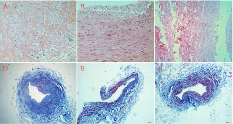
According to H&E staining, the saphenous veins exhibited proliferative medial smooth muscle cells with focal or diffuse disruption and severe intimal degenera-tion (Fig. 1A). The radial arteries exhibited smooth muscle
Figure 1 -Hematoxylin and eosin staining showing (A) proliferative medial smooth muscle cells with inflammatory cell infiltration in the saphenous veins, (B) focal intimal degeneration with slight disrupted media in the radial arteries, and (C) roughly normal vascular wall structures of the internal mammary arteries; H&E6200. Masson’s trichrome staining showing that (D) the saphenous veins showed
more collagen deposition but less muscular fibers, (E) the radial arteries showed less collagen deposition and more muscular tissues, and (F) the mammary arteries showed the least collagen accumulation in the vascular wall but the most muscular tissue; Masson6100.
Transforming growth factor-bin graft vessels
Yuan S-M et al. CLINICS 2011;66(5):895-901
cell proliferation, elastic fibrous degeneration and col-lagenization (Fig. 1B). Evenly distributed smooth muscle cells of the media with mild degenerative changes in the intima could be observed in the internal mammary arteries (Fig. 1C).
Based on Masson’s trichrome staining, the saphenous veins showed more collagen deposition but less muscular fibers (Fig. 1D), the radial arteries had less collagen deposition with more muscular tissues (Fig. 1E), and the
mammary arteries had the least collagen accumulation in the vascular wall but the most muscular tissues (Fig. 1F).
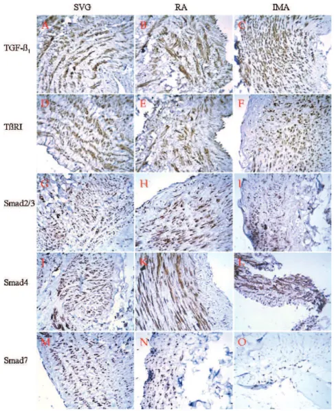
Immunostaining demonstrated that the five tested pro-teins were positive in the cytoplasm of the medial layers of all three types of grafts. The investigated signaling cytokines were the most intense in the saphenous vein, followed by the radial arterial grafts and then the internal mammary arterial grafts.
Figure 2 -Immunostaining showed that the five tested proteins (TGF-b1, TbRI, Smad2/3, Smad4, and Smad7) were positive, primarily in the
cytoplasm of the medial layers, in all three kinds of grafts. (A) (B) In the saphenous vein and radial arterial grafts, TGF-b1expression was
moderately positive (++) in the cytoplasm and interstices of the intima, intensely positive (+++) in the cytoplasm and interstices of the media, and weakly positive (+) in the cytoplasm and interstices of the adventitia. (C) In the intima (++), media (+++) and adventitia (+) of the internal mammary arterial grafts, TGF-b1staining was only observed in the cytoplasm and not in the interstices. In the saphenous vein
grafts, TbRI was moderately positive in the cytoplasm and interstices (+-++) of the intima, intensely positive (+++) primarily in the cytoplasm but also in the nuclei and interstices of the media, and weakly positive (+) or negative (-) in the cytoplasm or interstices, respectively, of the adventitia (D). This receptor was positive in the cytoplasm and interstices of the intima (+), media (++) and adventitia (+) of the radial arterial grafts (E), and positive in the cytoplasm of the intima (+), media (++) and adventitia (+) of the internal mammary arterial grafts (F). Smad2/3 positivity was more intense in the saphenous vein grafts (G) than in the internal mammary arterial grafts (I), whereas the radial artery (H) showed the least intense Smad2/3 staining. In the saphenous vein grafts, Smad4 was weakly positive (+) in the cytoplasm of the intima, intensely positive (+++) in the cytoplasm of the media, and weakly positive (--+) in the cytoplasm of the adventitia (J). In the radial arteries, it was weakly positive in the cytoplasm (+) and negative (-) in the intima, moderately positive (++) in the cytoplasm and interstices of the media, and weakly positive in the cytoplasm and interstices of the adventitia (K). In the internal mammary arteries, it was positive in the cytoplasm of the intima (+), media (++), and adventitia (+) (L). Smad7 was expressed most intensely in the saphenous veins (M), more intense in the radial arteries (N), and it was weaker in scattered nuclei and interstices in the internal mammary arterial grafts (O). IMA = internal mammary artery; RA = radial artery; SVG = saphenous vein graft; TGF-b1: transforming
TGF-b1 expression was moderately positive (++) in the
cytoplasm and interstices of the intima, intensely positive (+++) in the cytoplasm and interstices of the media, and
weakly positive (+) in the cytoplasm and interstices of the adventitia of the saphenous vein and radial arterial grafts. In the intima (++), media (+++), and adventitia (+) of the
internal mammary arterial grafts, TGF-b1staining was seen only in the cytoplasm and not in the interstices. TGF-b1
staining was the most intense in the internal mammary artery, less intense in the saphenous vein, and the least intense in the radial arterial grafts (Fig. 2A–2C).
TbRI was moderately positive in the cytoplasm and interstices (+-++) of the intima of the saphenous vein grafts,
intensely positive (+++) primarily in the cytoplasm but also
in the nuclei and interstices of the media, and weakly positive (+) or negative (-) in the cytoplasm and interstices of the adventitia. This receptor was positive in the cytoplasm and interstices of the intima (+), media (++) and adventitia
(+) of the radial arterial grafts. It was also positive in the
cytoplasm of the intima (+), media (++) and adventitia (+) of the internal mammary arterial grafts (Fig. 2D–2F).
Smad2/3 was virtually only present in the cytoplasm. Smad2/3 positivity was more intense in the saphenous vein grafts than in the internal mammary arterial grafts, whereas the radial arteries showed the least intense Smad2/3 staining (Fig. 2G–2I).
In the saphenous vein grafts, Smad4 was weakly positive (+) in the cytoplasm of the intima, intensely positive (+++) in
the cytoplasm of the media, and weakly positive (--+) in the
cytoplasm of the adventitia, and the positivity rate was 85.71% (6/7). For the radial arteries, Smad4 was weakly positive in the cytoplasm (+) and negative (-) in the intima,
moderately positive (++) in the cytoplasm and interstices of
the media, and weakly positive in the cytoplasm and interstices of the adventitia. In the internal mammary arteries, it was positive in the cytoplasm of the intima (+), media (++), and adventitia (+) (Fig. 2J–2L).
Smad7 was expressed in the cytoplasm and nucleus and was also present in the interstices in one of the radial arteries. Smad7 was the most intense in the saphenous vein, more intense in the radial artery, and weaker in scattered nuclei and interstices of the internal mammary arterial grafts. The positive rates were 62.5% (5/8), 75% (6/8) and 62.5% (5/8), respectively for the intima, media, and adventitia of the saphenous vein grafts. In the radial artery, it was negative in 1/3, 1/3 and 2/2 of the intima, media, and adventitia, respectively (Fig. 2M–2O) (Table 1).
DISCUSSION
Masson’s trichrome staining is a popular technique for observing collagen deposition between elastin layers, in which the reactivity and integrity of the vascular wall,8
vascular regeneration, and graft patency9 can all be
assessed. This technique stains the extracellular matrix blue and the cellular portion red.10 Using Masson’s trichrome staining, the saphenous veins that were endos-copically harvested with a no-touch technique showed well-preserved collagen fibers, whereas those harvested using conventional techniques showed more subendothe-lial collagen degradation.11 In comparison, the architec-tures of the radial arterial grafts were preserved with both endoscopic and conventional techniques,8indicating that
the wall structures of the vein grafts were more prone to
being damaged by surgical maneuvers. In addition, the observed higher durability of the left internal mammary arterial grafts may be due to their appropriate elastic tension and internal diameter12, as well as to the relatively limited atherosclerotic changes.13
TGF-b1 stimulates arteriogenesis, thereby contributing to
the occurrence of restenosis after neointimal damage caused by angioplasty or stenting. TGF-b1is upregulated rapidly in
the restenotic and injured vessels following balloon catheter injury along with associated increases in TGF-b3, activin
receptor-like kinase 5 (ALK-5), and transforming growth factor-breceptor II (TbRII) immunoreactive peptide levels.14
Smooth muscle cells and macrophages in the atherosclerotic lesions may be predisposed to the upregulation of TbRII and ALK5.15TGF-bantagonists may inhibit fibroblast differentia-tion and intimal injury following angioplasty,16and it may prevent adventitial fibrosis.17 Both TGF-b
1 and TGF-b2
upregulate type VII collagen gene expression.18 They may increase the expression of protease inhibitors, including inhibitors of matrix metalloproteinases and of tissue plasmi-nogen activator-1, and they may crosstalk with proteins of the Smad signaling pathway.19Plasminogen activator inhibitor-1
(PAI-1),20matrix metalloproteinases,21and vascular endothe-lial growth factor22 have been shown to be modulated by TGF-b1and are thus involved in the signal transduction.
In TGF-bsignal transduction, Smad2/3 are considered to be the major mediators of TGF-b-induced fibrotic pathogen-esis.23 Smad4 is implicated in the pathology of human
vascular disorders, with essential roles in vascular remodel-ing, maturation, and integrity. Smad4 deficiency may cause failures of remodeling and efficient sprouting in vivo.24 Smad7 is an inhibitor of TGF-b signaling, and it is usually expressed in human vascular endothelial cells that have been injured by shear stress.25 The ectopic expression of Smad7 inhibits TGF-bresponses in vascular smooth muscle cells, and the biological function of Smad7 can be reversed by Smad2.26 Conversely, Smad7 overexpression reduces
Smad2 phosphorylation in response to TGF-b1 via TbRI.27
Smad7 may induce the ubiquitination, degradation, and endocytosis of TbRI and, hence, play an important role in the crosstalk between different signaling pathways. Moreover, an alternative biological function of Smad7 is to mediate TGF-b-induced apoptosis.28In addition, it has been reported that a marked Smad7 deficiency may be respon-sible for TGF-b hyperresponsiveness.29The overexpression of Smad7 had been shown to counteract TGF-b, activin A, and bone morphogenetic protein-induced growth arrest and apoptosis in tumor B cell lines,30and the overexpression of Smad7 in the adventitia of the carotid arteries significantly attenuateda-smooth muscle actin expression in the adven-titia, media, and neointima, or, in other words, in areas of reduced lumen, after balloon injury.31
Increased flow and shear stress can mediate the release of TGF-b1 in rabbit arteries.32 Stress in the endothelial and
smooth muscle cells may alter the synthesis and secretion of collagen, elastin, and connective tissue proteases.33 Flow supply to the conduits could be a determinant of graft patency as evidenced by a series of observations;,50% or
,70% coronary stenosis may be associated with increased internal mammary or radial arterial graft occlusion, respec-tively, during follow-up.34,35TGF-b1may promote monocyte
adhesion to the endothelial cells and migration across the endothelium, probably due to interaction with CD44, which may link more to TbRI than to TbRII, thereby increasing
Transforming growth factor-bin graft vessels
Yuan S-M et al. CLINICS 2011;66(5):895-901
Cytokine SVG RA IMA
Intima Media adventitia Intima Media adventitia Intima Media adventitia
TGF-b1 Cytoplasm +-++ (n = 8)/ interstices ++(n = 1)
Cytoplasm +++(n = 8)/ interstices ++(n = 1)
Cytoplasm+ (n = 7)/ interstices ++(n = 1)/ -(n = 1)
Cytoplasm +-++(n = 3)/ interstices+(n = 1)
Cytoplasm+ ++(n = 2)/ interstices +(n = 1)
Cytoplasm ++(n = 2)/ interstices +(n = 1)
Cytoplasm +-++ (n = 3)
Cytoplasm +++(n = 3)
Cytoplasm - -+(n = 3)
TbRI Cytoplasm +(n = 8)/ interstices ++(n = 1)
Cytoplasm ++(n = 5)/ cytoplasm& interstices+ (n = 1)/ interstices ++(n = 1)/ nucleus
++(n = 2)/cytoplasm &nucleus +++(n = 1)
-(n = 1)/ interstices +(n = 3)/ cytoplasm +(n = 5)
Cytoplasm +(n = 2)/ interstices +(n = 1)
Cytoplasm ++(n = 2)/ interstices +(n = 1)
Cytoplasm +(n = 2)/ interstices+ (n = 1)
Cytoplasm +(n = 3)
Cytoplasm ++(n = 3)
Cytoplasm +(n = 3)
Smad2/3 Cytoplasm +-++(n = 7)
Cytoplasm + +-+++(n = 7)
Cytoplasm +(n = 6)/ interstices +(n = 1)
Cytoplasm +(n = 3)
Cytoplasm +-++(n = 3)
Cytoplasm +(n = 1)
Cytoplasm +(n = 3)
Cytoplasm ++(n = 3)
Cytoplasm +(n = 3)
Smad4 -(n = 1)/ cytoplasm +(n = 7)
-(n = 1)/ Cytoplasm +++(n = 7)
-(n = 1)/ Cytoplasm - -+(n = 7)
Cytoplasm +(n = 2)/ -(n = 1)
Cytoplasm +-++(n = 2)/ cytoplasm& interstices +-++(n = 1)
Interstices+ (n = 2)/ cytoplasm - -+(n = 1)
Interstices +(n = 1)/ cytoplasm +(n = 1)
Interstices +(n = 1)/ cytoplasm ++(n = 1)
Interstices +(n = 1)/ cytoplasm +(n = 1)
Smad7 -(n = 3)/ nucleus +-++ (n = 1)/ - -+(n = 4)
-(n = 2)/nucleus +-++(n = 1)/ cytoplasm + +-+++(n = 5)
-(n = 3)/interstices +(n = 1)/nucleus +-++(n = 1)/ cytoplasm ++(n = 4)
-(n = 1)/ cytoplasm& nucleus +(n = 1)
-(n = 1)/ interstices
= (n = 1)/ cytoplasm& interstices ++(n = 1)
-(n = 2) Cytoplasm +(n = 2)
Cytoplasm +(n = 2)/ nucleus - -+(n = 1)
Cytoplasm +(n = 2)/ nucleus - -+(n = 1)
IMA: internal mammary artery; RA: radial artery; SVG: saphenous vein graft; TGF-b1: transforming growth factor-b1; TbRI: transforming growth factor-breceptor I.
Smad2/3 phosphorylation.36TGF gene expression was found
to be increased in arterialized vein grafts from the coronary artery bypasses.37,38 Therefore, the ectopic implantation of either venous or arterial grafts into the coronary circulation may place these vessels in a state of increased stress, which may upregulate TGF-bsignaling cytokines.
We found that the internal mammary arteries showed a weak Smad7 expression. Therefore, the dual regulatory effects of TGF-b on the activation and phosphorylation of the Smad proteins may lead to the normal transcription of target genes. The most prominent difference in the signaling pathways between the three grafts may lie in the ectopic TGF-b1, TbRI, and Smad7 overexpression in the interstices was observed particularly in the saphenous veins and radial arteries relative to the internal mammary arteries. Therefore, the increased TGF-b signaling activity in the extracellular matrix of the saphenous vein and radial arterial grafts may lead to considerable proliferation of the intima and muscular layers of these the grafts.
CONCLUSION
In conclusion, severe vascular wall degeneration and collagen deposition together with overexpressed TGF-b
signaling cytokines may provide preliminary evidence for the failure (early or late) of the saphenous vein and radial arterial grafts. Weak Smad7 expression in the internal mammary arterial grafts with well-preserved structures may imply less matrix deposition, which may explain their superior durability. More comprehensive studies of the grafts are required to obtain more accurate information for the prevention of graft disorders.
ACKNOWLEDGEMENT
We are indebted to Miss Xiao-Hui Cao, Technician, and Gui-Mei Li, MD, Department of Pathology, The 81 Hospital of Nanjing, No. 34, Biao 34, Yanggongjing, Nanjing 210002, Jiangsu Province, People’s Republic of China, for their technical support.
REFERENCES
1. Kanzaki T, Tamura K, Takahashi K, Saito Y, Akikusa B, Oohashi H, et al. In vivo effect of TGF-b1. Enhanced intimal thickening by administration
of TGF-b1in rabbit arteries injured with a balloon catheter. Arterioscler
Thromb Vasc Biol. 1995;15:1951-7.
2. Friedl R, Li J, Schumacher B, Hanke H, Waltenberger J, Hannekum A, et al. Intimal hyperplasia and expression of transforming growth factor-b1in saphenous veins and internal mammary arteries before coronary
artery surgery. Ann Thorac Surg. 2004;78:1312-8, doi: 10.1016/j.athor-acsur.2004.02.066.
3. Sterpetti AV, Cucina A, Randone B, Palumbo R, Stipa F, Proietti P, et al. Growth factor production by arterial and vein grafts: relevance to coronary artery bypass grafting. Surgery. 1996;120:460-7, doi: 10.1016/ S0039-6060(96)80064-X.
4. Stracke S, Konner K, Ko¨stlin I, Friedl R, Jehle PM, Hombach V, et al. Increased expression of TGF-b1 and IGF-I in inflammatory stenotic
lesions of hemodialysis fistulas. Kidney Int. 2002;61:1011-9, doi: 10.1046/ j.1523-1755.2002.00191.x.
5. Widimsky P, Straka Z, Stros P, Jirasek K, Dvorak J, Votava J, et al. One-year coronary bypass graft patency: a randomized comparison between off-pump and on-pump surgery angiographic results of the PRAGUE-4 trial. Circulation. 2004;110:3418-23, doi: 10.1161/01.CIR.0000148139. 79580.36.
6. Khot UN, Friedman DT, Pettersson G, Smedira NG, Li J, Ellis SG. Radial artery bypass grafts have an increased occurrence of angiographically severe stenosis and occlusion compared with left internal mammary arteries and saphenous vein grafts. Circulation. 2004;109:2086-91, doi: 10. 1161/01.CIR.0000127570.20508.5C.
7. Thasler WE, Weiss TS, Schillhorn K, Stoll PT, Irrgang B, Jauch KW. Charitable state-controlled foundation Human Tissue and Cell Research: Ethic and legal aspects in the supply of surgically removed human tissue
for research in the academic and commercial sector in germany. Cell Tissue Bank. 2003;4:49-56, doi: 10.1023/A:1026392429112.
8. Medalion B, Tobar A, Yosibash Z, Stamler A, Sharoni E, Snir E, et al. Vasoreactivity and histology of the radial artery: comparison of open versus endoscopic approaches. Eur J Cardiothorac Surg. 2008;34:845-9, doi: 10.1016/j.ejcts.2008.06.015.
9. Cho SW, Lim SH, Kim IK, Hong YS, Kim SS, Yoo KJ, et al. Small-diameter blood vessels engineered with bone marrow-derived cells. Ann Surg. 2005;241:506-15, doi: 10.1097/01.sla.0000154268.12239.ed. 10. Sakaguchi T, Asai T, Belov D, Okada M, Pinsky DJ, Schmidt AM, et al.
Influence of ischemic injury on vein graft remodeling: role of cyclic adenosine monophosphate second messenger pathway in enhanced vein graft preservation. J Thorac Cardiovasc Surg. 2005;129:129-37, doi: 10. 1016/j.jtcvs.2004.04.014.
11. Silva VF, Ishigai MM, Freymu¨ller E, Branco JN, Gaia DF, Gabriel EA, et al. Microscopic and ultrastructural evaluation of the saphenous vein endothelium for CABG prepared by the no touch technique. Rev Bras Cir Cardiovasc. 2008;23:323-9, doi: 10.1590/S0102-76382008000300007. 12. Yamabuki K. Thickness of the muscle layer of the gastroepiploic artery
and the internal mammary artery--a presumable factor of flow instability in GEA during the perioperative period. Nippon Kyobu Geka Gakkai Zasshi. 1997;45:1725-32.
13. Mestres CA, Rives A, Igual A, Vehi C, Murtra M. Atherosclerosis of the internal mammary artery. Histopathological analysis and implications on its results in coronary artery bypass graft surgery. Thorac Cardiovasc Surg. 1986;34:356-8.
14. Ward MR, Agrotis A, Kanellakis P, Dilley R, Jennings G, Bobik A. Inhibition of protein tyrosine kinases attenuates increases in expression of transforming growth factor-bisoforms and their receptors following arterial injury. Arterioscler Thromb Vasc Biol. 1997;17:2461-70. 15. Bobik A, Agrotis A, Kanellakis P, Dilley R, Krushinsky A, Smirnov V,
et al. Distinct patterns of transforming growth factor-bisoform and receptor expression in human the atherosclerotic lesions. Colocalization implicates TGF-b in fibrofatty lesion development. Circulation. 1999;99:2883-91.
16. Khan R, Agrotis A, Bobik A. Understanding the role of transforming growth factor-b1in intimal thickening after vascular injury. Cardiovasc
Res. 2007;74:223-34, doi: 10.1016/j.cardiores.2007.02.012.
17. Schwarte-Waldhoff I, Schmiegel W. Smad4 transcriptional pathways and angiogenesis. Int J Gastrointest Cancer. 2002;31:47-59, doi: 10.1385/ IJGC:31:1-3:47.
18. Ryyna¨nen J, Sollberg S, Olsen DR, Uitto J. Transforming growth factor-beta up-regulates type VII collagen gene expression in normal and transformed epidermal keratinocytes in culture. Biochem Biophys Res Commun. 1991;180:673-80, doi: 10.1016/S0006-291X(05)81118-0. 19. Ryan ST, Koteliansky VE, Gotwals PJ, Lindner V. Transforming growth
factor-beta-dependent events in vascular remodeling following arterial injury. J Vasc Res. 2003;40:37-46, doi: 10.1159/000068937.
20. Ikedo H, Tamaki K, Ueda S, Kato S, Fujii M, Ten Dijke P, et al. Smad protein and TGF-bsignaling in vascular smooth muscle cells. Int J Mol Med. 2003;11:645-50.
21. Simionescu A, Philips K, Vyavahare N. Elastin-derived peptides and TGF-b1induce osteogenic responses in smooth muscle cells. Biochem
Biophys Res Commun. 2005;334:524-32, doi: 10.1016/j.bbrc.2005.06.119. 22. Padua D, Massague´ J. Roles of TGFbin metastasis. Cell Res.
2009;19:89-102, doi: 10.1038/cr.2008.316.
23. Jiang Z, Tao M, Omalley KA, Wang D, Ozaki CK, Berceli SA. Established neointimal hyperplasia in vein grafts expands via TGF-b-mediated progressive fibrosis. Am J Physiol Heart Circ Physiol. 2009;297:H1200-7, doi: 10.1152/ajpheart.00268.2009.
24. Lan Y, Liu B, Yao H, Li F, Weng T, Yang G, et al. Essential role of endothelial Smad4 in vascular remodeling and integrity. Mol Cell Biol. 2007;27:7683-92, doi: 10.1128/MCB.00577-07.
25. Topper JN, Cai J, Qiu Y, Anderson KR, Xu YY, Deeds JD, et al. Vascular MADs: two novel MAD-related genes selectively inducible by flow in human vascular endothelium. Proc Natl Acad Sci USA. 1997;94:9314–9. 26. Kato S, Ueda S, Tamaki K, Fujii M, Miyazono K, ten Dijke P, et al. Ectopic
expression of Smad7 inhibits transforming growth factor-bresponses in vascular smooth muscle cells. Life Sci. 2001;69:2641-52, doi: 10.1016/ S0024-3205(01)01350-9.
27. Hayashi H, Abdollah S, Qiu Y, Cai J, Xu YY, Grinnell BW, et al. The MAD-related protein Smad7 associates with the TGFbreceptor and functions as an antagonist of TGFbsignaling. Cell. 1997;89:1165-73, doi: 10.1016/S0092-8674(00)80303-7.
28. Yan X, Chen YG. Smad7: not only a regulator, but also a cross-talk mediator of TGF-bsignalling. Biochem J. 2011;434:1-10, doi: 10.1042/ BJ20101827.
29. Dong C, Zhu S, Wang T, Yoon W, Li Z, Alvarez RJ, et al. Deficient Smad7 expression: a putative molecular defect in scleroderma. Proc Natl Acad Sci U S A. 2002;99:3908-13, doi: 10.1073/pnas.062010399.
30. Li R, Rosendahl A, Brodin G, Cheng AM, Ahgren A, Sundquist C, et al. Deletion of exon I of SMAD7 in mice results in altered B cell responses. J Immunol. 2006;176:6777-84.
Transforming growth factor-bin graft vessels
Yuan S-M et al. CLINICS 2011;66(5):895-901
31. Siow RC, Mallawaarachchi CM, Weissberg PL. Migration of adventitial myofibroblasts following vascular balloon injury: insights from in vivo gene transfer to rat carotid arteries. Cardiovasc Res. 2003;59:212-21, doi: 10.1016/S0008-6363(03)00292-X.
32. Song RH, Kocharyan HK, Fortunato JE, Glagov S, Bassiouny HS. Increased flow and shear stress enhance in vivo transforming growth factor-b1 after experimental arterial injury. Arterioscler Thromb Vasc Biol. 2000;20:923-30.
33. Ohno M, Cooke JP, Dzau VJ, Gibbons GH. Fluid shear stress induces endothelial transforming growth factor beta-1 transcription and production. Modulation by potassium channel blockade. J Clin Invest. 1995;95:1363-9. 34. Seki T, Kitamura S, Kawachi K, Morita R, Kawata T, Mizuguchi K, et al.
A quantitative study of postoperative luminal narrowing of the internal thoracic artery graft in coronary artery bypass surgery. J Thorac Cardiovasc Surg. 1992;104:1532-8.
35. Maniar HS, Sundt TM, Barner HB, Prasad SM, Peterson L, Absi T, et al. Effect of target stenosis and location on radial artery graft patency. J Thorac Cardiovasc Surg. 2002;123:45-52, doi: 10.1067/mtc.2002. 118686.
36. Agrotis A, Kalinina N, Bobik A. Transforming growth factor-b, cell signaling and cardiovascular disorders. Curr Vasc Pharmacol. 2005;3:55-61, doi: 10.2174/1570161052773951.
37. Dattilo JB, Lust RM, Dattilo MP, Parker FM, Meadows WMJr, Sun YS, et al. The temporal expression of transforming growth factor-b1in early
aortocoronary vein grafts. J Surg Res. 1997;69:349-53, doi: 10.1006/jsre. 1997.5076.