Original Article
Artigo Original
Otoacoustic emissions in newborns with
mild and moderate perinatal hypoxia
Emissões otoacústicas em recém-nascidos
com hipóxia perinatal leve e moderada
Juliana Neves Leite1Vinicius Souza Silva1
Byanka Cagnacci Buzo1
Keywords
Newborn Hypoxia Hearing Auditory Hair Cells Hearing Loss
Descritores
Recém-nascido Hipóxia Audição Células Ciliadas Auditivas Perda Auditiva
Correspondence address:
Byanka Cagnacci Buzo Rua Dr. Cesário Mota Junior, 61, 10º andar, Vila Buarque, São Paulo (SP), Brazil, CEP: 01221-020. E-mail: byankacb@gmail.com
Received: April 01, 2015
Study carried out at the School of Medical Sciences of Santa Casa de São Paulo - São Paulo (SP), Brazil.
1Santa Casa de São Paulo, Faculdade de Ciências Médicas - São Paulo (SP), Brazil.
Financial support: Fundação de Amparo à Pesquisa do Estado de São Paulo – FAPESP – no. 2013/14739-8. Conlict of interests: nothing to declare.
ABSTRACT
Introduction: Severe neonatal hypoxia (as evidenced by the Apgar value) is currently considered the only risk for hearing loss. Hypoxia is one of the most common causes of injury and cell death. The deprivation of oxygen in mild or moderate cases of hypoxia, although smaller, occurs and could cause damage to the auditory system. Objective: To investigate the amplitude of otoacoustic emissions in neonates at term with mild to moderate hypoxia and no risk for hearing loss. Methods: We evaluated 37 newborns, divided into two groups: a control group of 25 newborns without hypoxia and a study group of 12 newborns with mild to moderate hypoxia. TEOAE and DPOAE were investigated in both groups. Results: The differences between groups
were statistically signiicant in the amplitude of DPOAE at the frequencies of 1000, 2800, 4000 and 6000 Hz. In TEOAE, statistically signiicant differences were found in all tested frequency bands. OAE of the study
group were lower than those in the control group. Conclusion: Although the occurrence of mild and moderate neonatal hypoxia is not considered a risk factor for hearing loss, deprivation of minimum oxygen during neonatal hypoxia seems to interfere in the functioning of the outer hair cells and, consequently, alter the response level of otoacoustic emissions. Thus, hese children need longitudinal follow-up in order to identify the possible impact of these results on language acquisition and future academic performance.
RESUMO
Introdução: Atualmente, somente a hipóxia neonatal grave (evidenciada pelo valor do Apgar) é considerada
risco para a deiciência auditiva. A hipóxia é uma das causas mais comuns de lesão e morte celular. Nos casos de
hipóxia leve ou moderada, embora menor, a privação da oxigenação está presente e, dessa forma, algum dano ao sistema auditivo pode ocorrer. Objetivo: Investigar as amplitudes das emissões otoacústicas em recém-nascidos
a termo sem risco para deiciência auditiva que apresentaram hipóxia leve ou moderada. Método: Foram selecionados 37 recém-nascidos de ambos os sexos, divididos em dois grupos: 25 do grupo controle, formado por recém-nascidos sem hipóxia, e 12 do grupo estudo, formado por recém-nascidos com hipóxia leve ou moderada.
Resultados: Foram pesquisadas as EOAT e EOAPD em ambos os grupos e comparados os seus resultados. Nas EOAPD foram encontradas diferenças estatísticas entre as amplitudes nas frequências 1.000, 2.800, 4.000 e 6.000 Hz. Nas EOAT foram encontradas diferenças estatísticas nas bandas de frequência de 1.000, 1.400, 2.000, 2.800 e 4.000 Hz, sendo as EOA do grupo estudo menores que as do grupo controle. Conclusão: Embora a ocorrência de hipóxia neonatal leve e moderada não seja considerada risco para perda auditiva, a mínima privação do oxigênio durante o momento de hipóxia neonatal parece interferir no funcionamento das células ciliadas externas e, consequentemente, no nível de respostas das emissões otoacústicas. Dessa forma, faz-se necessário
o acompanhamento longitudinal desses lactentes, a im de identiicar o possível impacto desses resultados na
INTRODUCTION
Some newborns are at risk of hearing loss. According to the criteria of the Joint Committee on Infant Hearing(1) and of Comusa(2), among other risks described, only severe neonatal hypoxia, as evidenced by the Apgar scale value(3), is considered a risk for hearing loss. However, some newborn infants may present mild or moderate neonatal hypoxia, indicating a need for special care, but without great risk to life. In such cases, damage to the auditory system can already be observed(4).
Perinatal hypoxia can be deined in various ways. One concerns
the metabolism and nutrition between mother and fetus, in which asphyxia leads to changes in the homeostasis of the fetus. The decrease in this relationship between metabolism and nutrition between the mother and the newborn can have two
causes. The irst refers to a reduction in the amount of oxygen
and the second to a reduction in the amount of blood circulating among the different organs and tissues(5).
The severity of hypoxia varies, and may cause the child’s death. The recovery will depend on the fetus’s ability to adapt. Hypoxia is one of the most common causes that lead to injury and cell death. Each cell resists oxygen deprivation for a certain time. If exposed to longer periods of deprivation, change processes will begin, leading to irreversible damage to the cell’s structure and cell death(6).
Previous studies have shown that hypoxia plays a signiicant
role in the death of hair cells and consequent hearing loss(7-10). The outer hair cells are the main recipients of acoustic signals. However, they are extremely sensitive to lack of oxygen caused by hypoxia. The death of these cells causes irreversible hearing loss, that is, sensorineural hearing loss(11). Recent studies
in guinea pigs showed a signiicant reduction in the cochlear blood low during periods of hypoxia(12,13).
Yoshikawa et al.(14) indicate that one of the hypotheses for the reversal of initial damage is due to the fact that the cochlea, due to being located very close to the brain and, consequently, to the central nervous system, is privileged in the redistribution of blood and oxygen to more vital organs. However, if systemic hypoxia continues, cochlear damage, as well as damage to the central nervous system, can occur(15).
Hearing loss due to hypoxia has been shown to be temporary in most cases. However, there are few studies in the area that demonstrate how this mechanism recovers. Studies show that there is a temporary change in the hearing threshold, but more research is needed to prove the dynamic changes to the threshold. Hearing variation and recovery time also vary according to the measured frequency range, suggesting that some cochlear regions are more sensitive to hypoxia(16).
Thus, although the occurrence of mild and moderate neonatal hypoxia is not considered a risk to hearing loss, it is known that oxygen deprivation may result in damage to the hair cells of the cochlea. Therefore, the slightest oxygen deprivation during the time of neonatal hypoxia may interfere with the functioning of the outer hair cells and, consequently, with the level of responses of otoacoustic emissions.
OBJECTIVES
This study aimed to investigate the amplitude of evoked otoacoustic emissions (EOAEs) in newborns at term, without
risk for hearing loss, who showed neonatal hypoxia classiied
by the medical team as mild or moderate.
METHOD
The study was approved by the research ethics committee of the respective service, under protocol no. 353.754. The sample was composed according to the occurrence of newborns at the Rooming-in of Maternidade da Irmandade da Santa Casa de
Misericórdia de São Paulo, who fulilled the inclusion criteria
described below. A total of 37 newborns of both genders were selected. Participants were divided into two groups: control group, made up of 25 newborns at term without auditory risk and without perinatal hypoxia (Apgar 8 to 10), and study group, made up of 12 newborns at term without auditory risk and mild or moderate perinatal hypoxia (Apgar 5 to 7).
All participants were volunteers. Those responsible for the newborns were invited to participate and were informed of the objectives and conditions of the study. Those who agreed to participate signed an informed consent.
The study included newborns at term (37-41 completed weeks) with or without mild and/or moderate neonatal hypoxia with up to 35 days of life, without risk factors for hearing loss according to JCIH(1). All participants had been evaluated by the Neonatal Hearing Screening program during the hospital stay in the Rooming-In with satisfactory results, i.e., with a “Pass.” The EOAE test for the study was conducted at the time of the
newborn’s discharge, during the irst consultation with the
pediatrician at the Pediatric Service or, ultimately, a return was scheduled for this purpose. Pre- or post-term newborns, over 35 days or with any risk factor for hearing loss were excluded from this study.
Table 1 shows the comparison between the groups regarding the mean age in days and Apgar scores for the 1st and 5th minute of life. The diagnosis of mild and/or moderate perinatal hypoxia and the Apgar scores considered by the investigators were determined by neonatologist doctors. There was no
statistically signiicant difference between the groups with
regard to the newborns’ age in days. With regard to Apgar scores, the difference between the control and study groups at the 1st
and 5th minutes were signiicant, with higher Apgar average values in the control group, conirming that the groups are in
fact statistically different.
in a 20-ms window. The levels of responses in ive frequency
ratios were studied:. 1,000, 1,400, 2,000, 2,800, and 4,000 Hz. The test was interrupted when 260 stimuli were reached. The level of stimulus was between 78 and 82 dBSPLpe.
Descriptive statistical analysis was conducted for the amplitudes of the DPOAE and TEOAE for the two groups. To check the comparison of the mean amplitudes of DPOAE and TEOAE, the ANOVA test was used. For this purpose, a
0.05 signiicance level (5%) was deined. Importantly, for all
analyses, the data for the right and left ears were used together.
RESULTS
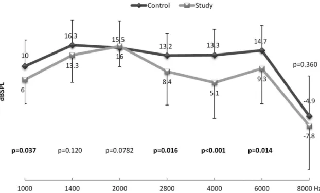
Figure 1 shows the mean amplitudes of DPOAE per frequency studied in newborns in the control group (without hypoxia) and in the study group (with mild or moderate hypoxia). Statistical difference can be observed in f2 for 1,000, 2,800, 4,000, and
6,000 Hz, when comparing the two groups, indicating a decrease in the amplitude of DPOAE in the study group, that is, in newborns with mild and/or moderate hypoxia.
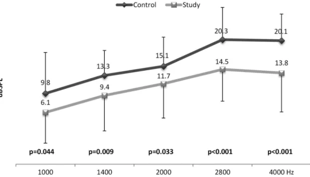
With respect to the amplitudes of TEOAEs, Figure 2 shows that the comparison of the control and study groups showed a statistical difference in all tested frequency ratios.
DISCUSSION
Studies indicate that the incidence of hypoxia in newborn infants can vary between 3 and 6 per 1,000 births(16,17). Although
the occurrence of hypoxia is signiicantly high, it is observed
that some of these newborns have other associated impairments.
It is important to report that there was some dificulty in
the composition of the study group’s sample (mild to moderate hypoxia) due to the occurrence of other risk factors for hearing loss, according to the criteria(1,2). According to the exclusion
Table 1. Descriptive analysis and p-value of the characterization data of the study population regarding their age in days, birth weight, and value of the Apgar score at the 1st and 5th minutes of birth
Group Mean Median Standard
Deviation Minimum Maximum N p-value
Age in days Control 9.7 7 7.1 2 33 25 0.782
Study 10.4 8.5 7.2 5 32 12
Birth weight (g) Control 3273.0 3040.0 389.9 2740 4030 25 0.658
Study 3467.5 3422.5 355.6 2755 3910 12
Apgar 1st min Control 8.7 9 0.6 8 10 25 <0.001
Study 5.8 7 1.6 4 7 12
Apgar 5th min Control 9.7 10 0.5 9 10 25 <0.001
Study 8.7 9 0.5 8 9 12
Caption: N = number of participants
criteria of this study, the presence of these indicators would hamper the tests, as they may alter the results of the examination. The present results could be more robust if the study group population was higher.
According to the publication by the JCIH(1), in 2007, a mild to moderate degree of hypoxia is not considered an indicative factor of hearing loss. However, the results of this study and of the DPOAE and TEOAE tests (Figures 1 and 2, respectively) indicate that there is a decrease in levels of OAE responses in newborns exposed to oxygen deprivation. The decrease in the level of OAE seems to indicate the presence of alterations in the functioning of the outer hair cells, the main receivers of acoustic signals in the inner ear.
Similar results were reported describing the difference when comparing cases with and without hypoxia(18-21). In a study that investigated the effects of hypoxia in the inner ear in adults through DPOAEs, the authors(18) observed a signiicant decrease in the amplitude of DPOAE during hypoxia in 5 of the 16 people tested. The destabilization of the level of DPOAE with
considerable luctuations during hypoxia was observed in nine
individuals. Thus, the authors concluded that the observed results seem to be characteristic of metabolic cochlear disturbances caused by hypoxia.
The decrease in the level of responses of OAEs demonstrated by this research indicates changes in the functioning of the outer hair cells resulting from hypoxia. Such changes show that the slightest oxygen deprivation can really affect the hearing of children(16).
The sensitivity of the outer hair cells to hypoxia and their death in the presence of such risk has been described previously(10). Temporary changes in the threshold can be found and are associated with the fact that the human body focuses on organs such as the brain during changes in the homeostasis of
the newborn. The fact that the cochlea is located near the brain may explain hearing preservation and recovery of the hearing threshold. The monitoring of the hearing of children exposed to such risks is necessary.
One study that explored the long-term effects of hypoxia in the hearing of animals using Auditory Brainstem Responses (ABRs)(18) and another study(20) that examined the DPOAEs in children with a history of hypoxia also showed results similar to this study. While hearing, evaluation methods used were different and different parts of the auditory pathway have been examined; the similarity of the results suggests that the effects of hypoxia can occur both at the level of the outer hair cells and of the brainstem.
It is important to note that the main differences observed were in the 2,800 and 4,000 (p < 0.001) frequency ratios for the TEOAE (Figure 2) and, for f2, were in the 4.000 (p < 0.001) frequency ratio for the DPOAE (Figure 1). Thus, the cochlear
region that differed between the groups was deined tonotopically
between 2,500 and 4,000 Hz.
It is also interesting to note that this frequency range has been extensively researched in studies investigating hearing loss induced
by noise, since the irst frequencies that present hearing loss on
the audiogram are 3,000, 4,000, and/or 6,000 Hz(22,23). Clearly, the paths that lead the exposure to noise to generate cochlear lesions are numerous and complex. Continuous exposure to excessive noise has been described for damaging the cochlear hair cells, initially in the outer hair cells in the most basal portion of the cochlea and, in more severe degrees, also affecting the inner hair cells(22). The partial oxygen pressure in the perilymph and
endolymph and the cochlear blood low were found decreased in
guinea pigs exposed to loud noises(24,25). Hsu et al.(26) suggested that cochlear injury induced by intense sound would be made by two different mechanisms: mechanical and metabolic(26).
The authors explain that the acoustic overstimulation could incite metabolic activity, resulting in depletion of energy reserves, which would be related to a decrease in enzyme activity and energy production, leading to dysfunction and morphological lesions in the cochlea. These metabolic pathways observed in noise-induced hearing loss can be closely related to cellular hypoxia and ischemia, since hair cells are highly energy demanding(24,25).
These changes can occur in the entire Corti, but the hair cells located in the basal portions of the cochlea seem to be the most vulnerable and seem to be affected initially(22). Considering the above and assuming cochlear vulnerability in this region, it is possible that the most robust differences in the 2,800 and 4,000 frequencies of infants with hypoxia may arise from similar processes directly related to the decreased oxygen available during mild to moderate deprivation.
In experiments with newborn mice(27), researchers found that cochlear alterations caused by hypoxia and/or ischemia jointly affected the outer and inner hair cells, but the change initially affected the inner hair cells, and then, with an increasing hypoxia degree, expanded to the outer ones. Cheng et al.(28) observed a decrease in the number of hair cells in mice after 10 hours of hypoxia in an in vitro experiment, and also warn that both the outer and the inner hair cells were considered equally susceptible to hypoxia, and the cell loss increased from the apex to the base.
If we consider the fact that inner hair cells can be damaged more easily than the outer in the presence of hypoxia, and noting that there is currently no clinical test that can measure small lesions of these cells, the monitoring of these infants is extremely necessary because these structures play a key role in auditory sensory transduction.
In this study, newborn infants exposed to mild and moderate hypoxia presented responses to the OAEs in lower amplitude than newborns without hypoxia, pointing to possible changes in the functioning of the outer hair cells resulting from oxygen deprivation. It is extremely important to consider that all newborns assessed in this study achieved a “Pass” result in the neonatal
hearing screening, that is, in a more supericial assessment, which
is the proposal of a screening program, these newborns were within normality criteria. However, given these results, whether the presence of mild to moderate hypoxia should therefore be considered as a predisposing factor for hearing impairment, requiring proper monitoring, aiming to monitor the auditory and language development, is up to discussion.
Obviously, as explained above, a limitation of the study was a reduced casuistry in the study group, since isolating the presence of mild and/or moderate perinatal hypoxia from other
hearing risks was very dificult. We intend to continue with
the study, in an attempt to make the current data more robust.
CONCLUSION
Based on the results, it can be concluded that the OAEs were lower in the study group than in the control group, with
the largest differences and statistical signiicance located in the
2,800-, 4,000-, and 6,000-Hz frequencies in the DPOAE and in all frequency rations in the TEOAE. Although the occurrence of
mild and moderate neonatal hypoxia is not considered a risk for hearing loss, the slightest oxygen deprivation during the time of neonatal hypoxia appears to interfere in the functioning of the outer hair cells and, consequently, in the response level of otoacoustic emissions.
Thus, the longitudinal follow-up of these infants is necessary in order to identify the potential impact of these results on language acquisition and, later, on academic performance.
REFERENCES
1. Joint Committee on Infant Hearing. Year 2007 position statement: principles and guidelines for early hearing detection and intervention program. Audiol Today. 2007;12:7-27.
2. Lewis DR, Marone SAM, Mendes BCA, Cruz OLM, Nóbrega M. Comitê multiprofissional em saúde auditiva - COMUSA. Braz J Otorhinolaryngol [Internet] 2010 [citado em 2013 Mar 15];76(1):121-8. Disponível em: www.audiologiabrasil.org.br
3. Crawford JS. Apgar score and neonatal asphyxia. Lancet. 1982;1(8273):684-5. PMid:6121993.
4. Nuttall A, Lawrence M. Endocochlear potential and scala media oxygen tension during partial anoxia. Am J Otolaryngol. 1980;1(2):147-53. http:// dx.doi.org/10.1016/S0196-0709(80)80008-1. PMid:7446837.
5. Zaconeta CAM. Asfixia perinatal. In: Margotto PR. Assistência ao recém nascido de risco. 2. ed. Rio de Janeiro: Anchieta; 2004. p. 1255. 6. Kumar V, Abbas AK, Fausto N, Aster JC. Respostas Celulares ao estresse
e aos estímulos tóxicos: adaptação, lesão e morte. In: Kumar V, Abbas AK, Fausto N, Aster JC. Patologia: bases patológicas das doenças. 8. ed. Rio de Janeiro: Elsevier; 2010. p. 6-42.
7. Kim JS, Lopez I, Dipatre PL, Liu F, Ishiyama A, Baloh RW. Internal auditory artery infarction:clinicopathologic correlation. Neurology. 1999;52(1):40-4. http://dx.doi.org/10.1212/WNL.52.1.40. PMid:9921846.
8. Tsuji S, Tabuchi K, Hara A, Kusakari J. Long-term observations on the reversibility of cochlear dysfunction after transient ischemia. Hear Res. 2002;166(1-2):72-81. http://dx.doi.org/10.1016/S0378-5955(02)00299-X. PMid:12062760.
9. Koga K, Hakuba N, Watanabe F, Shudou M, Nakagawa T, Gyo K. Transient cochlear ischemia causes delayed cell death in the organ of Corti: an experimental study in gerbils. J Comp Neurol. 2003.456:105-11. 10. Amarjargal N, Andreeva N, Gross J, Haupt H, Fuchs J, Szczepek AJ, et al.
Differential vulnerability of outer and inner hair cells during and after oxygen-glucose deprivation in organotypic cultures of newborn rats. Physiol Res. 2009;58(6):895-902. PMid:19093732.
11. Morales TM, Garcia MAP. Apoptose em otorrinolaringologia. In: Sih T, coordenador. VI Manual de otorrinolaringologia pediátrica da IAPO. 1. ed. São Paulo: Lis Gráfica e Editora; 2006. p. 40-4.
12. Reif R, Qin J, Shi L, Dziennis S, Zhi Z, Nuttall AL, et al. Monitoring hypoxia induced changes in cochlear blood flow and hemoglobin concentration using a combined dual-wavelength laser speckle contrast imaging and doppler optical microangiography system. PLoS One. 2012;7(12):e52041. http:// dx.doi.org/10.1371/journal.pone.0052041. PMid:23272205.
13. Dziennis S, Reif R, Zhi Z, Nuttall AL, Wang RK. Effects of hypoxia on cochlear blood flow in mice evaluated using Doppler optical microangiography. J Biomed Opt. 2012;17(10):106003. http://dx.doi. org/10.1117/1.JBO.17.10.106003. PMid:23224002.
14. Yoshikawa S, Ikeda K, Kudo T, Kobayashi T. The effects of hypoxia, premature birth, infection, ototoxic drugs, circulatory system and congenital disease on neonatal hearing loss. Auris Nasus Larynx. 2004;31:361-8. PMID: 15571908.
15. Sohmer H, Freeman S, Malachie S. Multi-modality evoked potentials in hypoxemia. Electroencephalogr Clin Neurophysiol. 1989;73:328-33. 16. Jiang ZD, Zang Z, Wilkinson AR. Cochlear function in 1-year-old term
17. Cruz ACS, Ceccon MEJ. Prevalência de asfixia perinatal e encefalopatia hipóxico-isquêmica em recém-nascidos de termo considerando dois critérios diagnósticos. Rev Bras Cresc Desenvol Hum. 2010;20:302-16.
18. Kisser U, Becker S, Feddersen B, Fischer R, Fesl G, Haegler K, et al. Complex level alterations of the 2f1–f2 distortion product due to hypoxia. Auris Nasus Larynx. 2014;41(1):37-40. http://dx.doi.org/10.1016/j. anl.2013.07.009. PMid:23921076.
19. Daniel SJ, Mcintosh M, Akinpelu OV, Rohlicek CV. Hearing outcome of early postnatal exposure to hypoxia in Sprague–Dawley rats. J Laryngol Otol. 2014;128(04):331-5. http://dx.doi.org/10.1017/S002221511300265X. PMid:24735907.
20. Zang Z, Wilkinson AR, Jiang ZD. Distorcion product otoacustic emissions at 6 months in term infants after prenatal hypoxia-ischaemia or with low Apgar score. Eur J Pediatr. 2008;167(5):575-8. http://dx.doi.org/10.1007/ s00431-007-0511-2. PMid:17541637.
21. Olzowy B, von Gleichenstein G, Canis M, Plesnila N, Strieth S, Deppe C, et al. Level alterations of the 2f (1)-f (2) distortion product due to hypoxia in the guinea pig depend on the stimulus frequency. Eur Arch Otorhinolaryngol. 2010;267(3):351-5. http://dx.doi.org/10.1007/s00405-009-1052-2. PMid:19629511.
22. Henderson D, Bielefeld EC, Harris KC, Hu BH. The role of oxidative stress in noise-induced hearing loss. Ear Hear. 2006;27(1):1-19. http:// dx.doi.org/10.1097/01.aud.0000191942.36672.f3. PMid:16446561. 23. Konings A, van Laer L, van Camp G. Genetic studies on noise-induced
hearing loss: a review. Ear Hear. 2009;30(2):151-9. http://dx.doi.org/10.1097/ AUD.0b013e3181987080. PMid:19194285.
24. Thorne PR, Nuttall AL. Laser Doppler measurements of cochlear blood flow during loud sound exposure in the guinea pig. Hear Res. 1987;7(1):1-10. http://dx.doi.org/1987;7(1):1-10.1016/0378-5955(87)90021-9. PMid:2953704. 25. Thorne PR, Nuttall AL. Alterations in oxygenation of cochlear endolymph
during loud sound exposure. Acta Otolaryngol. 1989;107(1-2):71-9. http:// dx.doi.org/10.3109/00016488909127481. PMid:2929318.
26. Hsu CJ, Shau WY, Chen YS, Liu TC, Lin-Shiau SY. Activities of Naþ,Kþ-ATPase and Ca2þ-Naþ,Kþ-ATPase in cochlear lateral wall after acoustic trauma. Hear Res. 2000;142:203-11. http://dx.doi.org/10.1016/S0378-5955(00)00020-4. PMid:10748339.
27. Mazurek B, Winter E, Fuchs J, Haupt H, Gross J. Susceptibility of the hair cells of the newborn rat cochlea to hypoxia and ischemia. Hear Res. 2003;182(1-2):2-8. http://dx.doi.org/10.1016/S0378-5955(03)00134-5. PMid:12948595.
28. Cheng AG, Huang T, Stracher A, Kim A, Liu W, Malgrange B, et al. Calpain inhibitors protect auditory sensory cells from hypoxia and neurotrophin-withdrawal induced apoptosis. Brain Res. 1999;850(1-2):234-43. http:// dx.doi.org/10.1016/S0006-8993(99)01983-6. PMid:10629769.
Author contributions
BCB participated in the study conception and design, drafting of the project, data analysis and interpretation, drafting, and reviewing of the manuscript; JNL participated in the study design and drafting of the project, data collection,

