Keywords
Myocardial ischemia, neovascularization, physiologic, endothelial cells, adult.
Mailing adress: Renato Abdala Karam Kalil •
Av. Princesa Isabel, 370 - 90620-000 – Santana, Porto Alegre, RS, Brazil E-mail: [email protected], [email protected] Manuscript received March 24, 2011; revised manuscript received June 13, 2011; accepted July 04, 2011.
Coronary Angiogenesis as an Endogenous Response to Myocardial
Ischemia in Adults
Gabriel Lorier
1, Cristina Touriño
1, Renato A. K. Kalil
2,3Universidad de La Republica, Montevideo, Uruguay (UDELAR)1; Instituto de Cardiologia do Rio Grande do Sul - Fundação Universitária de
Cardiologia - (IC/FUC)2; Universidade Federal de Ciências da Saúde de Porto Alegre - (UFCSPA)3, Porto Alegre, Rio Grande do Sul, Brazil
Abstract
The process of angiogenesis involves a complex sequence of stimuli and integrated responses, such as stimulation of endothelial cells (ECs) for their proliferation and migration, stimulation of the extracellular matrix (ECM) for the attraction of pericytes and macrophages, stimulation of smooth muscle cells for their proliferation and migration, and formation of new vascular structures.
Angiogenesis is mainly an adaptive response to tissue hypoxia and depends on the accumulation of the hypoxia-inducible factor (HIF-1α) in the ischemic myocardial area, which increases the transcription of the vascular endothelial growth factor (VEGF) and its receptors VEGF-R by the ECs undergoing ischemia.
Those steps involve enzymatic mechanisms and plasminogen activator proteases, metalloproteinases (MMP) of the ECM, and kinases that cause proteolytic molecular degradation of the ECM and activation and release of growth factors, such as: basic fibroblast growth factor (bFGF), VEGF, and insulin growth factor-1 (IGF-1). In the intermediate phase, stabilization of the immature neovascular sprout occurs. The final phase is characterized by vascular maturation of the physiological angiogenesis.
In conclusion, coronary angiogenesis in adults is fundamentally a paracrine response of the preexisting capillary network under pathophysiological condition of ischemia and inflammation.
Introduction
This review article approaches endogenous myocardial angiogenesis in the adult considering a unified view of several molecular mechanisms. Such view is based on the “dynamic” interpretation of Folkman’s definition:
“Angiogenesis is the generation and expansion of blood vessels from a preexisting vascular network under endogenous or exogenous stimuli”1. In the adult,
angiogenesis is mainly an adaptive response to tissue hypoxia and occurs in a wide variety of situations, ranging from embryonic development to tumor growth2.
Mediating the cascade of cell and molecular events is the activation of the hypoxia-inducible factor (HIF-1), which increases the transcription of vascular endothelial growth factors (VEGF) and their receptors (VEGF-R) by endothelial cells (ECs) undergoing ischemia3.
The preexisting capillary network is believed to be the common site of beginning in the development and generation of new capillaries, mediated by different signal systems that trigger the production of growth factors and that induce the proliferation of endothelial cells. Folkman’s definition of angiogenesis is based on solid anatomical and morphometric elements.
The normal capillary/myocardial fiber ratio in the adult heart is 1:1, meaning one capillary to one myocardial fiber. The maximum diameter of new vessels identified in the angiogenesis process is 200-μm4. If there is no
epicardial coronary circulation to feed that collateral circulation with adequate blood flow, gradual regression of the angiogenesis process is likely to occur. Myocardial fibers have diameters and lengths ranging from 10 to 25 μm and 50 to 100 μm, respectively, and uniform capillary distribution in the left ventricular wall from 3,000 to 4,000/cm2 5.
Thus, that definition has strong morphometric support, meaning that, in the normal adult heart at rest, a source of nutrients has to exist to maintain cell life. In other words, circulating endothelial progenitor cells (cEPCs) require preexisting coronary circulation to get to the site where myocardial ischemia occurs.
I. Regulation Phases of the Endogenous
Myocardial-Angiogenesis-Inducing
Mechanisms in the Adult:
1. Initial phase: myocardial ischemia as the initial stimulus
Angiogenesis is mainly an adaptive response to tissue hypoxia and depends on the accumulation of HIF-1α, which, under tissue hypoxia conditions, activates the expression of growth factors and initiates the process of angiogenesis in the ischemic zone from the preexisting coronary network6.
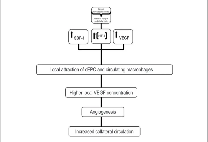
In addition, the attraction and recruitment of cEPCs and macrophages activate, in the paracrine form, angiogenesis in the area7. The VEGF-A expression indicates recruitment of
myeloid progenitors in the ischemic myocardium, while the perivascular cells of angiogenesis are mediated by stromal-cell-derived factors (SDF)-1 (Figure 1)8.
In ischemic heart disease, both HIF-1α and VEGF-A are present in the atherosclerotic plaque, suggesting that HIF, through angiogenic signaling, is directly involved in the atherosclerotic plaque growth9.
Severe myocardial ischemia leads to accumulation of HIF-1 and the expression of factors that stimulate angiogenic sprouts in the proximities of blood vessels in the ischemic myocardium. It is worth noting that severe and persistent ischemia causes
apoptosis of the ECs and myocardial fibers. In addition, myeloid and endothelial bone-marrow-derived progenitors are also recruited to actively support angiogenesis10.
In the myocardium, HIF-1α is clearly important for angiogenesis. However, little is known about HIF-2α. Cardiomyocytes deprived from specific HIF-1α led to a reduction in vascularization, alterations in the energetic metabolism, and abnormality in the contractile function. On the other hand, overexpression of the HIF-1α/VP16 fusion protein (fusion protein containing HIF-1α and herpes virus VP16 sequences) could promote angiogenesis and reduce the size of the infarction in a rat experimental model. Similarly to that which occurs in other hypoxic tissues, HIF-1α is essential to increase the concentration of EC precursors in the bone marrow. Even a partial deficiency of HIF-1α, due to aging, is associated with a significant reduction in the concentration of those cells and with poor vascularization in the ischemic muscle11.
Hypoxemia also regulates the VEGF production and expression, because of the increase in HIF-1 transcription and in mRNA region-3-dependent VEGF stability11.
The molecular mechanism starting angiogenesis can be triggered by deprivation of oxygen and nutrients, such as glucose, which regulate gene expression. In addition, hypoxia regulates TIE-2 receptors, modulators of the VEGF
Figure 1 – Illustration of the determinant molecular and pathophysiological mechanisms of myocardial angiogenesis secondary to ischemia.
Severe myocardial ischemia
Isquemic injury of endothelial cells
SDF-1
HIF-1VEGF
Local attraction of cEPC and circulating macrophages
Higher local VEGF concentration
Angiogenesis
activity, leading ECs to proliferate through the mechanisms of sprouting or division. According to the signal pattern of VEGF and TIE-2, angiogenesis can be induced by sprouts or intussusception12. An important fact regarding the
therapeutic application of VEGF is that ischemia stimulates its expression and that of its receptors13. (Figure 1)
1.1. Mechanism of angiogenesis through the attraction of circulating stem cells during myocardial acute infarction
During myocardial ischemia, the increase in expression and stabilization of HIF-1 transcription promotes local production and release of SDF-1 and VEGF-A by ischemic ECs14. Circulating HIF regulates several genes of angiogenesis,
and VEGF induction is probably the most remarkable. HIF increases the local VEGF concentration in up to 30 times in a few minutes. VEGF stimulates physiological and pathological angiogenesis in a strict dose-dependent response6. Those
mediators can increase the mobilization and recruitment of cEPCs in the ischemic area15.
The increase in the SDF-1 expression is essential to regulate the selective attraction, migration and retention, in the ischemic myocardium, of cEPCs or ECs with the surface chemokine receptor type 4, CXC-R416. This means
that the HIF-1 pathway is the unifying mechanism of the angiogenesis signaling system in the context of acute myocardial infarction (Figure 1).
Those cEPCs, similarly to that which occurs with bone marrow mesenchymal cells and others, are mobilized, attracted and redirected from the peripheral circulation to myocardial ischemic areas17.
There is evidence that SDF-1 along with signal receptor CXC-R4+ play an important role in the recruitment of circulating bone marrow cells in the context of severe myocardial ischemia. That signal is essential for the recruitment of stem cells in the heart. CXC-R4+ is a cell-surface receptor of SDF-1, expressed on cEPCs and on circulating hematopoeitic stem-cells18.
Circulating endothelial progenitor cells play an important role in endothelial vascular wall maintenance, aiding in reendothelialization (physiological change in ECs) and angiogenesis19.
However, endogenous SDF-1 levels decrease after an AMI, returning to normal four to seven days after the AMI20.
Circulating bone marrow-derived mesenchymal cells constitute a bridge to the interior of the ischemic myocardium, expressed by SDF-1, which can be a facilitating signal to the migration of circulating and bone marrow cells17.
Endothelial precursors were believed to exist only during embryonic life. Currently, precursors are known to exist in the bone marrow and peripheral vessels in adults. Some growth factors, such as the granulocyte colony stimulating factor (GCSF), basic fibroblast growth factor (bFGF), and insulin growth factor 1 (IGF-1), stimulate their differentiation and mobilization21. Those endothelial cell
precursors colonize new areas of angiogenic sprouts in adults, and, in the future, can be therapeutic targets.
Rehman et al22 have questioned the origin of those cEPCs
and their true incorporation into the wall of growing vessels. Those authors have reported that most cEPCs derive from monocytes/macrophages, with only a small population of CD-34+ stem cells/hematopoeitic progenitor cells originating
from stem cells/angioblasts. Those monocytes/macrophages secret multiple angiogenic growth factors, which promote, with their paracrine stimulation, neovascular growth22.
The concept of cEPCs is very attractive; however, more than one decade after their discovery, specific markers for cEPCs still lack, and the results of the clinical trials are still controversial. A recent study has shown, by use of a proteomic approach, that cells with a cEPC phenotype could be circulating mononuclear cells with the incorporation of platelet microparticles23.
Schmeisser et al24 have shown that CD-34+ monocytes
can develop in vitro an endothelial phenotype and acquire a tubular form, suggesting a potential role of monocytes in angiogenesis24.
Finally, Ziegelhoeffer et al25 have reported that, in an
adult organism, bone marrow-derived stem cells do not promote vascular growth through their incorporation into vascular walls, but rather through multiple paracrine effects of angiogenic cytokines25.
Briefly: angiogenesis is a physiological mechanism organized under strict regulation, with several factors influencing the active process at molecular level, including several soluble polypeptides, such as VEGF, angiopoietin (Ang), FGF, platelet-derived growth factor (PDGF), transforming growth factor β (TGF-β), tumor necrosis factor (TNF)-α, and GCSF26.
1.2. Mechanism of endogenous angiogenesis through resident cardiac stem cell activation
The true role of the mechanism of endogenous angiogenesis through resident cardiac stem cell activation and of the quantification of its angiogenic effect is uncertain. Resident cardiac stem cells were able to differentiate into cardiomyocytes, endothelial cells, and smooth muscle cells27. Those resident cardiac stem cells express receptors
for hepatocyte growth factor (HGF) and for IGF-1, which can lead to the recruitment and proliferation of ECs and restore some functions of the ischemically injured heart, similarly to that which occurs with circulating mesenchymal cells, which can secret HGF and IGF-1 in response to injury28.
1.3. Proliferation of endothelial cells as the common
inal response to myocardial ischemia: generation of the
angiogenic vascular sprout
In adults, that process is mainly due to hypoxemia and mediated by the activation of HIF-1α, that increases the transcription of VEGF and its receptors, VEGFRs2.
The VEGF stimulates physiological and pathological angiogenesis on a strict dose-dependent relation29. The
tyrosine-kinase receptors (RTKs), VEGFR-1 (Flt-1) and VEGFR-2 (KDR, Flk-1). The Flt-1 tyrosine-kinase receptors (VEGFR-1) are the second highest-affinity receptors for VEGF. Although not so specific, Flk-1 receptors do not have an affinity with cells of the inflammatory system, such as monocytes or cancer cells, unlike the Flt-1 receptor. A recent and elegant study of the Leuven University has made an important discovery with therapeutic implications for the future, which is the relative specificity of VEGF-B regarding its angiogenic activity in ischemic myocardial tissue31.
Regarding the mechanism of angiogenesis, because of the vascular growth in the form of a vascular sprout, the following steps could be determined:
a) initial vasodilation, a process that involves nitric oxide and VEGF32;
b) increased vascular permeability in response to VEGF, such as plasma protein extravasation, which functions as a support for EC migration. The increase in permeability is produced by the formation of organo-vesicular fenestrations and redistribution of platelet endothelial cell adhesion molecules (PECAMs)33;
c) proteolytic degradation of the extracellular matrix (ECM): proteinases expose proteins of the degraded ECM, such as collagen IV and fibrillar collagen monomer, which induce migration of ECs and smooth muscle cells (SMCs)34.
The three steps described involve enzyme mechanisms that include Ang-2, a TIE-2 inhibitor35, plasminogen
activator proteases, matrix metalloproteinases (MMP), and kinases or heparinase family.
All proteases influence angiogenesis through proteolytic molecular degradation of the ECM and activation and release of growth factors, such as bFGF, VEGF, and IGF1, sequestered in the ECM (box 8 of Figure 2)36. In addition,
proteolytic action is also present in the transforming growth factor (TGF) and proteolytic activation of angiogenic chemokines, such as IL-1. During vascular remodeling through sprouting, proteolytic degradation occurs, involving plasminogen activators, such as urokinase plasminogen activator (UPA) and its inhibitor PAI-1, MMPs, and inhibitors of tissue MMPs (TIMPs), heparinases, kinases, tyrosinases, and cadherins37.
Analyzing the critical role of the ECM proteolytic degradation in the growth and maintenance of the angiogenic sprout, the time limits of that process should be considered. In addition, the process should be balanced, because insufficient degradation prevents vascular cells from leaving their original positions. On the other hand, excessive degradation hinders the support and orientation for EC migration, and, consequently, also angiogenesis38.
In the angiogenesis process, the ECM proteolytic degradation is followed by fragmentation of the basal
Figure 2 – Formation of the primary vascular plexus of the mesoderm, during the embryonic phase. Hemangioblasts and the bipotentiality of their precursors represent the intermediate step of endothelial precursors (angioblasts); in the embryonic phase, they form the primitive vascular network called vasculogenesis, and, in adult life, they expand and remodel, leading to angiogenesis (Boxes 3 and 4). EC: endothelial cells. Box 5 shows the stabilization phase, represented by endothelial cell binding under the action of VEGF and PDGF-β, which recruit pericytes and smooth muscle cells, covering endothelial cells during vessel stabilization. Angiopoietin-1 (Ang-1) and TGF-β1 stabilize the emerging vessel. Ang-2, in the absence of growth factors, causes regression of the emerging sprout. Box 6 illustrates intussusceptive and sprouting angiogenesis. Boxes 7 and 8 illustrate the remodeling and maturation phases of the emerging vessel. SMC: smooth muscle cells; PCT: pericytes. Binding of VEGF and endothelial cells, PDGF-β recruits pericytes. Adapted from Carmeliet.
1. Mesodermal precursor
Mesodermal inductors
VEGF-R2• VEGF-R2•
b-FGF
VEGF-R2 Ang1 VEGF
2. Hemangioblast 3. Angioblast 4. EC grouping 5. Stabilization
6. Angiogenesis
Vascular sprout 1. Vascular sprout:
Bridges SMC/PCT
Mature vascular
system
PDGFβ-R TGFβ-R
EC
7. Remodeling 8. Maturation
SMC
Mc Tie-2
PDGFR-B PDGF-8B
INTUSCEPÇÃO INTUSCEPÇÃO:
VEGF-R2 VEGF-R1 TIE-2/TIE-1
TGF βR
VEGF-R2 VEGF-R1
CML
TGF-βR
membrane, and then by chemotactic migration of the ECs through the basal membrane. That requires cleavage of endothelial intercellular connections. When the ECs migrate to form new sprouts, the ECM undergoes proteolytic degradation and changes in its composition (Figure 3).
Migration of the ECs occurs 24 hour before the following processes:
a) proliferation;
b) adhesion and reestablishment of intracellular contacts; c) formation of the vascular lumen;
d) functional maturation of the endothelium.
As shown, proteases play a critical role in the angiogenesis process39.
The VEGF stimulation of the tyrosine-kinase membrane receptors of the ECs is expressed through Src genes of the tyrosine-kinase family, such as Src, Fyn, and Yes, which regulate multiple intracellular functions. Each of the Src, Fyn and Yes genes plays a unique role in the VEGF mitogenic signaling
system. They contribute to VEGF modulation in cell migration. The Fyn gene plays a unique role in VEGF induction for the formation of the neovascular tube, having a negative regulatory effect on the migration and stabilization of the VEGF-induced neovascular tube. Those results provide direct evidence that the Src, Fyn and Yes genes play an important role in VEGF regulation in EC-mediated events40.
The existence of tubes with pericytes reflects the early beginning of the participation of pericytes in intussusceptive angiogenesis (box 4 of Figure 2). The creation of a functional vascular network requires that emerging vascular sprouts mature and remain functional over time. The association of pericytes and SMC with the recently formed EC sprouts regulates their proliferation, survival, migration, differentiation, vascular branching, blood flow, and vascular permeability. The early participation of the pericytes in the development of the microvasculature has important implications for the therapeutic intervention in several diseases41.
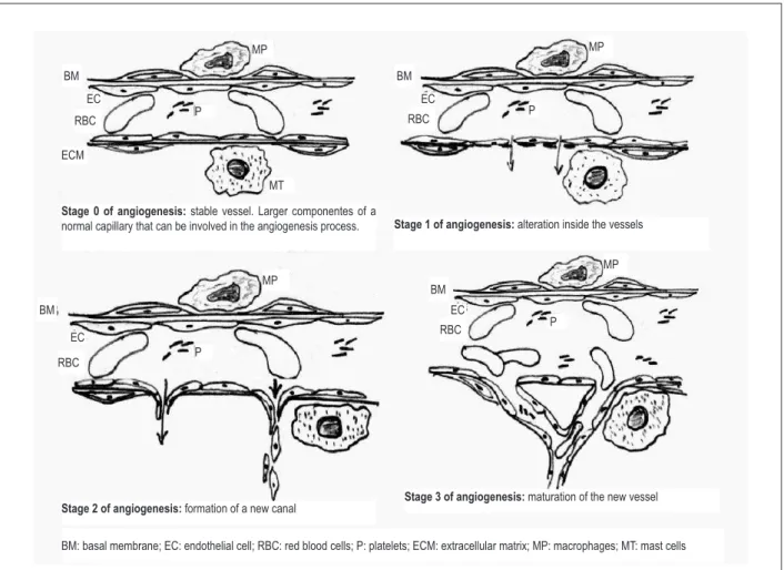
Figure 3 – Detailed draft of angiogenesis showing: stage 1: vasodilation, activation of endothelial cells, activation of platelets, secretion of plasminogen activators and proteolytic enzymes, degranulation of mast cells, activation of macrophages, rupture of the basal membrane, and increased permeability with passage of ibrin and other proteins; stage 2: formation of pseudopodia, degradation of the extracellular matrix, migration of endothelial cells to the extravascular space with their proliferation and formation of vascular sprouts; and stage 3: new basal membrane and maturation of the new vascular wall for establishing blood low, formation of tubes, connections, and new vessels.
Stage 0 of angiogenesis: stable vessel. Larger componentes of a
normal capillary that can be involved in the angiogenesis process. Stage 1 of angiogenesis: alteration inside the vessels
Stage 2 of angiogenesis: formation of a new canal Stage 3 of angiogenesis: maturation of the new vessel
BM: basal membrane; EC: endothelial cell; RBC: red blood cells; P: platelets; ECM: extracellular matrix; MP: macrophages; MT: mast cells
BM BM
BM
BM
EC
EC ECM
MT
MP MP
MP MP
RBC RBC
RBC
RBC
EC EC
P P
P
Blood flow, exerting pressure on the new vascular wall, interacts in an integrated and dynamic way with the cytoskeleton and ECM. Continuous blood flow stimulates the proliferation of ECs and regulates VEGF, integrin aVb3, PECAM-1, and VE-cadherin. Shear stress stimulates EC proliferation, and, thus, increases the diameter of the vessel42.
The ECM provides the necessary contacts between the ECs and surrounding tissues, preventing the degeneration of the neovascular sprouts. A matrix of interstitial collagen and elastin between the vascular cells provide viscoelasticity and resistance to the vascular wall. The ECM also regulates the formation of the new vascular sprout. When the ECs migrate to form new vascular sprouts, the matrix is not only proteolytically cleaved, but its composition is altered as well. Proteinases expose new epitopes in the ECM proteins (for example, collagen IV) or alter their structure (fibrillar collagen monomers), leading to migration of ECs and SMCs34. In
addition, fibronectin, fibrin and other components of the matrix provide support and orientation for ECs.
Integrins are specific cell surface receptors of the ECM, which, through transmission of bidirectional information between the exterior and interior of the vascular cells, help in constructing new vascular sprouts coordinately. Integrins
α5β3 and α5β5 have long been considered a positive regulator of angiogenesis, because their pharmacological antagonists suppress pathological angiogenesis43.
Platelet-derived growth factor-β and its receptor (PDGF-Rβ) play an essential role in the stabilization of new blood vessels, recruiting PDGF-Rβ-positive mesenchymal cells. The lack of EC recruitment results in pathological neovascular growth, which causes: neovascular permeability changes; neovascular fragility; bleeding; insufficient perfusion; and hypoxia in embryos lacking PDGF-β44. However, a combination of
PDGF-β and VEGF results in the formation of more mature vascular sprouts than that obtained with monotherapy with any of the factors. That is extremely important for the future development of therapeutic strategies of angiogenesis.
Another signaling system involved in the maintenance, growth, and stabilization of the neovascular sprout is the TIE-2 receptor, which binds Ang-1 and Ang-TIE-2. Differently from Ang-2, which activates TIE-2 in some cells, but blocks TIE-2 in other cells, Ang-1 consistently activates the TIE-2 receptor. On its turn, Ang-1 compacts the neovascular sprouts through binding molecules and causes the interaction of mural ECs, as a protein adhesive, and the contraction of pericytes. The angiogenic activity of Ang-2 is synergic with VEGF to stimulate cardiac angiogenesis, but when the signals are not sufficient, it causes the death of the ECs and regression of the neovascular sprout. The TIE-2 activation signs should function as a precision scale45.
2. Intermediate phase: stabilization of the
immature neovascular sprout
The neovascular sprout is stabilized through the recruitment of mural cells and generation of a new ECM. At least four molecular pathways are involved in the regulation of that process: PDGF and PDGF-Rβ; sphingosine-1-phosphate-1
(S1P1); endothelial differentiation factor [sphingosine G-1 protein receptor (EDG1)]; Ang-1-TIE-2; and TGF. The PDGF-β is secreted by the EC, probably in response to VEGF. Although PDGF-β is expressed by ECs and mural cells, the latter are responsible during the maturation phase of angiogenesis. In addition, the TIE-1 and TIE-2 receptors and the two TIE-2 ligands, Ang-1 and Ang-2, are essential for the formation and stabilization of the neovascular sprout. The major sources of Ang-1 and Ang-2 are the mural and specific organ ECs, respectively. Angiopoietin-1 is known due to its ability to stabilize the emerging vascular sprouts and to make them resistant to evasion between intercellular connections. In the absence of VEGF, Ang-2 acts as an Ang-1 antagonist and destabilizes the emerging neovascular sprout, eventually leading to its regression. In the presence of VEGF, Ang-2 facilitates angiogenesis. The TGF-β1 is a multifunctional cytokine that promotes the maturation cycle of the developing neovascular sprout through ECM stimulation, by stimulating the induction and differentiation of mesenchymal cells into mural ECs46.
3. Final phase: vascular maturation of
physiological angiogenesis
The molecular determinants of neovascular maturation can be grouped into three categories: I) growth factor binding to a cell receptor type, and its corresponding effect; II) molecular regulation of cell interactions; III) molecular regulation of the interactions between ECs and ECM.
I) Growth factor binding to a cell receptor type, and its corresponding effect:
a) EC receptors Flt-1 and Flk-1 (VEGF-R1, VEGF-R2): 1) Regulation of proteases in the ECM organization; 2) Generation of temporary ECM, which allows permeability increase; 3) Regulation of PDGF-β, which recruits cells to stabilize the emerging vessel wall; 4) Suppression of apoptosis in emerging vessels; 5) Stabilization of ECs.
b) PDGF-Rβ; Ang-1/TIE-2: 1) Promotion of proliferation, migration and recruitment of mural cells; 2) Stabilization of emerging vessels, which facilitates the interactions between intercellular junctions (EC-EC, EC-ECM); 3) Suppression of apoptosis of ECs.
c) Ang-2/TIE-1: induce apoptosis of ECs, in the absence of VEGF.
d) Ang-1/TIE-1: coordinate vascular polarity. e) TGF-β1/TGF-βRII:
1) Promote the production of proteases and ECM. 2) Promote the differentiation of fibroblasts into myofibroblasts.
f) TGF-β1/ALK1: regulate the proliferation and migration of ECs.
g) TGF-β1/ALK5: regulate the maturation of new vessels. h) TGF-β1/ALK1 and endoglin: promote arterial and venous specialization.
b) N-cadherin: connect the cells of the vascular wall to ECs. c) Connectins: establish connections between ECs and ECs and mural cells.
III) Molecular regulation of the interactions between ECs and ECM:
a) α5β1, α1β1, α2β1, αvβ3, αvβ5: suppression of EC apoptosis.
b) Proteases: their enzymatic action promotes the production of the ECM, such as cleavage of the collagen XVIII to endostatin, plasminogen, angiotensin and proteases (for example, MMP2 and PEX).
c) Inhibitory proteases: prevent the ECM proteolytic cleavage to stabilize the vessel.
Such data consistently support the hypothesis that Ang-2 and VEGF lead to the formation of neovascular sprouts, while Ang-1 is involved in stabilizing the sprout through the interaction of ECs and mural cells46.
3.1. Formation of the vascular lumen
During the angiogenesis process, EC migration and proliferation occur for approximately 24 hours, a period during which proteases play a crucial role46. Proliferation is recorded
at the level of ECs and SMCs47.
The process of the vascular lumen formation is a coordinate phase of molecular-cellular adhesion during blood vessel extension. The specific molecules responsible for vessel extension are not well known48.
The receptor of TIE-2 and EC is necessary for the development of blood vessels, as well as for their maintenance and repair. Angiopoietin was identified as a specific ligand for the TIE-2 receptor. As already cited, Ang-1 is an agonist of TIE-2 receptors that promotes EC survival and reduces vascular permeability49.
Unlike Ang-1, Ang-2 has been initially described as an antagonist that blocks TIE-2/Ang-1 and activates TIE-2, which results in EC apoptosis. In the signaling system of ECs, for their survival and migration, TIE-2 is an essential component for the development of embryonic vasculogenesis, as well as for angiogenesis in the adult. Recent studies have identified protein tyrosine phosphatase beta (HPTP-β) as a new specific EC-phosphatase that regulates the Ang-1-TIE-2 signaling of ECs. Thus, the strategies oriented to HPTP-β can benefit Ang-1 signaling, and, thus, improve the formation and maturation of neovessels in the context of therapeutic angiogenesis. In addition, one can conclude that hypoxia increases HPTP-β expression and decreases TIE-2 phosphorylation, shedding new light on the potential role of HPTP-β on angiogenesis. The induction of HPTP-β due to hypoxia can represent an important regulatory mechanism of TIE-2 activation in pathological angiogenesis50.
Briefly, ECs have a selective tyrosine-kinase receptor; TIE-2 and its ligands, Ang-1 and Ang-2, are essential for the maintenance and repair of blood vessels. Ang-1 is an agonist of the activation of TIE-2 receptors. However, Ang-2 is context-dependent, which means, it can be either antagonist or agonist. In the survival of ECs, HPTP-β plays a relevant role modulating Ang-1-TIE-2 signaling.
3.2. Neovascular permeability
In the angiogenesis process, VEGF leads to proliferation and migration of ECs. It was initially identified as a vascular permeability factor (VPF). There is plenty of evidence suggesting that angiogenesis is preceded and/or accompanied by increased permeability4.
The mechanisms by which VEGF/VPF increases vascular permeability (VP) remain unknown. Murohara et al51 have
identified nitric oxide (NO) in VP regulation, while van der Zee et al52 have identified, in vitro, that VEGF/VPF stimulates
NO production in the macrovascular endothelium.
Isner4 has shown the synergic action between NO and
prostacyclins produced by the interaction of VEGF/VPF with its receptor Flk-1/KDR/VEGF-R2 as a mediator of the VEGF/ VPF-induced vascular permeability, constituting a unique property of VEGF/VPF among the angiogenic cytokines. When the proteases were assessed based on their capacity to increase permeability by the Miles test, VEGF was the only to increase permeability – the mechanism of action, however, is unknown. Nitric oxide has been implicated as a permeability regulator factor, depending on its local concentration4.
There are three different types of vascular permeability: 1) vascular basal membrane permeability (VBMP), present in normal tissues;
2) increased vascular permeability in response to a single and brief exposure to VEGF-A or other agents;
3) chronic increase in vascular permeability, which characterizes pathological angiogenesis.
Finally, VEGF-A varies significantly in different rat strains, and the permeability response, both basal and induced by some factors, is likely to differ in mice with different genetic structures, although systematic investigation still lacks53.
II. Responses of Vascular Cells to Hypoxia
Hypoxia leads to increased signaling through HIF-1transcription.1. Endothelial cells
There are two mechanisms identified in hypoxemia that are linked to angiogenesis or its block: one that promotes proliferation and survival of ECs through the activation of VEGF receptors and eNOS signaling and expression; and another that initiates apoptosis. The results depend on the severity of hypoxia. Moderate hypoxemia mainly promotes proliferation and survival, while severe hypoxemia, close to anaerobic conditions, can cause significant apoptosis. In vivo, the accumulation of HIF resulting from EC apoptosis has not been reported6.
2) Vascular smooth muscle cells
might act as an accessory molecule of the proteolytic activity associated with the PDGF-β receptor45.
3) Macrophages
In response to tissue hypoxemia, a large number of monocytes are recruited from the circulation to hypoxic tissues, where they differentiate into macrophages. Several factors and proteins in hypoxic tissues contribute to the recruitment of monocytes as follows: monocyte chemotactic protein-1 (MCP-1); TNF-a; colony stimulating factor-1 (CSF-1); SDF-1, and VEGF-A. Macrophages promote angiogenesis by stimulating the secretion of MMPs and several angiogenic factors, such as VEGF-A, FGF, interleukin-2, and TNF. In addition, macrophages also secret a short peptide with 39 amino acid residues (PR39), which can easily cross the plasma membrane, enter the cells, and inhibit HIF-α cleavage. That PR39 function can improve the capacity of the resident cells of hypoxic tissue to secret angiogenic factors, because of the increase in the local HIF-1 concentration6.
Finally, macrophages respond to hypoxemia through the production of an oxygen-regulated protein (ORP) by the endoplasmic reticulum and the secretion of VEGF-A by the same endoplasmic reticulum6.
Conclusion
In general, coronary angiogenesis in adults is fundamentally a potent paracrine response of the preexisting capillary network to pathophysiological conditions of ischemia and/ or inflammation. Basic research and understanding the mechanisms are required to fill in the knowledge gaps and enable the establishment of effective strategies in the field of coronary angiogenic therapy.
Potential Conflict of Interest
No potential conflict of interest relevant to this article was reported.
Sources of Funding
There were no external funding sources for this study.
Study Association
This article is part of the thesis of Doctoral submitted by Gabriel Lorier, from IC/FUC.
References
1. Kornowski R, Epstein SE, Leon MB. Handbook of myocardial revascularization and angiogenesis. London: Martin Dunitz Ltd; 1999.
2. Folkman J, Shing Y. Angiogenesis. J Biol Chem. 1992;267(16):10931-4. 3. Schaper W, Buschmann I. Arteriogenesis, the good and bad of it. Cardiovasc
Res. 1999;43(4):835-7.
4. Isner JM. Angiogenesis. In: Topol EJ, ed. Textbook of cardiovascular medicine. Philadelphia: Lippincott Williams & Wilkins; 1998. p. 2491-518.
5. Gottschall C. Função cardíaca: da normalidade à insuficiência. São Paulo: BYK; 1995.
6. Pugh CW, Ratcliffe PJ. Regulation of angiogenesis by hypoxia: role of the HIF system. Nat Med. 2003;9(6):677-84.
7. Kocher AA, Schuster MD, Szabolcs MJ, Takuma S, Burkhoff D, Wang J, et al. Neovascularization of ischemic myocardium by human bone-marrow-derived angioblasts prevents cardiomyocyte apoptosis, reduces remodeling and improves cardiac function. Nat Med. 2001;7(4):430-6.
8. Grunewald M, Avraham I, Dor Y, Bachar-Lustig E, Itin A, Jung S, et al. VEGF-induced adult neovascularization: recruitment, retention, and role of accessory cells. Cell. 2006;124(1):175-89.
9. Vink A, Schoneveld AH, Lamers D, Houben AJ, van der Groep P, van Diest PJ, et al. HIF-1 alpha expression is associated with an atheromatous inflammatory plaque phenotype and upregulated in activated macrophages. Atherosclerosis. 2007;195(2):e69-75.
10. Banai S, Shweiki D, Pinson A, Chandra M, Lazarovici G, Keshet E. Upregulation of vascular endothelial growth factor expression induced by myocardial ischaemia: implications for coronary angiogenesis. Cardiovasc Res. 1994;28(8):1176-9.
11. Semenza GL. Transcriptional regulation by hypoxia-inducible factor 1 molecular mechanisms of oxygen homeostasis. Trends Cardiovasc Med. 1996;6(5):151-7. 12. Risau W. Mechanisms of angiogenesis. Nature. 1997;386(6626):671-4. 13. H e n r y T D. C a n w e r e a l l y g r o w n e w b l o o d v e s s e l s ? L a n c e t .
1998;351(9119):1826-7.
14. Ceradini DJ, Kulkarni AR, Callaghan MJ, Tepper OM, Bastidas N, Kleinman ME, et al. Progenitor cell trafficking is regulated by hypoxic gradients through HIF-1 induction of SDF-1. Nat Med. 2004;10(8):858-64. 15. Kalka C, Tehrani H, Laudenberg B, Vale PR, Isner JM, Asahara T, et al.
VEGF gene transfer mobilizes endothelial progenitor cells in patients with inoperable coronary disease. Ann Thorac Surg. 2000;70(3):829-34. 16. Kleinman ME, Greives MR, Churgin SS, Blechman KM, Chang EI, Ceradini
DJ, et al. Hypoxia-induced mediators of stem/progenitor cell trafficking are increased in children with hemangioma. Arterioscler Thromb Vasc Biol. 2007;27(12):2664-70.
17. Tang YL, Zhao Q, Qin X, Shen L, Cheng L, Ge J, et al. Paracrine action enhances the effects of autologous mesenchymal stem cell transplantation on vascular regeneration in rat model of myocardial infarction. Ann Thorac Surg. 2005;80(1):229-36.
18. Yamaguchi J, Kusano KF, Masuo O, Kawamoto A, Silver M, Murasawa S, et al. Stromal cell-derived factor-1 effects on ex vivo expanded endothelial progenitor cell recruitment for ischemic neovascularization. Circulation. 2003;107(9):1322-8.
19. Jo DY, Hwang JH, Kim JM, Yun HJ, Kim S. Human bone marrow endothelial cells elaborate non-stromal-cell-derived factor-1 (SDF-1)-dependent chemoattraction and SDF-1-dependent transmigration of haematopoietic progenitors. Br J Haematol. 2003;121(4):649-52.
20. Abbott JD, Huang Y, Liu D, Hickey R, Krause DS, Giordano FJ. Stromal cell-derived factor-1alpha plays a critical role in stem cell recruitment to the heart after myocardial infarction but is not sufficient to induce homing in the absence of injury. Circulation. 2004;110(21):3300-5.
21. Pettersson A, Nagy JA, Brown LF, Sundberg C, Morgan E, Jungles S, et al. Heterogeneity of the angiogenic response induced in different normal adult tissues by vascular permeability factor/vascular endothelial growth factor. Lab Invest. 2000;80(1):99-115.
23. Prokopi M, Pula G, Mayr U, Devue C, Gallagher J, Xiao Q, et al. Proteomic analysis reveals presence of platelet microparticles in endothelial progenitor cell cultures. Blood. 2009;114(3):723-32.
24. Schmeisser A, Garlichs CD, Zhang H, Eskafi S, Graffy C, Ludwig J, et al. Monocytes coexpress endothelial and macrophagocytic lineage markers and form cord-like structures in Matrigel under angiogenic conditions. Cardiovasc Res. 2001;49(3):671-80.
25. Ziegelhoeffer T, Fernandez B, Kostin S, Heil M, Voswinckel R, Helisch A, et al. Bone marrow-derived cells do not incorporate into the adult growing vasculature. Circ Res. 2004;94(2):230-8.
26. Papetti M, Herman IM. Mechanisms of normal and tumor-derived angiogenesis. Am J Physiol Cell Physiol. 2002;282(5):C947-70.
27. Beltrami AP, Barlucchi L, Torella D, Baker M, Limana F, Chimenti S, et al. Adult cardiac stem cells are multipotent and support myocardial regeneration. Cell. 2003;114(6):763-76.
28. Linke A, Muller P, Nurzynska D, Casarsa C, Torella D, Nascimbene A, et al. Stem cells in the dog heart are self-renewing, clonogenic, and multipotent and regenerate infarcted myocardium, improving cardiac function. Proc Natl Acad Sci USA. 2005;102(25):8966-71.
29. Ferrara N, Kerbel RS. Angiogenesis as a therapeutic target. Nature. 2005;438(7070):967-74.
30. Ferrara N, Davis-Smyth T. The biology of vascular endothelial growth factor. Endocr Rev. 1997;18(1):4-25.
31. Ferrara N, Gerber HP, LeCouter J. The biology of VEGF and its receptors. Nat Med. 2003;9(6):669-76.
32. Carmeliet P. Mechanisms of angiogenesis and arteriogenesis. Nat Med. 2000;6(4):389-95.
33. Taipale J, Makinen T, Arighi E, Kukk E, Karkkainen M, Alitalo K. Vascular endothelial growth factor receptor-3. Curr Top Microbiol Immunol. 1999;237:85-96.
34. Hangai M, Kitaya N, Xu J, Chan CK, Kim JJ, Werb Z, et al. Matrix metalloproteinase-9-dependent exposure of a cryptic migratory control site in collagen is required before retinal angiogenesis. Am J Pathol. 2002;161(4):1429-37.
35. Carmeliet P. Developmental biology: controlling the cellular brakes. Nature. 1999;401(6754):657-8.
36. Carmeliet P. Fibroblast growth factor-1 stimulates branching and survival of myocardial arteries: a goal for therapeutic angiogenesis? Circ Res. 2000;87(3):176-8.
37. Pepper MS. Extracellular proteolysis and angiogenesis. Thromb Haemost. 2001;86(1):346-55.
38. Luttun A, Dewerchin M, Collen D, Carmeliet P. The role of proteinases in angiogenesis, heart development, restenosis, atherosclerosis, myocardial ischemia, and stroke: insights from genetic studies. Curr Atheroscler Rep. 2000;2(5):407-16.
39. Isner JM, Takayuki A. Therapeutic angiogenesis. Front Biosci. 1998;3:e49-69. 40. Werdich XQ, Penn JS. Src, Fyn and Yes play differential roles in
VEGF-mediated endothelial cell events. Angiogenesis. 2005;8(4):315-26. 41. Ozerdem U, Stallcup WB. Early contribution of pericytes to angiogenic
sprouting and tube formation. Angiogenesis. 2003;6(3):241-9. 42. Tomanek RJ. Formation of the coronary vasculature during development.
Angiogenesis. 2005;8(3):273-84.
43. Hynes RO. A reevaluation of integrins as regulators of angiogenesis. Nat Med. 2002;8(9):918-21.
44. Montrucchio G, Alloatti G, Camussi G. Role of platelet-activating factor in cardiovascular pathophysiology. Physiol Rev. 2000;80(4):1669-99. 45. Werner S, Grose R. Regulation of wound healing by growth factors and
cytokines. Physiol Rev. 2003;83(3):835-70.
46. Jain RK. Molecular regulation of vessel maturation. Nat Med. 2003;9(6):685-93.
47. Bauters C, Asahara T, Zheng LP, Takeshita S, Bunting S, Ferrara N, et al. Physiological assessment of augmented vascularity induced by VEGF in ischemic rabbit hindlimb. Am J Physiol. 1994;267(4 Pt 2):H1263-71. 48. Chang MW, Barr E, Lu MM, Barton K, Leiden JM. Adenovirus-mediated
over-expression of the cyclin/cyclin-dependent kinase inhibitor, p21 inhibits vascular smooth muscle cell proliferation and neointima formation in the rat carotid artery model of balloon angioplasty. J Clin Invest. 1995;96(5):2260-8.
49. Isner JM, Walsh K, Symes J, Pieczek A, Takeshita S, Lowry J, et al. Arterial gene therapy for therapeutic angiogenesis in patients with peripheral artery disease. Circulation. 1995;91(11):2687-92.
50. Yacyshyn OK, Lai PF, Forse K, Teichert-Kuliszewska K, Jurasz P, Stewart DJ. Tyrosine phosphatase beta regulates angiopoietin-Tie2 signaling in human endothelial cells. Angiogenesis. 2009;12(1):25-33.
51. Murohara T, Horowitz JR, Silver M, Tsurumi Y, Chen D, Sullivan A, et al. Vascular endothelial growth factor/vascular permeability factor enhances vascular permeability via nitric oxide and prostacyclin. Circulation. 1998;97(1):99-107.
52. van der Zee R, Murohara T, Luo Z, Zollmann F, Passeri J, Lekutat C, et al. Vascular endothelial growth factor/vascular permeability factor augments nitric oxide release from quiescent rabbit and human vascular endothellium. Circulation. 1997;95(4):1030-7.